. 1998 Feb 16;187(4):547-60.
doi: 10.1084/jem.187.4.547.

Association of phosphorylated serine/arginine (SR) splicing factors with the U1-small ribonucleoprotein (snRNP) autoantigen complex accompanies apoptotic cell death

Affiliations

Association of phosphorylated serine/arginine (SR) splicing factors with the U1-small ribonucleoprotein (snRNP) autoantigen complex accompanies apoptotic cell death

P J Utz et al. J Exp Med. .

Abstract

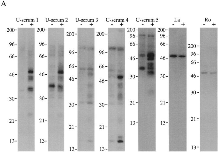
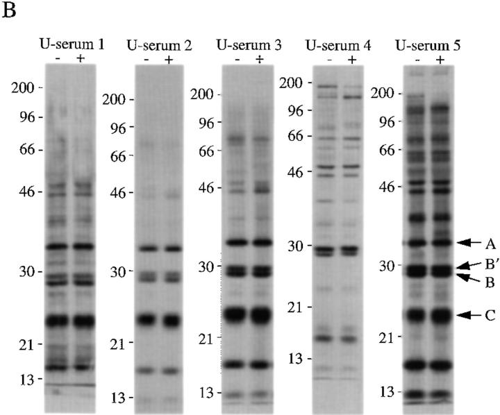
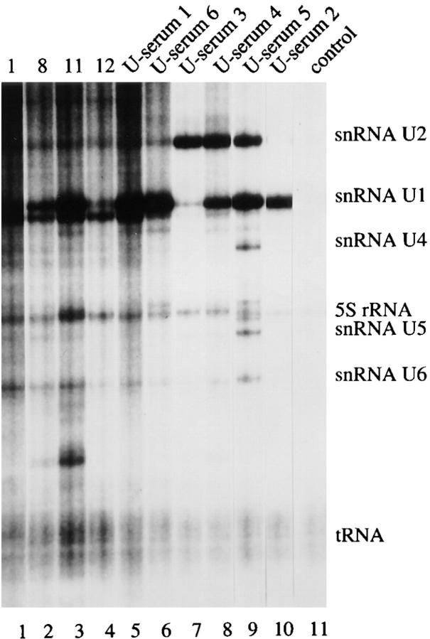
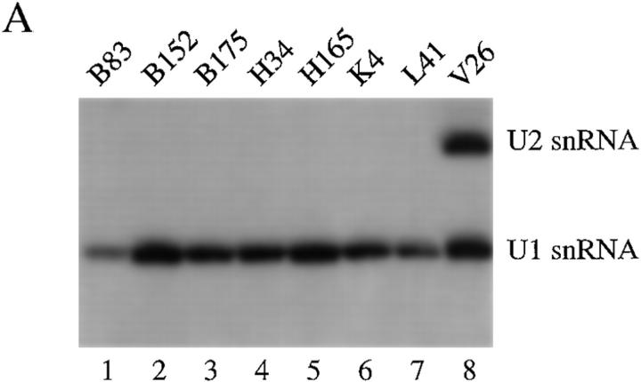
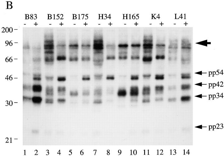
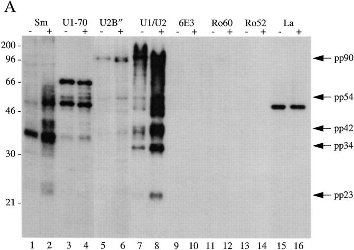
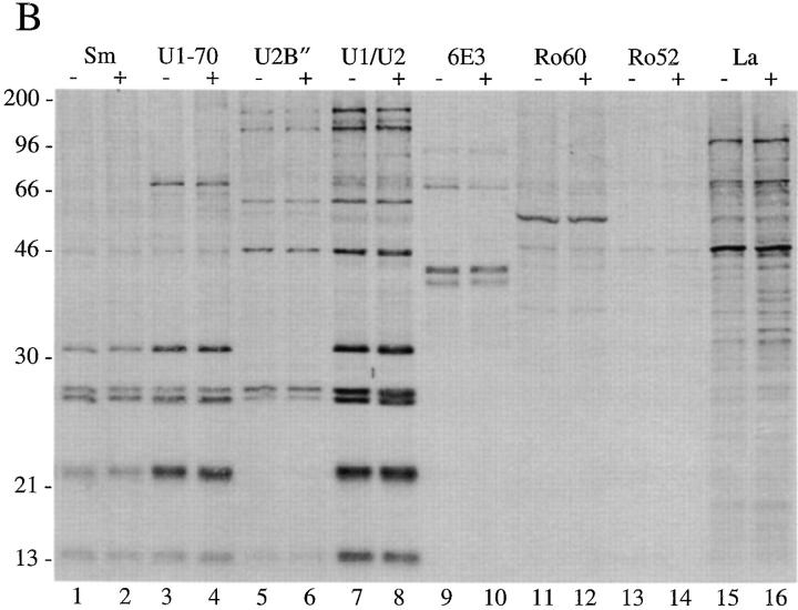
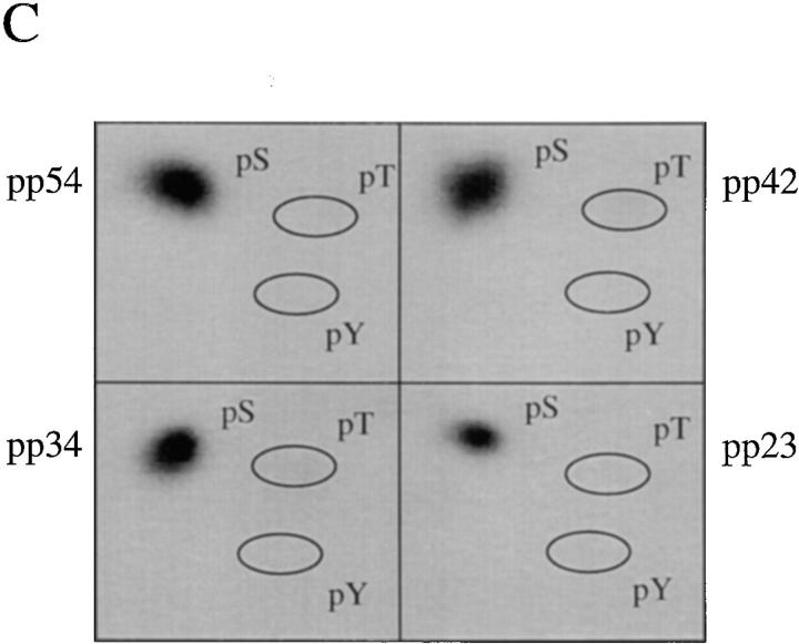
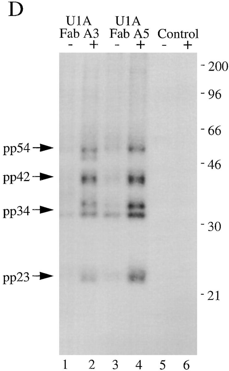
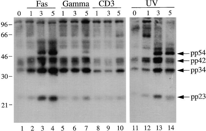
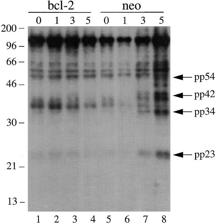
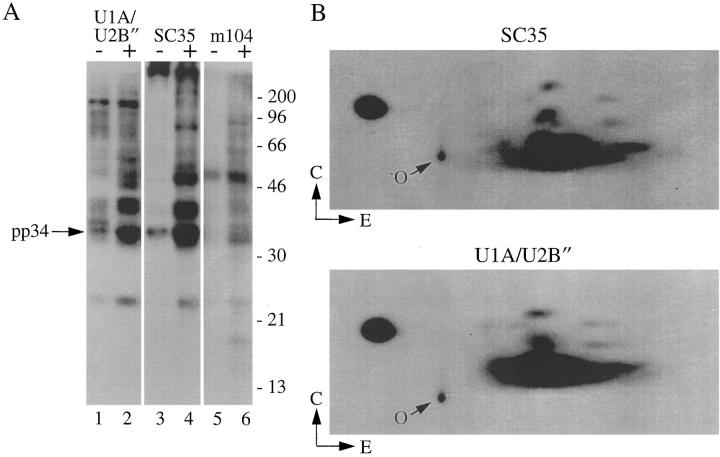
Proteins subject to proteolysis or phosphorylation during apoptosis are commonly precipitated by autoantibodies found in the serum of patients with systemic lupus erythematosus (SLE). We screened a panel of murine monoclonal and human monospecific sera reactive with known autoantigens for their ability to selectively precipitate phosphoproteins from apoptotic Jurkat T cell lysates. Sera known to recognize the U1-small nuclear ribonucleoprotein (snRNP) complex (confirmed by their ability to precipitate U1-snRNA) selectively precipitated a phosphoprotein complex (pp54, pp42, pp34, and pp23) from apoptotic lysates. Monoclonal antibodies reactive with U1-snRNP proteins precipitated the same phosphoprotein complex from apoptotic lysates. The phosphorylation and/or recruitment of these proteins to the U1-snRNP complex is induced by multiple apoptotic stimuli (e.g., Fas ligation, gamma irradiation, or UV irradiation), and is blocked by overexpression of bcl-2. The U1-snRNP-associated phosphoprotein complex is immunoprecipitated by monoclonal antibodies reactive with serine/arginine (SR) proteins that comprise a structurally related family of splicing factors. The association of phosphorylated SR proteins with the U1-snRNP complex in cells undergoing apoptosis suggests a mechanism for regulation of alternative splicing of apoptotic effector molecules.


Figures

Figure 1
Figure 1
Human autoimmune sera specific for U–snRNP complexes precipitate phosphoproteins from apoptotic Jurkat cell lysates. (A) Jurkat cells were labeled with 32P orthophosphate and lysed either before (−) or 3 h after (+) the addition of anti-Fas 7C11. Proteins were then precipitated using the indicated autoimmune serum, separated on a 12% SDS–polyacrylamide gel, transferred to nitrocellulose, and exposed for autoradiography. Individual sera are described in detail in Materials and Methods. U-serum 1, Immunovision antihistone/RNP; U-serum 2, CDC/AF reference serum 4 (anti-U1–RNP); U-serum 3, serum Ga; U-serum 4, serum Ya; U-serum 5, CDC/AF reference serum 5 (anti-Sm). The relative migration of molecular mass markers in kilodaltons is indicated on the left side of the gel. (B) Immunoprecipitation from 35S-labeled Jurkat cells. Jurkat cells were labeled with 35S-methionine and cysteine and lysed either before (−) or 3 h after (+) the addition of anti-Fas 7C11 before immunoprecipitation using sera derived from the indicated patient. Immunoprecipitates were separated on a 12% SDS–polyacrylamide gel, transferred to nitrocellulose, and then subjected to autoradiographic analysis. The relative migration of molecular mass markers in kilodaltons is indicated on the left side of each panel. Bands corresponding to the U–snRNP proteins A, B, B′, and C are indicated on the right side of the panel.
Figure 1
Figure 1
Human autoimmune sera specific for U–snRNP complexes precipitate phosphoproteins from apoptotic Jurkat cell lysates. (A) Jurkat cells were labeled with 32P orthophosphate and lysed either before (−) or 3 h after (+) the addition of anti-Fas 7C11. Proteins were then precipitated using the indicated autoimmune serum, separated on a 12% SDS–polyacrylamide gel, transferred to nitrocellulose, and exposed for autoradiography. Individual sera are described in detail in Materials and Methods. U-serum 1, Immunovision antihistone/RNP; U-serum 2, CDC/AF reference serum 4 (anti-U1–RNP); U-serum 3, serum Ga; U-serum 4, serum Ya; U-serum 5, CDC/AF reference serum 5 (anti-Sm). The relative migration of molecular mass markers in kilodaltons is indicated on the left side of the gel. (B) Immunoprecipitation from 35S-labeled Jurkat cells. Jurkat cells were labeled with 35S-methionine and cysteine and lysed either before (−) or 3 h after (+) the addition of anti-Fas 7C11 before immunoprecipitation using sera derived from the indicated patient. Immunoprecipitates were separated on a 12% SDS–polyacrylamide gel, transferred to nitrocellulose, and then subjected to autoradiographic analysis. The relative migration of molecular mass markers in kilodaltons is indicated on the left side of each panel. Bands corresponding to the U–snRNP proteins A, B, B′, and C are indicated on the right side of the panel.
Figure 2
Figure 2
Coprecipitation of U1–snRNA using selected autoantisera. Jurkat cells were labeled with 32P-orthophosphate and solubilized in NP-40 lysis buffer. After immunoprecipitation with the indicated serum, RNA was extracted and separated on 6% sequencing gels before drying and autoradiographic exposure. The relative migration of known RNA moieties is depicted on the right side of the figure. The serum specificity is indicated above each sample. Lanes are numbered at the bottom of the panel. Lanes 1–4, patients 1, 8, 11, 12 (19); U-serum 1, Immunovision antihistone/RNP; U-serum 2, CDC/AF reference serum 4 (anti-U1–RNP); U-serum 3, serum Ga; U-serum 4, serum Ya; U-serum 5, CDC/AF reference serum 5 (anti-Sm); U-serum 6, anti–U1-70 kD serum (gift of A. Rosen).
Figure 3
Figure 3
U1-specific autoantisera coprecipitate the U1–snRNA molecule and pp54, pp42, pp34, and pp23 from apoptotic extracts. (A) Jurkat cells were labeled with 32P-orthophosphate and lysed in NP-40 lysis buffer. After immunoprecipitation with the indicated serum, RNA was extracted and separated on 6% sequencing gels before drying and autoradiographic exposure. Patient sera specific for the U1–snRNP complex were used in lanes 1–7. A patient serum (V26, lane 8) capable of precipitating both the U1– and U2–snRNPs is shown for comparison. The relative migration of the U1– and U2–snRNAs is depicted on the right side of the figure. (B) Jurkat cells were labeled with 32P-orthophosphate and lysed either before (−) or 3 h after (+) the addition of anti-Fas (7C11) before immunoprecipitation using sera derived from the indicated patient. Immunoprecipitates were separated on a 12% SDS–polyacrylamide gel, transferred to nitrocellulose, and subjected to autoradiographic analysis. Sera correspond to the seven U1-specific autoantisera shown in Fig. 3 A. The relative migration of molecular size markers in kilodaltons is indicated on the left side of the figure. Bands corresponding to pp54, pp42, pp34, and pp23 are shown on the right side of the panel. A high molecular mass complex is indicated with a large arrowhead. Lanes are numbered at the bottom of the figure.
Figure 3
Figure 3
U1-specific autoantisera coprecipitate the U1–snRNA molecule and pp54, pp42, pp34, and pp23 from apoptotic extracts. (A) Jurkat cells were labeled with 32P-orthophosphate and lysed in NP-40 lysis buffer. After immunoprecipitation with the indicated serum, RNA was extracted and separated on 6% sequencing gels before drying and autoradiographic exposure. Patient sera specific for the U1–snRNP complex were used in lanes 1–7. A patient serum (V26, lane 8) capable of precipitating both the U1– and U2–snRNPs is shown for comparison. The relative migration of the U1– and U2–snRNAs is depicted on the right side of the figure. (B) Jurkat cells were labeled with 32P-orthophosphate and lysed either before (−) or 3 h after (+) the addition of anti-Fas (7C11) before immunoprecipitation using sera derived from the indicated patient. Immunoprecipitates were separated on a 12% SDS–polyacrylamide gel, transferred to nitrocellulose, and subjected to autoradiographic analysis. Sera correspond to the seven U1-specific autoantisera shown in Fig. 3 A. The relative migration of molecular size markers in kilodaltons is indicated on the left side of the figure. Bands corresponding to pp54, pp42, pp34, and pp23 are shown on the right side of the panel. A high molecular mass complex is indicated with a large arrowhead. Lanes are numbered at the bottom of the figure.
Figure 4
Figure 4
Monoclonal antibodies directed against U1–snRNP components precipitate pp54, pp42, pp34, and pp23 from extracts prepared from apoptotic Jurkat cells. (A) Jurkat cells were labeled with 32P-orthophosphate and lysed either before (−) or 3 h after (+) the addition of anti-Fas 7C11. Proteins were then precipitated using the indicated autoimmune serum, separated on a 12% SDS–polyacrylamide gel, transferred to nitrocellulose, and then exposed for autoradiography. The relative migration of molecular mass markers in kilodaltons is indicated on the left side of the gel. Bands corresponding to pp90, pp54, pp42, pp34, and pp23 are shown on the right side of the panel. Lanes are numbered at the bottom of the panel. (B) The identical experiment in 35S-labeled Jurkat cells. The relative migration of molecular size markers in kilodaltons is indicated on the left side of the gel. Lanes are numbered at the bottom of the figure. (C) Phosphoamino acid analysis of pp54, pp42, pp34, and pp23. Jurkat cells were labeled with 32P-orthophosphate, treated with the anti-Fas monoclonal antibody 7C11, and solubilized using NP-40 lysis buffer after 3 h. Proteins were then precipitated with the anti-U1A/U2B′′ monoclonal antibody 9A9, separated on a 12% SDS–polyacrylamide gel, transferred to PVDF, and exposed for autoradiography. Individual phosphoproteins were localized on the membrane, excised, and then subjected to acid hydrolysis. Phosphoamino acids were separated by two-dimensional electrophoresis in pH 1.9 buffer in the horizontal dimension, followed by pH 3.5 buffer in the vertical dimension before autoradiographic analysis. Individual proteins are labeled on the side of each panel. Migration of phosphoamino acid standards are labeled with circles as follows: phosphoserine (pS), phosphothreonine (pT), phosphotyrosine (pY). (D) Anti-U1A antibody fragments coprecipitate pp54, pp42, pp34, and pp23 from apoptotic Jurkat cell lysates. Labeled Jurkat cell extracts were prepared as above. Proteins were precipitated using the indicated anti-U1A antibody fragments, separated on a 12% SDS–polyacrylamide gel, transferred to nitrocellulose, and then exposed for autoradiography. The relative migration of molecular mass markers in kilodaltons is indicated on the right side of the gel. Bands corresponding to pp54, pp42, pp34, and pp23 are shown on the left side of the panel.
Figure 4
Figure 4
Monoclonal antibodies directed against U1–snRNP components precipitate pp54, pp42, pp34, and pp23 from extracts prepared from apoptotic Jurkat cells. (A) Jurkat cells were labeled with 32P-orthophosphate and lysed either before (−) or 3 h after (+) the addition of anti-Fas 7C11. Proteins were then precipitated using the indicated autoimmune serum, separated on a 12% SDS–polyacrylamide gel, transferred to nitrocellulose, and then exposed for autoradiography. The relative migration of molecular mass markers in kilodaltons is indicated on the left side of the gel. Bands corresponding to pp90, pp54, pp42, pp34, and pp23 are shown on the right side of the panel. Lanes are numbered at the bottom of the panel. (B) The identical experiment in 35S-labeled Jurkat cells. The relative migration of molecular size markers in kilodaltons is indicated on the left side of the gel. Lanes are numbered at the bottom of the figure. (C) Phosphoamino acid analysis of pp54, pp42, pp34, and pp23. Jurkat cells were labeled with 32P-orthophosphate, treated with the anti-Fas monoclonal antibody 7C11, and solubilized using NP-40 lysis buffer after 3 h. Proteins were then precipitated with the anti-U1A/U2B′′ monoclonal antibody 9A9, separated on a 12% SDS–polyacrylamide gel, transferred to PVDF, and exposed for autoradiography. Individual phosphoproteins were localized on the membrane, excised, and then subjected to acid hydrolysis. Phosphoamino acids were separated by two-dimensional electrophoresis in pH 1.9 buffer in the horizontal dimension, followed by pH 3.5 buffer in the vertical dimension before autoradiographic analysis. Individual proteins are labeled on the side of each panel. Migration of phosphoamino acid standards are labeled with circles as follows: phosphoserine (pS), phosphothreonine (pT), phosphotyrosine (pY). (D) Anti-U1A antibody fragments coprecipitate pp54, pp42, pp34, and pp23 from apoptotic Jurkat cell lysates. Labeled Jurkat cell extracts were prepared as above. Proteins were precipitated using the indicated anti-U1A antibody fragments, separated on a 12% SDS–polyacrylamide gel, transferred to nitrocellulose, and then exposed for autoradiography. The relative migration of molecular mass markers in kilodaltons is indicated on the right side of the gel. Bands corresponding to pp54, pp42, pp34, and pp23 are shown on the left side of the panel.
Figure 5
Figure 5
Phosphoprotein components of the U1–snRNP complex are precipitated after multiple apoptotic stimuli but not an activation stimulus. Jurkat cells were labeled with 32P-orthophosphate, treated with the indicated stimulus, and then solubilized using NP-40 lysis buffer at the indicated times. Proteins were then precipitated with anti-U1A/U2B′′, separated on a 12% SDS–polyacrylamide gel, transferred to nitrocellulose, and then exposed for autoradiography. The apoptotic stimulus is indicated above each panel. The time in hours after each stimulus is indicated above each lane. The relative migration of molecular mass markers in kilodaltons is indicated on the left side of the panel. Bands corresponding to pp54, pp42, pp34, and pp23 are shown on the right side of the panel. Lanes are numbered at the bottom of each panel.
Figure 6
Figure 6
In vivo phosphorylation of U1–snRNP components is inhibited in gamma-irradiated Jurkat cells overexpressing bcl-2. Jurkat (bcl-2) transformants (lanes 1–4) or Jurkat (neo) control transformants (lanes 5–8) were labeled with 32P-orthophosphate, subjected to gamma irradiation, solubilized in NP-40 lysis buffer, precipitated using anti-U1A/U2B′′ antibodies, separated on a 12% SDS–polyacrylamide gel, transferred to nitrocellulose, and then exposed for autoradiography. The relative migration of molecular size markers in kilodaltons is indicated on the left side of the figure. Bands corresponding to pp54, pp42, pp34, and pp23 are shown on the right side of the figure. The time, in hours, from initial exposure to gamma irradiation is indicated at the top of each lane. Lane numbers appear at the bottom of the figure.
Figure 7
Figure 7
Monoclonal antibodies specific for SR proteins precipitate pp54, pp42, pp34, and pp23 from apoptotic Jurkat cell extracts. (A) Jurkat cells were labeled with 32P-orthophosphate and lysed either before (−) or 3 h after (+) the addition of anti-Fas 7C11. Proteins were then precipitated using the indicated monoclonal antibody, separated on a 12% SDS–polyacrylamide gel, transferred to nitrocellulose, and exposed for autoradiography. The relative migration of pp34 is indicated on the left side of the gel. Lanes are numbered at the bottom of the panel. The relative migration of molecular size markers in kilodaltons is indicated on the right side of the figure. (B) Two-dimensional phosphotryptic map of pp34. The 34-kD band from the U1A/U2B′′ immunoprecipitate (lane 2) and the anti-SC35 immunoprecipitate (lane 4) were excised and digested with trypsin. Phosphopeptides were separated electrophoretically at pH 1.9 in the first dimension, and by thin-layer chromatography in the second dimension before autoradiographic exposure. Direction of separation is shown by arrows. E, electrophoresis; C, chromatography; O, origin.

Similar articles

Cited by

References

    1. Astaldi-Ricotti G, Bestagno M, Cerino A, Negri C, Caporali R, Cobianchi F, Longhi M, Montecucco C. Antibodies to hnRNP core protein A1 in connective tissue diseases. J Cell Biochem. 1989;40:43–47. - PubMed
    1. Van Venrooij W, Sillekens P. Small-nuclear RNA-associated proteins: autoantigens in connective tissue diseases. Clin Exp Rheum. 1989;7:635–639. - PubMed
    1. von Muhlen CA, Tan EM. Autoantibodies in the diagnosis of systemic rheumatic diseases. Semin Arthritis Rheum. 1995;24:323–358. - PubMed
    1. Montecucco M, Caporali R, Negri C, deGennaro F, Cerino A, Bestagno M, Cobianchi F, Astaldi-Ricotti G. Antibodies from patients with rheumatoid arthritis and systemic lupus erythematosus recognize different epitopes of a single heterogeneous nuclear RNP core protein. Arthritis Rheum. 1990;33:180–186. - PubMed
    1. LeFeber WP, Norris DA, Ryan SR, Huff JC, Lee LA, Kubo M, Boyce ST, Kotzin BL, Weston WL. Ultraviolet light induces binding of antibodies to selected nuclear antigens on cultured keratinocytes. J Clin Invest. 1984;74:1545–1551. - PMC - PubMed

Publication types

MeSH terms