. 1998 Feb 16;187(4):655-60.
doi: 10.1084/jem.187.4.655.

B cell-attracting chemokine 1, a human CXC chemokine expressed in lymphoid tissues, selectively attracts B lymphocytes via BLR1/CXCR5

Affiliations

B cell-attracting chemokine 1, a human CXC chemokine expressed in lymphoid tissues, selectively attracts B lymphocytes via BLR1/CXCR5

D F Legler et al. J Exp Med. .

Abstract

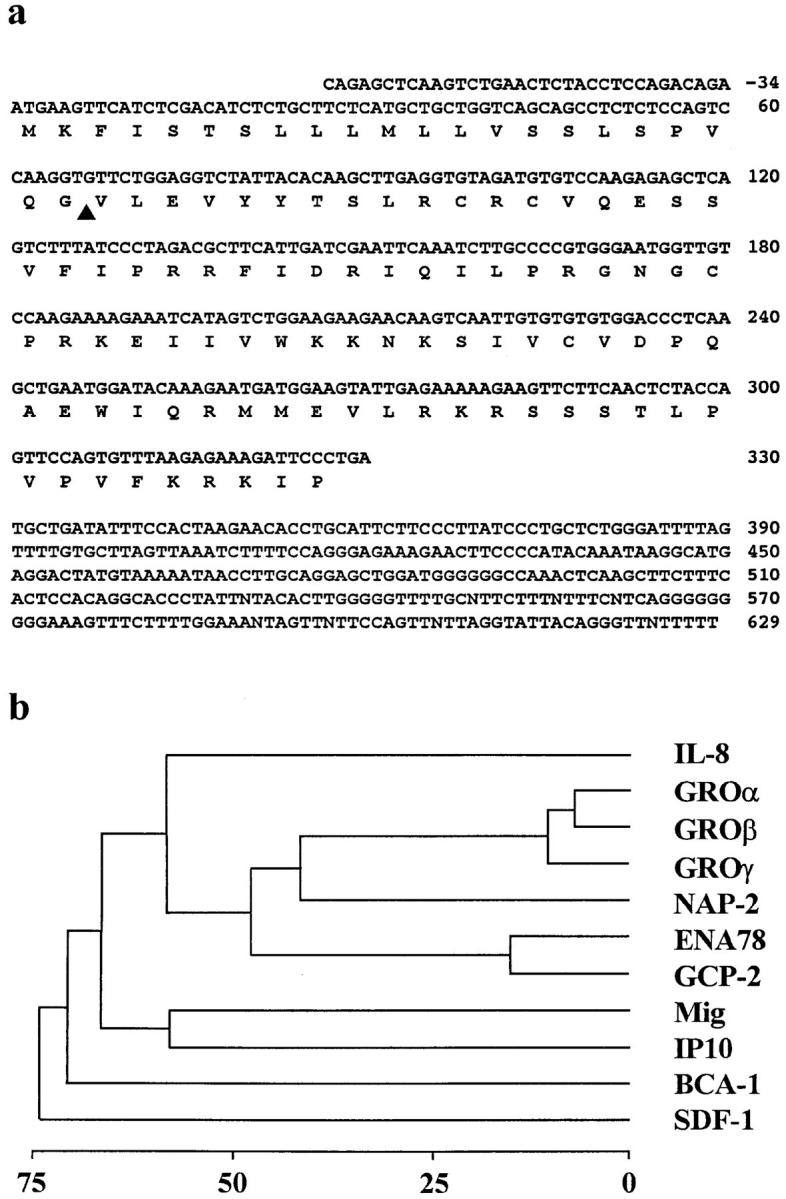
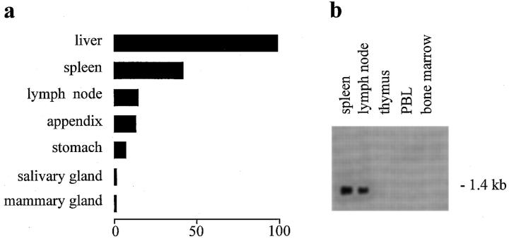
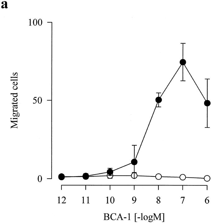
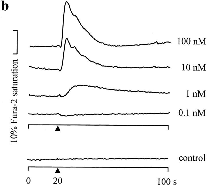
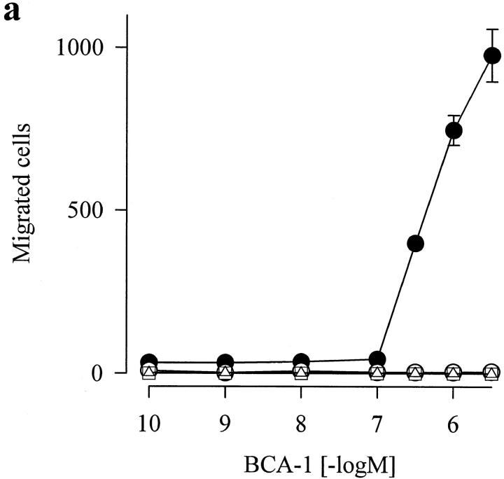
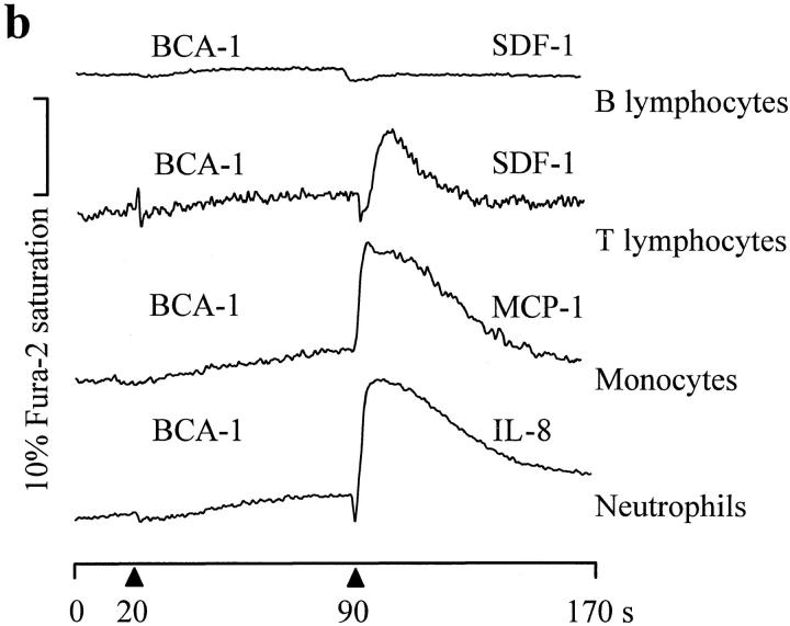
Although most leukocytes, T lymphocytes in particular, respond to several different chemokines, there is virtually no information on chemokine activities and chemokine receptors in B lymphocytes. A putative chemokine receptor, BLR1, that is expressed in Burkitt's lymphoma cells and B lymphocytes was cloned a few years ago. Deletion of the gene for BLR1 yielded mice with abnormal primary follicles and germinal centers of the spleen and Peyer's patches, reflecting the inability of B lymphocytes to migrate into B cell areas. By screening expressed sequence tag DNA sequences, we have identified a CXC chemokine, termed B cell-attracting chemokine 1 (BCA-1), that is chemotactic for human B lymphocytes. BCA-1 cDNA encodes a protein of 109 amino acids with a leader sequence of 22 residues. The mature protein shares 23-34% identical amino acids with known CXC chemokines and is constitutively expressed in secondary lymphoid organs. BCA-1 was chemically synthesized and tested for activity on murine pre-B cells 300-19 transfected with BLR1 and on human blood B lymphocytes. In transfected cells, BCA-1 induced chemotaxis and Ca2+ mobilization demonstrating that it acts via BLR1. Under the same conditions, no activity was obtained with 10 CXC and 19 CC chemokines, lymphotactin, neurotactin/fractalkine and several other peptide ligands. BCA-1 was also a highly effective attractant for human blood B lymphocytes (which express BLR1), but was inactive on freshly isolated or IL-2-stimulated T lymphocytes, monocytes, and neutrophils. In agreement with the nomenclature rules for chemokine receptors, we propose the term CXCR5 for BLR1. Together with the observed disturbance of B cell colonization in BLR1/ CXCR5-deficient mice, the present results indicate that chemotactic recruitment by locally produced BCA-1 is important for the development of B cell areas of secondary lymphoid tissues.


Figures

Figure 1
Figure 1
(a) Nucleotide and deduced amino acid sequences of BCA-1. Numbering starts at the first nucleotide of the start codon. The predicted open reading frame is indicated below the nucleotide sequence, and the putative signal peptide cleavage site is marked by an arrowhead. These sequence data are available from EMBL/Genbank/DDBJ under accession number AJ002211. (b) Phylogenetic tree diagram of human CXC chemokines. Similarity scores of the proteins were determined by average linkage cluster analysis (33). Gap penalty, window size, filtration level, and K-tulpe size parameters for pairwise alignments were set at 3, 10, 2.5, and 1, respectively. Distance to the branching points indicates percent sequence divergence.
Figure 2
Figure 2
Expression of BCA-1 in human tissues. (a) Dot blot analysis using a DNA probe spanning the whole open reading frame of BCA-1. The strongest signal, obtained with liver RNA (140,575 cpm), was taken as 100%, and the relative expression in other tissues was calculated. The lowest significant signal (2,759 cpm) was detected in the mammary gland. Most tissues yielded counts around background (∼50 cpm). Tissue samples that did not express BCA-1 transcripts included total brain, amygdala, caudate nucleus, cerebellum, cerebral cortex, frontal lobe, hippocampus, medulla oblongata, occipital lobe, putamen, substantia nigra, temporal lobe, thalamus, subthalamic nucleus, spinal cord, heart, aorta, skeletal muscle, colon, bladder, uterus, prostate, testis, ovary, pancreas, pituitary gland, adrenal gland, thyroid gland, kidney, small intestine, thymus, blood leukocytes, bone marrow, lung, trachea, placenta, fetal brain, fetal heart, fetal kidney, fetal liver, fetal thymus, and fetal lung. (b) Northern blot of human tissue RNAs hybridized with the same DNA probe.
Figure 3
Figure 3
BCA-1 responses of murine pre–B cells 300-19 stably transfected with BLR1/ CXCR5. (a) Chemotaxis of parental (open circles) and BLR1/ CXCR5-expressing cells (closed circles). Migrated cells (mean counts ± SD of triplicate assays per five high power fields) from one experiment that is representative of a total of six. (b) [Ca2+]i changes in BLR1/CXCR5-expressing 300-19 cells. BCA-1 was added at 0.1, 1, 10, and 100 nM to fura-2–loaded cells (arrowheads), and [Ca2+]i-dependent fluorescence changes were recorded. Parental 300-19 cells (control) were stimulated with 100 nM BCA-1. One out of five independent experiments with identical results is shown.
Figure 3
Figure 3
BCA-1 responses of murine pre–B cells 300-19 stably transfected with BLR1/ CXCR5. (a) Chemotaxis of parental (open circles) and BLR1/ CXCR5-expressing cells (closed circles). Migrated cells (mean counts ± SD of triplicate assays per five high power fields) from one experiment that is representative of a total of six. (b) [Ca2+]i changes in BLR1/CXCR5-expressing 300-19 cells. BCA-1 was added at 0.1, 1, 10, and 100 nM to fura-2–loaded cells (arrowheads), and [Ca2+]i-dependent fluorescence changes were recorded. Parental 300-19 cells (control) were stimulated with 100 nM BCA-1. One out of five independent experiments with identical results is shown.
Figure 4
Figure 4
Functional responses of human blood leukocytes to BCA-1. (a) In vitro chemotaxis of human B lymphocytes (closed circles), T lymphocytes cultured for 19 d in the presence of 250 U/ml IL-2 (open circles), monocytes (open squares), and neutrophils (open triangles) in response to increasing concentrations of BCA-1. (b) [Ca2+]i changes in human blood leukocytes. Fura-2–loaded cells were stimulated sequentially with 100 nM BCA-1 followed by another chemokine, and changes in [Ca2+]i were recorded. The data are representative for four independent experiments.
Figure 4
Figure 4
Functional responses of human blood leukocytes to BCA-1. (a) In vitro chemotaxis of human B lymphocytes (closed circles), T lymphocytes cultured for 19 d in the presence of 250 U/ml IL-2 (open circles), monocytes (open squares), and neutrophils (open triangles) in response to increasing concentrations of BCA-1. (b) [Ca2+]i changes in human blood leukocytes. Fura-2–loaded cells were stimulated sequentially with 100 nM BCA-1 followed by another chemokine, and changes in [Ca2+]i were recorded. The data are representative for four independent experiments.

References

    1. Baggiolini M, Dewald B, Moser B. Human chemokines: an update. Annu Rev Immunol. 1997;15:675–705. - PubMed
    1. Loetscher P, Seitz M, Baggiolini M, Moser B. Interleukin-2 regulates CC chemokine receptor expression and chemotactic responsiveness in T lymphocytes. J Exp Med. 1996;184:569–577. - PMC - PubMed
    1. Imai T, Yoshida T, Baba M, Nishimura M, Kakizaki M, Yoshie O. Molecular cloning of a novel T cell–directed CC chemokine expressed in thymus by signal sequence trap using Epstein-Barr virus vector. J Biol Chem. 1996;271:21514–21521. - PubMed
    1. Yoshida R, Imai T, Hieshima K, Kusuda J, Baba M, Kitaura M, Nishimura M, Kakizaki M, Nomiyama H, Yoshie O. Molecular cloning of a novel human CC chemokine EBI1-ligand chemokine that is a specific functional ligand for EBI1, CCR7. J Biol Chem. 1997;272:13803–13809. - PubMed
    1. Rossi DL, Vicari AP, Franz-Bacon K, McClanahan TK, Zlotnik A. Identification through bioinformatics of two new macrophage proinflammatory human chemokines–MIP-3α and MIP-3β. J Immunol. 1997;158:1033–1036. - PubMed

Publication types

MeSH terms

Associated data