. 1998 Feb;64(2):555-63.
doi: 10.1128/AEM.64.2.555-563.1998.

Molecular characterization and heterologous expression of the gene encoding a low-molecular-mass endoglucanase from Trichoderma reesei QM9414

Affiliations

Molecular characterization and heterologous expression of the gene encoding a low-molecular-mass endoglucanase from Trichoderma reesei QM9414

H Okada et al. Appl Environ Microbiol. 1998 Feb.

Abstract

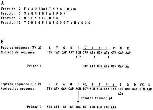
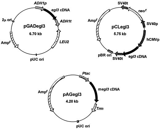
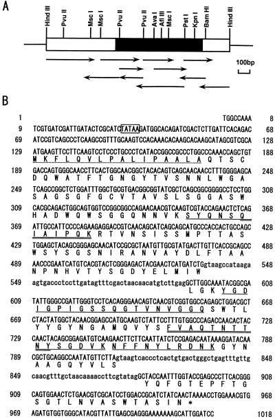
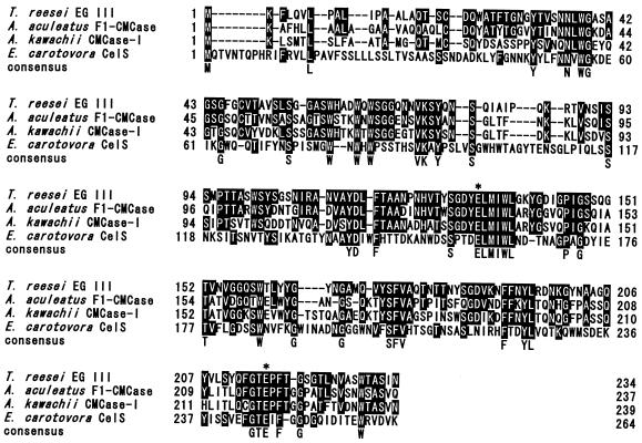
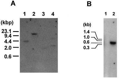
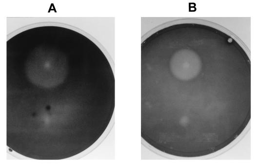
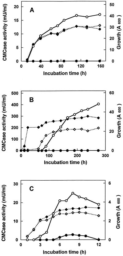
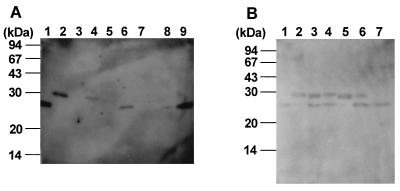
We have isolated the genomic and cDNA clones encoding EG III (a low-molecular-mass endo-beta-1,4-glucanase) gene from Trichoderma reesei QM9414. The nucleotide sequence of the cDNA fragment was verified to contain a 702-bp open reading frame that encodes a 234-amino-acid propeptide. The deduced protein sequence has significant homologies with family H endo-beta-1,4-glucanases. The 16-amino-acid N-terminal sequence was shown to function as a leader peptide for possible secretion. Northern blot analysis showed that the EG III gene transcript, with a length of about 700 bp, was expressed markedly by cellulose but not by glucose. The protein has been expressed as a mature form in Escherichia coli and as secreted forms in Saccharomyces cerevisiae and Schizosaccharomyces pombe under the control of tac, alcohol dehydrogenase (ADH1), and human cytomegalovirus promoters, respectively. The S. cerevisiae and Schizosaccharomyces pombe recombinant strains showed strong cellulolytic activities on agar plates containing carboxymethyl cellulose. The E. coli strain expressed small amounts of EG III in an active form and large amounts of EG III in an inactive form. The molecular masses of the recombinant EG IIIs were estimated to be 25, 28, and 29 kDa for E. coli, S. cerevisiae, and Schizosaccharomyces pombe, respectively, by immunoblot analysis following sodium dodecyl sulfate-polyacryl-amide gel electrophoresis. Parts of the yeast recombinant EG IIIs decreased their molecular masses to 25 kDa after treatment with endoglycosidase H and alpha-mannosidase, suggesting that they are N glycosylated at least partly.


Figures

FIG. 1
FIG. 1
Partial amino acid sequences of the EG III purified from T. reesei and the design of the oligonucleotide primers used in PCR. (A) Amino acid sequences from the peptides obtained by lysylendopeptidase digestion. Fraction numbers indicate the peak numbers of the digested peptides eluted by high-performance liquid chromatography. The uncertain amino acids are in parentheses. (B) Designs of oligonucleotide primers based on the fraction 2 and fraction 3 peptides. The letter N denotes a mixture of all four bases. I, P, and Y denote an inosine, a mixture of adenine and guanine, and cytosine and thymine, respectively. Fr., fraction.
FIG. 2
FIG. 2
Plasmids pGADegl3, pCLegl3, and pAGegl3. Relevant gene locations are indicated. See the text for details on the construction of plasmids. ADH1p and ADH1t, promoter and terminator of the S. cerevisiae ADH1 gene, respectively; LEU2, LEU2 gene of S. cerevisiae; 2μ ori, origin of replication of the 2μm plasmid; pUC ori, replication origin of the E. coli pUC18 plasmid; hCMVp, promoter of the hCMV gene; SV40p and SV40t, promoter and terminator of the simian virus 40 (SV40) gene, respectively; neor, neomycin resistance gene of Tn5 conferring G418 resistance in Schizosaccharomyces pombe; pBR ori, origin of replication of the E. coli pBR322 plasmid; Ptac, tac promoter; Trrn, rrnB terminator; Ampr, ampicillin resistance gene.
FIG. 3
FIG. 3
Restriction map and sequencing strategy of the genomic DNA for T. reesei egl3 (A) and the complete nucleotide sequence of the gene and deduced amino acid sequence of the EG III protein (B). (A) The HindIII fragment of the egl3 genomic clones is shown as a bar, and the egl3 structural gene region is shown as a filled box. The orientations and lengths of coverage of sequencing primers are shown as horizontal arrows. (B) Intron sequences are in lowercase type. The standard one-letter amino acid code is used. The presumed signal sequence is indicated by the dotted underline. The internal amino acid sequences determined for the lysylendopeptidase-digested peptides of the purified T. reesei EG III are underlined. The amino acid sequences for the design of PCR primers are double underlined.
FIG. 4
FIG. 4
Alignment of the EG III sequence with sequences of family H cellulases, namely, A. aculeatus F1-CMCase (29), A. kawachii CMCase-I (37), and Erwinia carotovora subsp. carotovora CelS (35). The standard one-letter amino acid code is used. Amino acid residues identical to those of EG III are indicated by white letters in black boxes, whereas the consensus indicates amino acid residues that are identical in all sequences. Hyphens indicate gaps. Putative catalytic amino acid residues are indicated by asterisks.
FIG. 5
FIG. 5
Southern hybridization analysis of T. reesei genomic DNA (A) and Northern hybridization analysis of T. reesei RNA (B). (A) Aliquots (20 μg) of T. reesei genomic DNA were digested with each of the following restriction enzymes: BamHI, EcoRI, HindIII, and PstI (lanes 1 to 4, respectively). The resulting fragments were fractionated by agarose gel electrophoresis and then transferred to a nylon membrane for hybridization. The probe used was the egl3 cDNA. The fragments of lambda DNA digested with HindIII were used as molecular size markers. (B) Total RNA samples (10 μg each) were isolated from cells grown in medium containing glucose (lane 1) and Avicel (lane 2). The positions of migration of RNA molecular standards are shown on the left.
FIG. 6
FIG. 6
Hydrolysis halos produced on CMC plates by S. cerevisiae SC-adh-egl3 (A) and Schizosaccharomyces pombe SP-cmv-egl3 (B) expressing the egl3 gene of T. reesei. The control strains, S. cerevisiae and Schizosaccharomyces pombe containing only the pGAD10α (A) and pCL2M (B) vectors, respectively, were on the lower halves of the plates.
FIG. 7
FIG. 7
Time courses of cell growth (◊ and ⧫) and CMCase activity (○ and •) with S. cerevisiae (A), Schizosaccharomyces pombe (B), and E. coli (C) transformants. (A) ○ and ◊, S. cerevisiae SC-adh-egl3; • and ⧫, the control S. cerevisiae; (B) ○ and ◊, S. pombe SP-cmv-egl3; • and ⧫, the control Schizosaccharomyces pombe; (C) E. coli 105-AG-egl3 with IPTG addition (○ and ◊) and no addition (• and ⧫).
FIG. 8
FIG. 8
SDS-PAGE and immunostaining of extracellular EG IIIs secreted by S. cerevisiae and Schizosaccharomyces pombe and intracellular EG IIIs produced by E. coli. (A) Purified T. reesei EG III (lane 1), culture supernatants of Schizosaccharomyces pombe SP-cmv-egl3 (lane 2), the control Schizosaccharomyces pombe (lane 3), S. cerevisiae SC-adh-egl3 (lane 4), the control S. cerevisiae (lane 5), crude extracts of E. coli 105-AG-egl3 (lane 6), the control E. coli (lane 7), and the soluble fraction (lane 8) and the cell debris fraction (lane 9) of the crude extract of E. coli 105-AG-egl3. (B) Purified T. reesei EG III (lane 1), recombinant EG III from Schizosaccharomyces pombe (lanes 2 to 4), and recombinant EG III from S. cerevisiae (lanes 5 to 7). Lanes 2 and 5 were untreated, lanes 3 and 6 were treated with endo H, and lanes 4 and 7 were treated with endo H and α-mannosidase.

References

    1. Beldman G, Leeuwen M F S-V, Rombouts F M, Voragen F G J. The cellulase of Trichoderma viride. Eur J Biochem. 1985;146:301–308. - PubMed
    1. Claeyssens M, Tomme P. Structure-function relationships of cellulolytic proteins from Trichoderma reesei. In: Kubicek C P, Eveleigh D E, Esterbauer H, Steiner W, Kubicek-Prnz E M, editors. Trichoderma reesei cellulases: biochemistry, genetics, physiology and application. Cambridge, United Kingdom: The Royal Society of Chemistry; 1990. pp. 1–11.
    1. Farkas V, Liskova M, Biely P. Novel media for detection of microbial producers of cellulase and xylanase. FEMS Microbiol Lett. 1985;28:137–140.
    1. Gietz D, St. Jean A, Wood R A, Schiestl R H. Improved method for high efficiency transformation of intact yeast cells. Nucleic Acids Res. 1992;20:1425. - PMC - PubMed
    1. Giga-Hama Y, Tohda H, Okada H, Owada M K, Okayama H, Kumagai H. High-level expression of human lipocortin I in the fission yeast Schizosaccharomyces pombe using a novel expression vector. Bio/Technology. 1994;12:400–404. - PubMed

Publication types

LinkOut - more resources