. 1998 Mar 1;18(5):1633-41.
doi: 10.1523/JNEUROSCI.18-05-01633.1998.

Extracellular signal-regulated kinase and p38 subgroups of mitogen-activated protein kinases regulate inducible nitric oxide synthase and tumor necrosis factor-alpha gene expression in endotoxin-stimulated primary glial cultures

Affiliations

Extracellular signal-regulated kinase and p38 subgroups of mitogen-activated protein kinases regulate inducible nitric oxide synthase and tumor necrosis factor-alpha gene expression in endotoxin-stimulated primary glial cultures

N R Bhat et al. J Neurosci. .

Abstract

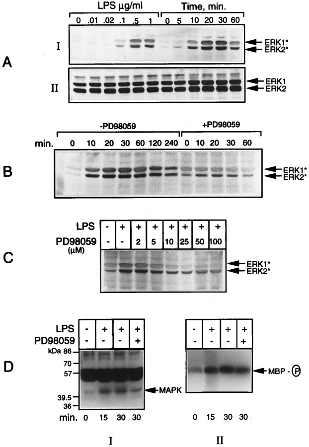
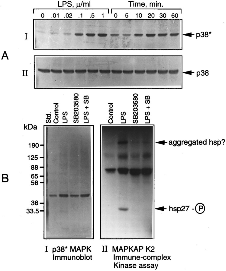
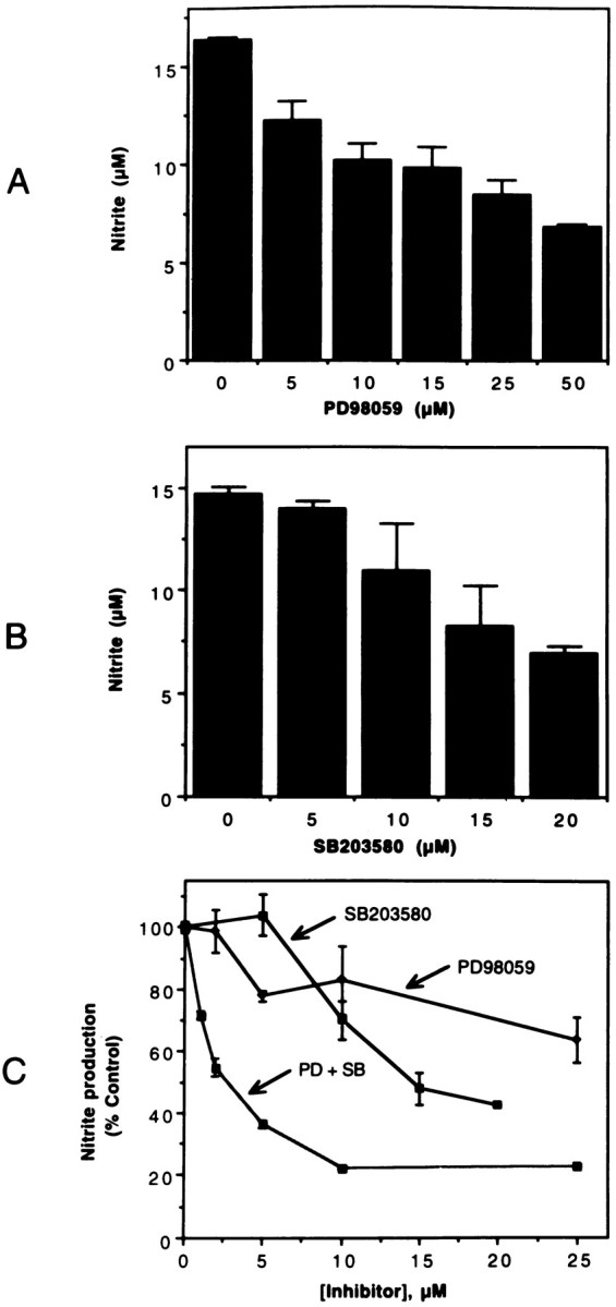
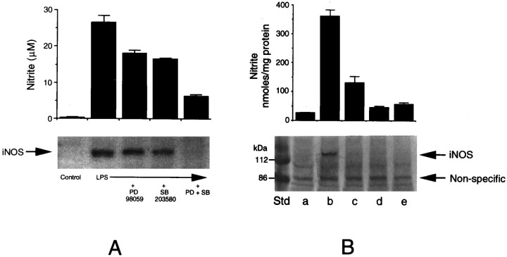
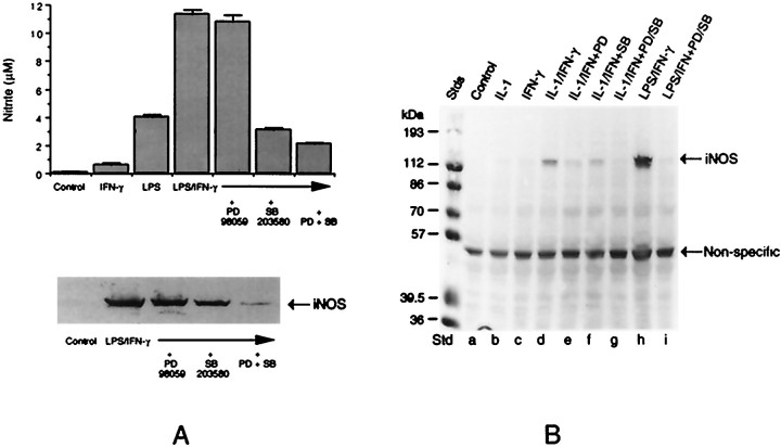
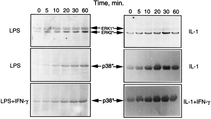
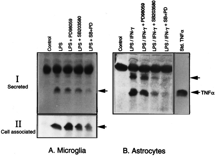
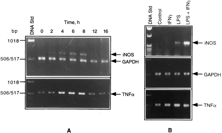
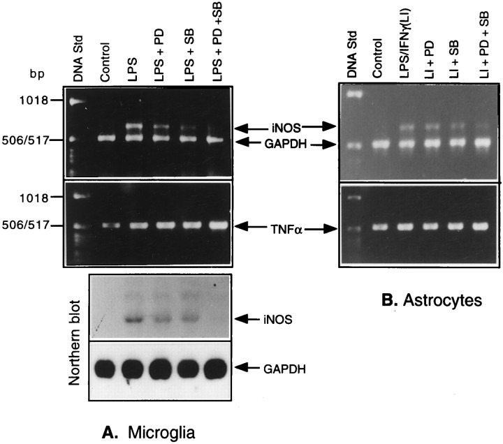
Tumor necrosis factor-alpha (TNFalpha) and nitric oxide (NO), the product of inducible NO synthase (iNOS), mediate inflammatory and immune responses in the CNS under a variety of neuropathological situations. They are produced mainly by "activated" astrocytes and microglia, the two immune regulatory cells of the CNS. In this study we have examined the regulation of TNFalpha and iNOS gene expression in endotoxin-stimulated primary glial cultures, focusing on the role of mitogen-activated protein (MAP) kinase cascades. The bacterial lipopolysaccharide (LPS) was able to activate extracellular signal-regulated kinase (ERK) and p38 kinase subgroups of MAP kinases in microglia and astrocytes. ERK activation was sensitive to PD98059, the kinase inhibitor that is specific for ERK kinase. The activity of p38 kinase was inhibited by SB203580, a member of the novel class of cytokine suppressive anti-inflammatory drugs (CSAIDs), as revealed by blocked activation of the downstream kinase, MAP kinase-activated protein kinase-2. The treatment of glial cells with either LPS alone (microglia) or a combination of LPS and interferon-gamma (astrocytes) resulted in an induced production of NO and TNFalpha. The two kinase inhibitors, at micromolar concentrations, individually suppressed and, in combination, almost completely blocked glial production of NO and the expression of iNOS and TNFalpha, as determined by Western blot analysis. Reverse transcriptase-PCR analysis showed changes in iNOS mRNA levels that paralleled iNOS protein and NO while indicating a lack of effect of either of the kinase inhibitors on TNFalpha mRNA expression. The results demonstrate key roles for ERK and p38 MAP kinase cascades in the transcriptional and post-transcriptional regulation of iNOS and TNFalpha gene expression in endotoxin-activated glial cells.


Figures

Fig. 1.
Fig. 1.
LPS activation of ERK in microglia. Aliquots of cell extracts prepared from microglial cultures treated either with 1 μg/ml LPS for the indicated times or with different concentrations of LPS for 15 min were subjected to immunoblot analysis by using antibodies raised against the active forms of ERK* (A, I). Parallel blots were run, using the antibodies recognizing the total ERK (A, II) proteins. Incubations also were run with LPS added in the presence and absence of 15 μm PD98059 for various times (B) and in the presence of different concentrations of the inhibitor for 15 min (C). In another set of experiments, the cell lysates were used for in-gel (D, I) and immune complex kinases assays (D, II) of activated ERK, using MBP as the substrate, as described in Materials and Methods. The results shown here and elsewhere are representatives from replicate experiments.
Fig. 2.
Fig. 2.
LPS activation of p38 MAPK and its inhibition by SB203580. Cultures of microglial cells treated either with a constant amount (1 μg/ml) of LPS for various times or with different concentrations of LPS for 15 min were lysed and subjected to immunoblot analysis by using antibodies specific for the active (p38*) and total p38 MAPK, as described in Materials and Methods (A). In another set of experiments (B), cultures treated with LPS in the presence and absence of SB203580 (15 μm) were lysed, and the lysates were subjected to immune complex kinase assay of MAPKAP kinase-2, using hsp27 as the substrate (B,II), as described in Materials and Methods. Aliquots of cell lysates also were immunoblotted with anti-Phosphoplus p38 antibodies (B, I).
Fig. 3.
Fig. 3.
MAPK inhibitors inhibit LPS-induced nitrite production. Microglial cultures were treated with 0.1 μg/ml of LPS in the presence of various concentrations of PD98059 (A), SB203580 (B), and combinations of the two inhibitors (C). After 48 hr, aliquots of the culture medium were mixed with Greiss reagent for the estimation of nitrite produced. The optical density of the colored product was measured at 570 nm. Untreated samples showed minimal (<0.5 μm) nitrite levels. The values shown are mean ± SD of data from triplicate determinations.
Fig. 4.
Fig. 4.
Immunoblot analysis of iNOS induction in microglia and its inhibition by MAPK inhibitors. A, Microglial cultures were treated with LPS in the presence of MAPK inhibitors, i.e., PD98059 (25 μm) and SB203580 (15 μm), individually or in combination for 48 hr. Control cultures, cultures treated with LPS alone, and those treated with LPS with the kinase inhibitors were analyzed for iNOS expression by immunoblot, using anti-iNOS antibodies (bottom). Aliquots of the culture medium were analyzed for nitrite production (top). B, In another experiment, cultures were exposed to LPS plus the inhibitor combination for 2 hr, washed, re-fed without the additives, and divided into two sets: one receiving no further treatment (d) and the other reexposed to LPS (e). These two sets of cultures, untreated controls (a), and those treated with LPS with the inhibitors (c) and without the inhibitors (b) were analyzed after 48 hr for nitrite production (top panel) and iNOS expression by immunoblot (bottom panel). The values for nitrite synthesis are mean ± SD of data from triplicate determinations.
Fig. 5.
Fig. 5.
Immunoblot analysis of iNOS induction in astrocytes and its inhibition by MAPK inhibitors. A, Triplicate cultures of astrocytes were treated with LPS (100 ng/ml) plus IFN-γ (50 U/ml) in the presence of MAPK inhibitors, i.e., PD98059 (25 μm) and SB203580 (15 μm), individually or in combination for 48 hr. Control cultures, cultures treated with LPS plus IFN-γ alone, and those treated with LPS plus IFN-γ with the kinase inhibitors were analyzed for iNOS expression by immunoblot analysis, using anti-iNOS antibodies (bottom). Aliquots of the culture medium were analyzed for nitrite production (top). B, Cultures also were treated with IL-1 (50 ng/ml) plus IFN-γ either in the presence or absence of PD98059, SB203580, or a combination of the two for 48 hr. These cultures, along with untreated controls and those exposed to individual cytokines, were analyzed for iNOS expression by immunoblot.
Fig. 6.
Fig. 6.
LPS activation of ERK and p38 MAPK in astrocytes. Astrocyte cultures were treated with LPS and IL-1 individually or in combination with IFN-γ for various time periods, and the cell lysates were subjected to immunoblot analysis, using antibodies specific for the active (phosphorylated) forms of ERK and p38 MAPK. IFN-γ by itself had no effect on kinase activation.
Fig. 7.
Fig. 7.
Immunoblot analysis of TNFα production in glia and its inhibition by MAPK inhibitors. Aliquots of the medium from cultures of microglia and astrocytes treated with LPS and LPS plus IFN-γ, respectively, in the presence and absence of the two kinase inhibitors were resolved on a 15% SDS gel and immunoblotted with antibodies specific for TNFα (I). Aliquots of the cell extracts from microglial cultures also were subjected to immunoblot to determine the cell-associated TNFα (II). The 17 kDa form of TNF comigrates with the standard cytokine. The top arrow in Bprobably indicates the 26 kDa form of TNFα, and the thick band above that is nonspecific.
Fig. 8.
Fig. 8.
PCR analysis of iNOS and TNFα gene expression in glial cultures stimulated with LPS. RNA samples of isolated microglia treated with LPS for different times (A) and astrocytes treated with LPS, IFN-γ, and a combination of the two for 4 hr (B) were subjected to RT-PCR, using the primers specific for iNOS, TNFα, and GAPDH, as described in Materials and Methods.
Fig. 9.
Fig. 9.
Effects of MAPK inhibitors on iNOS and TNFα levels in LPS-treated microglia and astrocytes. RNA samples isolated from glial cultures treated with either LPS or LPS plus IFN-γ in the presence and absence of the kinase inhibitors for 4 hr were analyzed by RT-PCR, using the primer pairs specific for iNOS, TNFα, and GAPDH. The products were run on a 1% agarose gel impregnated with ethidium bromide. The bands were visualized under UV. RNA samples extracted from a set of microglial cultures treated as above also were analyzed by Northern blot for iNOS expression.

Similar articles

Cited by

References

    1. Adamson DC, Wildemann B, Sasaki M, Glass JD, McArthur JC, Christov VI, Dawson TD, Dawson VL. Immunologic nitric oxide synthase: elevation in severe AIDS dementia and induction by HIV-1 gp41. Science. 1996;274:1917–1921. - PubMed
    1. Badger AM, Bradbeer JN, Votta B, Lee JC, Adams JL, Griswold DE. Pharmacological profile of SB 203580, a selective inhibitor of cytokine suppressive binding protein/p38 kinase, in animal models of arthritis, bone resorption, endotoxin shock, and immune function. J Pharmacol Exp Ther. 1996;279:1453–1461. - PubMed
    1. Bagasra O, Michaels FH, Zheng YM, Borroski LE, Spitsin SV, Fu ZF, Tawadros R, Koprowski H. Activation of the inducible form of nitric oxide synthase in the brains of patients with multiple sclerosis. Proc Natl Acad Sci USA. 1995;92:12041–12045. - PMC - PubMed
    1. Benveniste EN. Role of cytokines in multiple sclerosis, autoimmune encephalitis, and other neurological disorders. In: Aggarwal B, Puri R, editors. Human cytokines: their role in research and therapy. Blackwell Scientific; Boston: 1995. p. 195.
    1. Beutler B. Regulation of cachectin biosynthesis occurs at multiple levels. Prog Clin Biol Res. 1990;349:229–240. - PubMed

Publication types

MeSH terms

Substances

LinkOut - more resources