. 1998 Mar 1;18(5):1642-9.
doi: 10.1523/JNEUROSCI.18-05-01642.1998.

Myelin galactolipids are essential for proper node of Ranvier formation in the CNS

Affiliations

Myelin galactolipids are essential for proper node of Ranvier formation in the CNS

J L Dupree et al. J Neurosci. .

Abstract

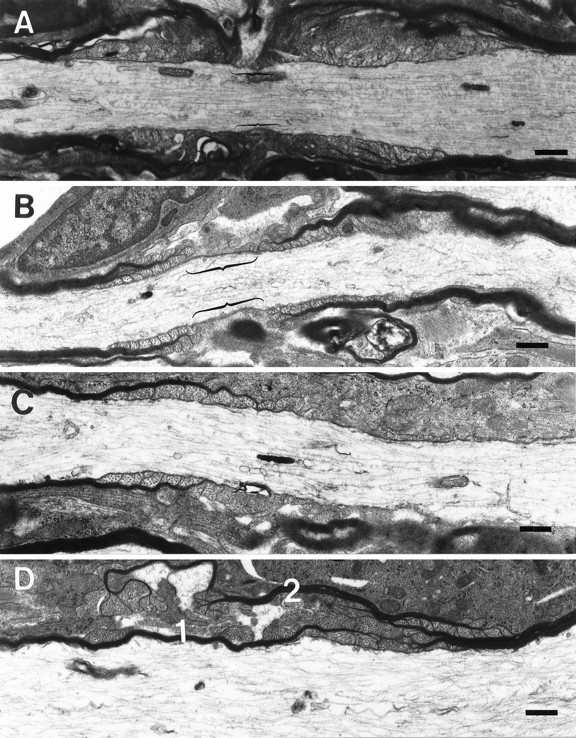
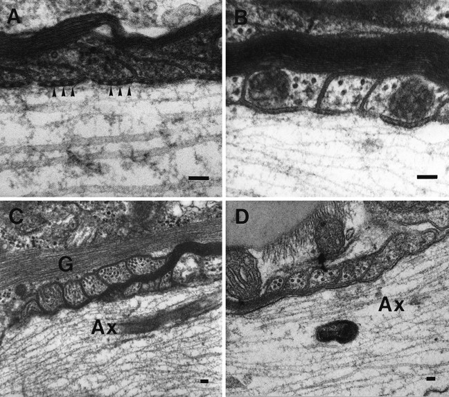
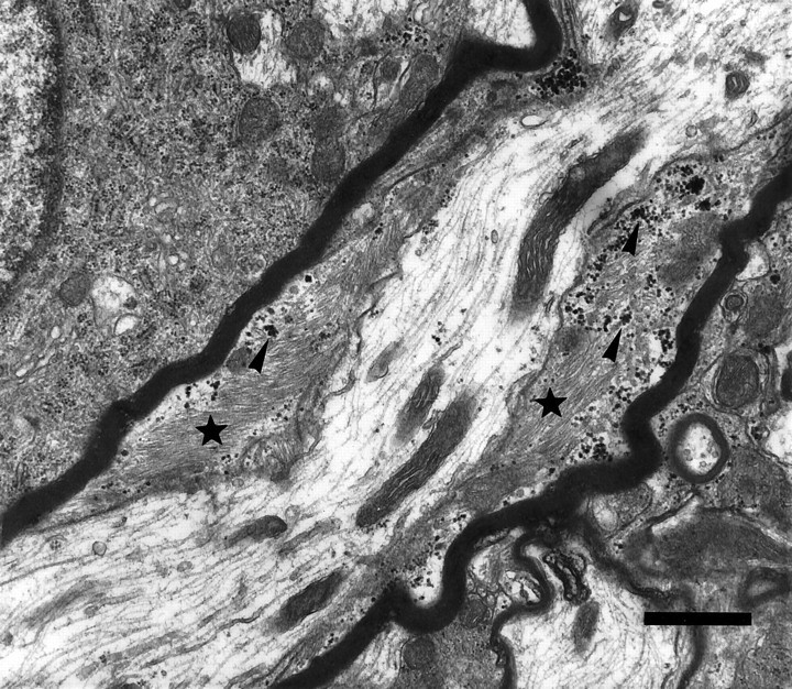
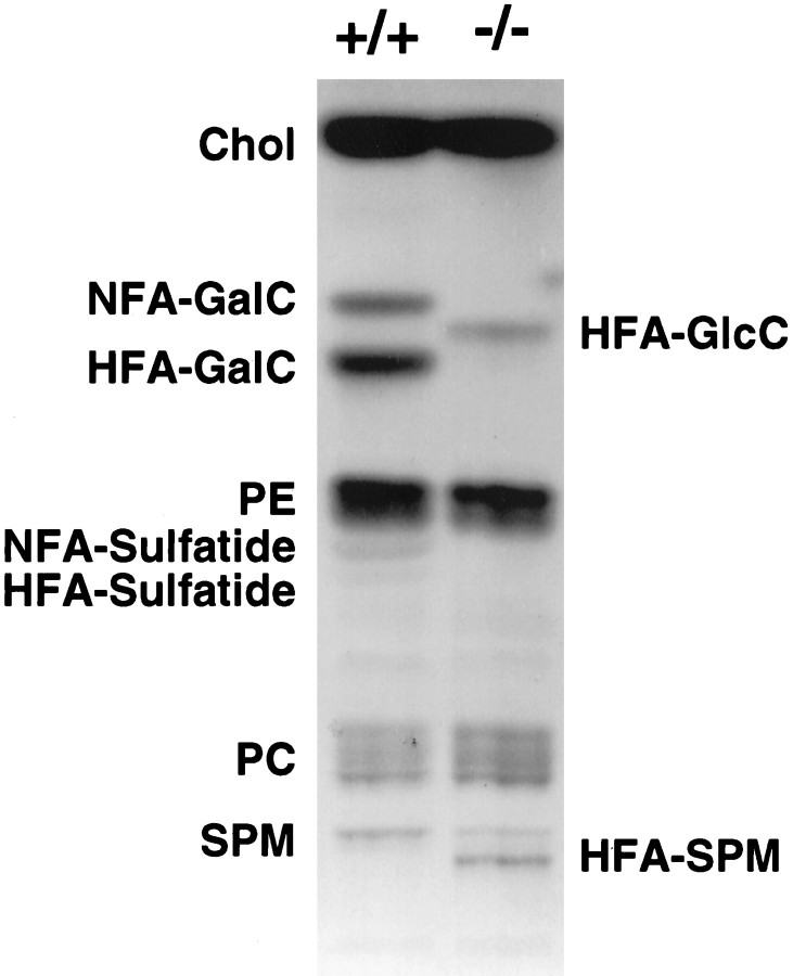
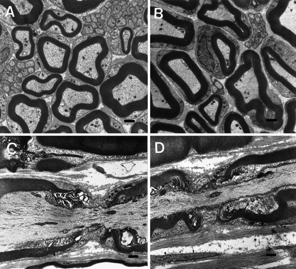
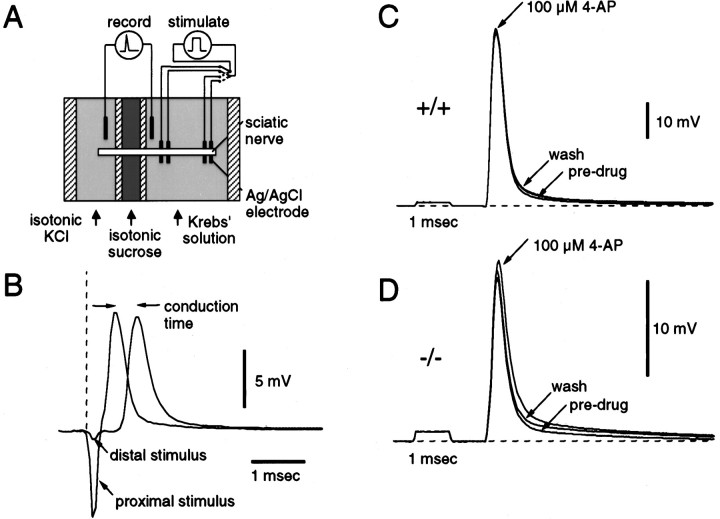
The vertebrate myelin sheath is greatly enriched in the galactolipids galactocerebroside (GalC) and sulfatide. Mice with a disruption in the gene that encodes the biosynthetic enzyme UDP-galactose:ceramide galactosyl transferase (CGT) are incapable of synthesizing these lipids yet form myelin sheaths that exhibit major and minor dense lines with spacing comparable to controls. These CGT mutant mice exhibit a severe tremor that is accompanied by hindlimb paralysis. Furthermore, electrophysiological studies reveal nerve conduction deficits in the spinal cord of these mutants. Here, using electron microscopic techniques, we demonstrate ultrastructural myelin abnormalities in the CNS that are consistent with the electrophysiological deficits. These abnormalities include altered nodal lengths, an abundance of heminodes, an absence of transverse bands, and the presence of reversed lateral loops. In contrast to the CNS, no ultrastructural abnormalities and only modest electrophysiological deficits were observed in the peripheral nervous system. Taken together, the data presented here indicate that GalC and sulfatide are essential in proper CNS node and paranode formation and that these lipids are important in ensuring proper axo-oligodendrocyte interactions.


Figures

Fig. 1.
Fig. 1.
Nodal and paranodal abnormalities in the CGT-deficient mice. Myelinated processes from the cervical region of the spinal cord of 30-d-old CGT-deficient mice demonstrating ultrastructurally normal (A) and abnormal nodes of Ranvier (B–D). B, An aberrantly formed node with lateral loops of the outermost lamellae forming farther from the node than the more medial lamellae, and these loops are facing away from the axon. Furthermore, the length of the nodes (bracketed) in the CGT-deficient mice is typically longer than in the age-matched wild types (bracketed inA). C, The most commonly observed aberrant nodal structure was the heminode, a myelinated segment adjacent to a nonmyelinated region of the axon. In addition, heminodes are the extreme example of node elongation. D, Less frequently observed was the overlapping of paranodal regions. Two myelin sheaths (1, 2) failed to form a node of Ranvier, because the regions of lateral loop formation overlap, thus excluding the nodal region. Scale bar, 1.0 μm.
Fig. 2.
Fig. 2.
High magnification of aberrant paranodal structures in the CGT mutant animals. A, A paranodal region from a 30-d-old littermate control displays a periaxonal space with regularly arrayed densities known as transverse bands (arrowheads). These structures are prominent features of the junctional complex normally formed between the axolemma and the myelin sheath. B, Transverse bands were not found in age-matched CGT-deficient mice. Lateral loops in both the spinal cord (C) and brain (D) frequently are reversed and face away from the axon. Furthermore, the outer most lamella terminates farther from the node. Ax, Axon; G, glial fibrillary acid protein. Scale bar, 0.1 μm.
Fig. 3.
Fig. 3.
Astrocytic intrusion between the myelin sheath and the axon. Myelinated process from the spinal cord of a CGT mutant animal demonstrating the intrusion of an astrocytic process, as identified by the presence of glycogen particles (arrowheads) and bundles of filaments (stars), between the myelin sheath and the axolemma. Scale bar, 1.0 μm.
Fig. 4.
Fig. 4.
Northern and Western blot analyses of myelin proteins. A, Message levels of MBP and PLP are similar between CGT-deficient mice and littermate controls at 30 and 45 d of age. B, Analysis of both total brain and isolated myelin revealed no difference in the levels of MBP and PLP at 30 and 45 d of age.
Fig. 5.
Fig. 5.
Lipid analysis of the PNS. Both NFA- and HFA-containing GalC and sulfatide were absent in the sciatic nerves of the CGT−/− mouse. Bands with mobility patterns consistent with HFA–glucocerebroside and sphingomyelin were prominent in the mutant but not in the wild-type. Chol, Cholesterol;HFA-GalC, hydroxy fatty acid galactocerebroside;NFA-GalC, normal fatty acid galactocerebroside;HFA-GlcC, hydroxyglucocerebroside; PE, phosphatidyl ethanolamine; PC, phosphatidyl choline;SPM, sphingomyelin; HFA-SPM, hydroxy fatty acid sphingomyelin.
Fig. 6.
Fig. 6.
Structural analysis of the PNS. Sciatic nerve myelin sheaths revealed no differences between CGT+/+ (A, C) and CGT−/− (B, D) mice with respect to thickness (A, B) or node formation (C, D). Scale bar, 1.0 μm.
Fig. 7.
Fig. 7.
Electrophysiological analysis of the PNS.A, Diagram of the recording apparatus used for measurement of conduction velocity. The isolated nerve was placed in a Plexigal chamber and superfused with oxygenated Krebs’ solution, and one end was isolated by a flowing sucrose gap in a separate chamber slowly perfused with isotonic KCl solution (120 mm). Silver–silver electrodes were used to record the potential across the gap and to stimulate the nerve at two points, 5.5 mm apart in the main chamber. An example of compound potentials recorded in response to stimulation of the nerve at proximal and distal electrode pairs is illustrated in B. The conduction time for the length of nerve between the stimulating electrodes was derived from the time between the compound potential peaks in each recording.C, Three recordings are superimposed showing the effect of superfusing a nerve from a CGT+/+ mouse with Krebs’ solution containing 100 μm 4-AP. There was a slight increase in amplitude of the response and the depolarizing afterpotential. The compound action potential increase reversed more readily with 10 min of washing in normal Krebs’ solution. D, Recordings, similar to those in C, show a slightly more pronounced effect of 4-AP on the amplitude of the compound potential and depolarizing afterpotential in a nerve from a CGT−/− mouse.

Similar articles

Cited by

References

    1. Benjamins JA, Studzinski DM, Skoff RP. Analysis of myelin proteolipid protein and F0 ATPase subunit 9 in normal and jimpy CNS. Neurochem Res. 1994;19:1013–1022. - PubMed
    1. Bosio A, Binczek E, Stoffel W. Functional breakdown of the lipid bilayer of the myelin membrane in central and peripheral nervous system by disrupted galactocerebroside synthesis. Proc Natl Acad Sci USA. 1996;93:13280–13285. - PMC - PubMed
    1. Coetzee T, Fujita N, Dupree J, Shi R, Blight A, Suzuki K, Popko B. Myelination in the absence of galactocerebroside and sulfatide: normal structure and abnormal function and regional instability. Cell. 1996;86:209–219. - PubMed
    1. Dentinger MP, Barron KD, Csiza CK. Ultrastructure of the central nervous system in a myelin deficient rat. J Neurocytol. 1982;11:671–691. - PubMed
    1. Duncan ID, Griffiths IR, Munz M. “Shaking pup”: a disorder of central myelination in the spaniel dog. III. Quantitative aspects of the glia and myelin in the spinal cord and optic nerve. Neuropathol Appl Neurobiol. 1983;9:355–368. - PubMed

Publication types

MeSH terms

LinkOut - more resources