. 1998 Mar 1;18(5):1693-703.
doi: 10.1523/JNEUROSCI.18-05-01693.1998.

Segregation of different GABAA receptors to synaptic and extrasynaptic membranes of cerebellar granule cells

Affiliations

Segregation of different GABAA receptors to synaptic and extrasynaptic membranes of cerebellar granule cells

Z Nusser et al. J Neurosci. .

Abstract

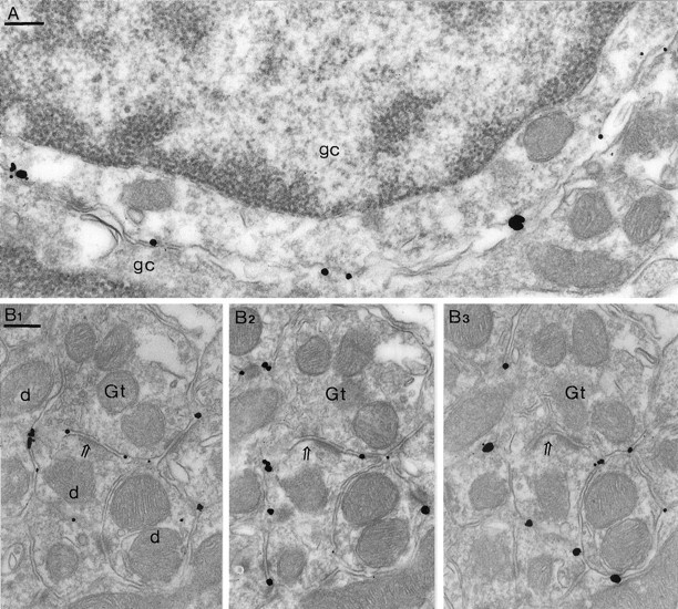
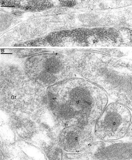
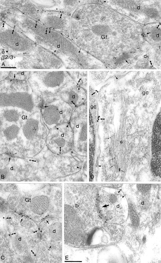
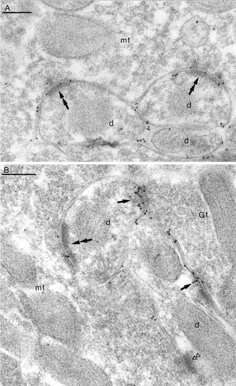
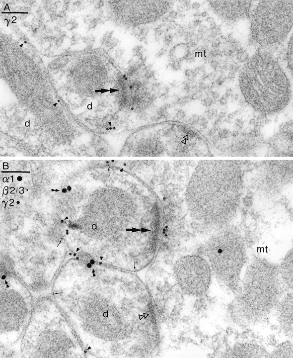
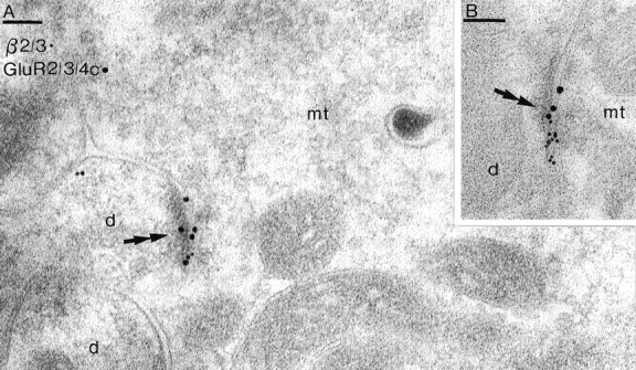
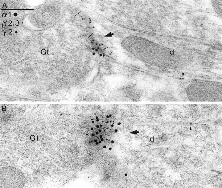
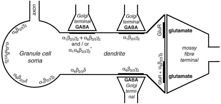
Two types of GABAA receptor-mediated inhibition (phasic and tonic) have been described in cerebellar granule cells, although these cells receive GABAergic input only from a single cell type, the Golgi cell. In adult rats, granule cells express six GABAA receptor subunits abundantly (alpha1, alpha6, beta2, beta3, gamma2, and delta), which are coassembled into at least four to six distinct GABAA receptor subtypes. We tested whether a differential distribution of GABAA receptors on the surface of granule cells could play a role in the different forms of inhibition, assuming that phasic inhibition originates from the activation of synaptic receptors, whereas tonic inhibition is provided mainly by extrasynaptic receptors. The alpha1, alpha6, beta2/3, and gamma2 subunits have been found by immunogold localizations to be concentrated in GABAergic Golgi synapses and also are present in the extrasynaptic membrane at a lower concentration. In contrast, immunoparticles for the delta subunit could not be detected in synaptic junctions, although they were abundantly present in the extrasynaptic dendritic and somatic membranes. Gold particles for the alpha6, gamma2, and beta2/3, but not the alpha1 and delta, subunits also were concentrated in some glutamatergic mossy fiber synapses, where their colocalization with AMPA-type glutamate receptors was demonstrated. The exclusive extrasynaptic presence of the delta subunit-containing receptors, together with their kinetic properties, suggests that tonic inhibition could be mediated mainly by extrasynaptic alpha6beta2/3delta receptors, whereas phasic inhibition is attributable to the activation of synaptic alpha1beta2/3gamma2, alpha6beta2/3gamma2, and alpha1alpha6beta2/3gamma2 receptors.


Figures

Fig. 1.
Fig. 1.
Distribution of immunoreactivity for the δ subunit of the GABAA receptor in the granule cell layer of mouse cerebellum as revealed by a preembedding, silver-intensified immunogold reaction. A, Immunoparticles are present along the nonsynaptic somatic membrane of granule cells (gc).B1B3, Serial ultrathin sections of a glomerulus showing that a synapse (open arrow) between a Golgi cell terminal (Gt) and a granule cell dendrite (d) is immunonegative for the δ subunit, although particles are present at the extrasynaptic dendritic membrane. Note that, when the membranes are cut at right angle (e.g., inB1), most of the particles are seen at the external face of the plasma membrane corresponding to the extracellular location of epitope(s) recognized by the antibody δ(1–44)R5. Scale bars, 0.2 μm.
Fig. 2.
Fig. 2.
Immunoreactivity for the δ subunit in the granule cell layer of rat cerebellum as revealed by a postembedding immunogold technique (10-nm gold). A, Immunoparticles are present along the extrasynaptic somatic membrane of granule cells (gc), including areas in which two cells are directly apposed. B, Immunogold particles are associated with the extrasynaptic membranes of granule cell dendrites (d), but no particles are seen in a synapse (open arrow) made by a Golgi cell terminal (Gt) and a granule cell dendrite. Scale bars:A, 0.2 μm; B, 0.1 μm.
Fig. 3.
Fig. 3.
Electron micrographs showing double labeling for the β2/3 (10 nm particles) and the δ (A, 18 nm;B–E, 20 nm particles) subunits. Postembedding immunogold reactions are shown for rat (A andE) and mouse (B–D) cerebella.AC, Synapses (arrows) made by Golgi cell terminals (Gt) with granule cell dendrites (d) are not labeled for the δ subunit, although the enrichment of immunoparticles for the β2/3 subunits shows that receptor immunoreactivity is well preserved in these GABAergic synapses. In addition, the presence of immunoparticles for the δ subunit (double arrowheads) at the extrasynaptic dendritic membranes demonstrates that the method is sensitive enough to visualize this subunit. Note that immunoparticles for the β2/3 subunits also are associated with the extrasynaptic dendritic membranes (e.g., single arrowheads).D, Immunoparticles for both the β2/3 (arrowheads) and the δ (double arrowheads) subunits are present at the somatic membrane of granule cells (gc). A Golgi apparatus (G) shows immunoreactivity for both of these subunits (small arrows). E, In the molecular layer symmetrical synapses (arrow) on interneuron dendrites (d) or on Purkinje cells and extrasynaptic (arrowheads) membranes showed immunoreactivity for the β2/3 subunits, but never for the δ subunit. b, Bouton. A–D have the same magnification; scale bars, 0.2 μm.
Fig. 4.
Fig. 4.
Immunoreactive β2/3 subunits are present in both GABAergic and glutamatergic synapses on granule cells. Postembedding immunogold reactions are shown on mouse cerebellum with 10-nm gold particles. A, B, Gold particles are present in asymmetrical synapses (double arrows) between mossy fiber terminals (mt) and granule cell dendrites (d) and in synapses (arrows inB) made by a Golgi cell terminal (Gt) with granule cell dendrites. One of the asymmetrical synapses (double open triangles) is immunonegative for these subunits. Usually a higher density of immunoparticles is found in Golgi synapses (single arrows) than in mossy fiber to granule cell synapses (synapses in B). Scale bars, 0.2 μm.
Fig. 5.
Fig. 5.
Immunoreactive γ2 subunits are present in mossy fiber to granule cell synapses. Postembedding immunogold reactions are shown on Lowicryl resin-embedded rat cerebellum. A, An asymmetrical synapse (double arrows) made by a mossy fiber terminal (mt) with a granule cell dendrite (d) is immunopositive for the γ2 subunit. Particles also are present on the extrasynaptic dendritic membranes (arrowheads). B, A triple-labeling experiment shows the presence of gold particles for the γ2 subunit in an asymmetrical synapse (double arrow) made by a mossy fiber terminal with a granule cell dendrite (d) and the presence of the α1 (double arrowheads), β2/3 (small arrows), and γ2 (arrowheads) subunits on the extrasynaptic membranes. In this example the immunoparticles for the β2/3 subunits are not detected in mossy synapses. One of the asymmetrical mossy synapses (double open triangles) is immunonegative for all of the subunits. Scale bars, 0.1 μm.
Fig. 6.
Fig. 6.
Synaptic colocalization of GABAA(antibody to the β2/3 subunits; 10-nm gold) and AMPA-type glutamate receptors (antibody to the GluR2/3/4c subunits; 20-nm gold). Asymmetrical synapses (double arrows) between mossy fiber terminals (mt) and granule cell dendrites (d) are immunopositive for both the GABAA receptor and the AMPA-type glutamate receptor. Postembedding reactions are shown in rat (A) and mouse (B) cerebella. Scale bars, 0.1 μm.
Fig. 7.
Fig. 7.
Colocalization of the α1 (20-nm gold), β2/3 (5-nm gold), and γ2 (10-nm gold) subunits of the GABAAreceptor in Golgi synapses of the mouse cerebellum. A,B, Labeling for each subunit is present in synapses (large arrows) between Golgi cell terminals (Gt) and granule cell dendrites (d). Small arrows point to 5-nm particles, indicating immunoreactive β2/3 subunits. Some extrasynaptic particles are shown by arrowheads. The synapse in B is cut tangentially; thus the receptor immunoreactivity is shown en face. A andB have the same magnification. Scale bar, 0.2 μm.
Fig. 8.
Fig. 8.
Schematic representation of the differential distribution of GABAA receptor subtypes on cerebellar granule cells, assuming that every receptor subtype is expressed by a single cell. The α6, β2/3, and γ2 subunits (α6β2/3γ2 receptors) are present in Golgi synapses, on the extrasynaptic membranes, and in some of the mossy fiber to granule cell synapses. Immunoreactive δ subunits (α6β2/3δ receptors) are found only on the extrasynaptic somatic and dendritic membranes. Immunoreactivity for the α1 subunit (α1β2/3γ2 receptors) is found in some Golgi cell to granule cell synapses and on the extrasynaptic membranes. *The α1 and α6 subunits are found colocalized in some Golgi synapses, suggesting either that some of these synapses contain both α1β2/3γ2 and α6β2/3γ2 receptors or that a receptor population with α1α6β2/3γ2subunit composition exists (see Discussion) in these synapses and in the extrasynaptic membranes. The β2/3 subunits were found colocalized with AMPA-type glutamate receptors (GluR) in some mossy fiber to granule cell synapses, but others were labeled only for AMPA receptors. Some of the data are from Nusser et al. (1995b, 1996b) andJones et al. (1997).

Similar articles

Cited by

References

    1. Andersen P, Eccles JC, Loyning Y. Recurrent inhibition in the hippocampus with identification of the inhibitory cell and its synapses. Nature. 1963;198:540–542. - PubMed
    1. Angelotti TP, Macdonald RL. Assembly of GABAA receptor subunits: α1β1 and α1β1γ2s subunits produce unique ion channels with dissimilar single-channel properties. J Neurosci. 1993;13:1429–1440. - PMC - PubMed
    1. Baude A, Nusser Z, Roberts JDB, Mulvihill E, McIlhinney RAJ, Somogyi P. The metabotropic glutamate receptor (mGluR1α) is concentrated at perisynaptic membrane of neuronal subpopulations as detected by immunogold reaction. Neuron. 1993;11:771–787. - PubMed
    1. Baude A, Nusser Z, Molnar E, McIlhinney RAJ, Somogyi P. High-resolution immunogold localization of AMPA type glutamate receptor subunits at synaptic and non-synaptic sites in rat hippocampus. Neuroscience. 1995;69:1031–1055. - PubMed
    1. Benke D, Honer M, Michel C, Mohler H. GABAA receptor subtypes differentiated by their γ-subunit variants: prevalence, pharmacology, and subunit architecture. Neuropharmacology. 1996;35:1413–1423. - PubMed

Publication types

Substances