Comparative Study
. 1998 Mar 1;18(5):1713-24.
doi: 10.1523/JNEUROSCI.18-05-01713.1998.

Role of the Jun kinase pathway in the regulation of c-Jun expression and apoptosis in sympathetic neurons

Affiliations
Comparative Study

Role of the Jun kinase pathway in the regulation of c-Jun expression and apoptosis in sympathetic neurons

A Eilers et al. J Neurosci. .

Abstract

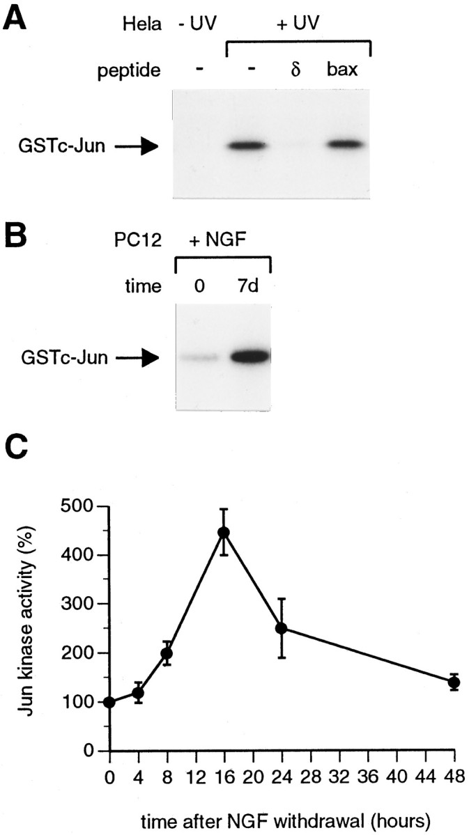
When deprived of nerve growth factor (NGF), developing sympathetic neurons die by apoptosis. This death is associated with an increase in the level of c-Jun protein and is blocked by expression of a c-Jun dominant negative mutant. Here we have investigated whether NGF withdrawal activates Jun kinases, a family of stress-activated protein kinases that can stimulate the transcriptional activity of c-Jun by phosphorylating serines 63 and 73 in the transactivation domain and which can activate c-jun gene expression. We found that sympathetic neurons contained high basal levels of Jun kinase activity that increased further after NGF deprivation. In contrast, p38 kinase, another stress-activated protein kinase that can also stimulate c-jun gene expression, was not activated after NGF withdrawal. Consistent with Jun kinase activation, we found using a phospho-c-Jun-specific antibody that c-Jun was phosphorylated on serine 63 after NGF withdrawal. Furthermore, expression of a constitutively active form of MEK kinase 1 (MEKK1), which strongly activates the Jun kinase pathway, increased c-Jun protein levels and c-Jun phosphorylation and induced apoptosis in the presence of NGF. This death could be prevented by co-expression of SEKAL, a dominant negative mutant of SAPK/ERK kinase 1 (SEK1), an activator of Jun kinase that is a target of MEKK1. In contrast, expression of SEKAL alone did not prevent c-Jun expression, increases in c-Jun phosphorylation, or cell death after NGF withdrawal. Thus, activation of Jun kinase and increases in c-Jun phosphorylation and c-Jun protein levels occur at the same time after NGF withdrawal, but c-Jun levels and phosphorylation are regulated by an SEK1-independent pathway.


Figures

Fig. 1.
Fig. 1.
Transcriptional activation of the c-jun promoter in NGF-deprived sympathetic neurons requires the jun1 and jun2 TRE elements and AP-1 activity.A, Sympathetic neurons were injected with pCDFLAGΔ169 (0.2 mg/ml) or the empty CMV expression vector pcDNA1 (0.2 mg/ml) together with guinea pig IgG (2.5 mg/ml). After injection, the cells were rinsed with medium lacking NGF and then were refed with −NGF medium supplemented with neutralizing anti-NGF antibody. Twenty-four hours later, the percentage of injected cells that expressed c-Jun protein was determined as described in Materials and Methods. Cells were only considered to be expressing c-Jun if nuclear staining with the c-Jun antibody was more intense than the background cytoplasmic staining. In each experiment 200 cells were injected per construct. Uninjected cells on the same coverslips were also scored for comparison. Coverslips were scored blind. The data shown represent the average of three independent experiments. Error bars indicate SE.B, Reporter gene structure. c-jun CAT contains wild-type c-jun promoter sequences from −1600 to +170 cloned upstream of the bacterial CAT gene. The position of the jun1 and jun2 TRE elements is indicated (j1j2). In j1j2 CAT, these have been mutated so that they are nonfunctional. 6xjun2 SVeCAT was constructed by cloning six copies of the jun2 TRE element upstream of the SV40 early promoter in SVeCAT (= pCAT3 promoter). C, c-jun CAT or j1j2 CAT was microinjected into sympathetic neurons at a concentration of 0.01 mg/ml together with guinea pig IgG (2.5 mg/ml). The injected cells were refed with medium containing (+NGF) or lacking (−NGF) NGF. Eighteen hours later, the percentage of cells expressing CAT was determined in immunofluorescence experiments with an anti-CAT antibody as described in Materials and Methods. The data shown represent the average of three independent experiments. Error bars indicate SE. D, 6xjun2 SVeCAT or SVeCAT was microinjected into sympathetic neurons at a concentration of 0.005 mg/ml together with guinea pig IgG. The injected cells were treated as described in C.
Fig. 2.
Fig. 2.
Jun kinase activity increases during PC12 differentiation and when differentiated PC12 cells are deprived of NGF.A, Control immune complex kinase assays were performed with extracts from quiescent (−UV) and UV-treated (+UV) HeLa cells as described in Materials and Methods. One hundred micrograms of extract were used for each immunoprecipitation. Competition with the c-Jun δ and Bax peptides was performed as described in Materials and Methods. The products of the Jun kinase assay were separated on a 10% SDS-polyacrylamide gel, which was fixed and dried, and autoradiography was performed. The position of phosphorylated GSTc-Jun[1–169] is shown. B, Jun kinase activity increases during PC12 differentiation. PC12 cells were treated with NGF, and extracts were prepared immediately after NGF addition (time 0) or 7 d later (7d). Jun kinase assays were performed using 100 μg of extract, as described in Materials and Methods. A representative result is shown. On average (7 experiments), Jun kinase activity increased fourfold during PC12 differentiation.C, Jun kinase is activated when differentiated PC12 cells are deprived of NGF. Differentiated PC12 cells were refed with defined medium lacking NGF, which had been supplemented with anti-NGF antibody. Extracts were made at the times indicated, and immune complex kinase assays were performed, using 100 μg of extract/immunoprecipitation. The resulting autoradiograph was scanned on a densitometer to determine the relative levels of Jun kinase activity at different times. The level of Jun kinase activity at time0 was set as 100%. The results shown are the average values for three independent experiments. Error bars indicate SE.
Fig. 3.
Fig. 3.
Jun kinase is activated in sympathetic neurons after NGF withdrawal. A, Jun kinase assays were performed with extracts prepared from sympathetic neurons that had been withdrawn from NGF for 4, 8, or 16 hr (−NGF) or that had been refed with fresh NGF-containing medium (4 hr, +NGF), as described in Materials and Methods. Fifty micrograms of extract were used per immunoprecipitation. Relative Jun kinase activity was determined by scanning autoradiographs on a densitometer. The level of Jun kinase activity at time 0was set as 100%. The results shown are the average of three independent experiments. Error bars indicate SE. B,Survival agents prevent the NGF withdrawal-induced activation of Jun kinase in sympathetic neurons. Sympathetic neurons were refed with fresh NGF-containing medium (+NGF) or were deprived of NGF in the absence (−NGF) or presence of 100 μm CPTcAMP (−NGF +100 μm CPTcAMP) or 30 mmN-acetylcysteine (−NGF + 30 mm NAC). Four hours later, extracts were prepared, and immune complex kinase assays were performed using 50 μg of extract/immunoprecipitation. The results shown are the average of three independent experiments. Error bars indicate SE.
Fig. 4.
Fig. 4.
c-Jun is phosphorylated on serine 63 when sympathetic neurons are deprived of NGF. A, The specificity of the phospho-c-Jun monoclonal antibody was verified in an immunoblotting experiment by performing peptide competition. Extracts were from untreated (−UV) or UV-irradiated (+UV) Rat 1 cells. The position of the phosphorylated forms of c-Jun (ph-c-Jun) is indicated. As predicted, UV treatment caused an increase in c-Jun phosphorylation. This signal was competed away by preincubating the antibody with the phosphopeptide used to generate it (phos) but not by an equal amount of the corresponding nonphosphorylated peptide (non-phos).B, Sympathetic neurons were isolated from neonatal rats and cultured for 7 d in vitro in the presence of NGF. The cells were then refed with medium lacking NGF, which had been supplemented with neutralizing anti-NGF antibody (−NGF), or with fresh NGF-containing medium (+NGF). Twenty-four hours later, the cells were fixed and stained with the phospho-c-Jun antibody and Hoechst dye as described in Materials and Methods. In the presence of NGF, weak background staining of the cell body was observed with the phospho-c-Jun antibody. After NGF withdrawal, nuclear phospho-c-Jun staining was apparent, indicating that the c-Jun protein in these cells was phosphorylated on serine 63. Phospho-c-Jun staining could be seen as early as 4 or 8 hr after NGF withdrawal (data not shown). Representative cells are shown.
Fig. 5.
Fig. 5.
p38 kinase is activated after NGF withdrawal in differentiated PC12 cells but not sympathetic neurons.A, Differentiated PC12 cells were withdrawn from NGF, and extracts were prepared at the times indicated. p38 kinase was immunoprecipitated from extracts using a Xenopusmpk2/p38 antibody that recognizes mammalian p38 (Rouse et al., 1994), and kinase assays were performed using GSTATF-2[1–96] as substrate. Relative p38 kinase activity was determined by scanning autoradiographs on a densitometer. The results of a representative experiment are shown. B, p38 kinase is not activated in NGF-deprived sympathetic neurons. p38 kinase assays were performed with extracts prepared from sympathetic neurons (SCG) or differentiated PC12 cells that had been deprived of NGF for the times indicated. Fifty micrograms of extract were used for each immunoprecipitation. A typical result is shown. The position of phosphorylated GSTATF2[1–96] is shown. C, p38 and phospho-p38 immunoblots were performed as described in Materials and Methods. Extracts were from sympathetic neurons (SCG) that had been deprived of NGF for 0, 4, 8, or 16 hr or were from control PC12 cells (−) or PC12 cells that had been exposed to anisomycin (A) or UV radiation (UV). p38 protein levels were 10-fold lower in SCG extracts compared with PC12 extracts. Activated p38 phosphorylated on tyrosine 182 (ph-p38) was readily detected in PC12 cell extracts (a 30 sec ECL exposure) but not in SCG extracts. Anisomycin or UV treatment increased phospho-p38 levels sixfold to sevenfold. Prolonged exposure of the same blot (a 30 min ECL exposure) revealed a low level of p38 phosphorylation in sympathetic neurons, which did not increase after NGF withdrawal.
Fig. 6.
Fig. 6.
Overexpression of a constitutively active form of MEKK1 in sympathetic neurons increases c-Jun protein levels and induces apoptosis. A, Morphology of neurons 4 d after injection with pSG5 or MEKK1. Sympathetic neurons were microinjected with the empty vector pSG5 or an MEKK1 expression vector together with Texas Red–dextran (5 mg/ml) to mark the injected cells. The injected cells were left for 4 d in NGF-containing medium and then were examined on an inverted fluorescence microscope. The majority of cells injected with pSG5 had a normal morphology on phase, and the injected Texas Red–dextran was still retained within the nucleus. A representative cell is shown. In contrast, the majority of cells injected with MEKK1 had a clearly apoptotic morphology on phase (two cells injected with MEKK1 are shown), and the dextran marker was no longer retained within the nucleus. B, Kinetics of cell death after microinjection of MEKK1. Sympathetic neurons were microinjected with the MEKK1 vector or the control vector, pSG5, each at 0.1 mg/ml, together with Texas Red–dextran (5 mg/ml). The percentage of viable injected cells that remained at different times after injection was determined as described in Materials and Methods. The results shown are the average for four independent experiments. Error bars indicate SE. In each experiment, 200 neurons were injected per construct. C, Co-expression of SEKALprevents cell death induced by MEKK1 overexpression. Sympathetic neurons were microinjected with expression vectors for MEKK1 (0.1 mg/ml), SEKAL (0.4 mg/ml), or the control vector pSG5 (0.1–0.5 mg/ml) in the combinations indicated. Where necessary, the total DNA concentration was adjusted to 0.5 mg/ml by addition of pSG5. Texas Red–dextran was included in the injection mixes at 5 mg/ml. Several hours after injection, the number of Texas Red-containing cells was determined, and the cells were left in NGF-containing medium. Three days later, the number of viable injected cells that remained was determined. The results shown are the average of three independent experiments. Error bars indicate SE. D, Overexpression of MEKK1 increases c-Jun protein levels. Sympathetic neurons were injected with the vectors shown at the concentrations described inC. Forty-eight hours after injection, the percentage of surviving injected cells that expressed c-Jun protein was determined as described in Materials and Methods. The results shown are the average of seven independent experiments. Error bars indicate SE.
Fig. 7.
Fig. 7.
Expression of SEKAL does not prevent NGF withdrawal-induced death or the increase in c-Jun protein levels that occurs after NGF withdrawal. A, Expression of SEKAL does not protect sympathetic neurons against NGF withdrawal-induced death. Neurons were injected with pcDNA1 (0.05 mg/ml), pCDBcl-2 (0.05 mg/ml), pSG5 (0.4 mg/ml), or the SEKAL vector (0.4 mg/ml) together with Texas Red–dextran. pcDNA1 and pSG5 were control vectors for pCDBcl-2 and SEKAL, respectively. After injection, the cells were deprived of NGF. Seventy-two hours later, the percentage of viable injected neurons was determined as described in Materials and Methods. The results shown are the average of three independent experiments. Error bars indicate SE. B, SEKAL does not inhibit expression of c-Jun protein after NGF withdrawal. Sympathetic neurons were injected with pCDFLAGΔ169 (0.2 mg/ml), the SEKAL expression vector (0.4 mg/ml), or the empty vector pSG5 (0.4 mg/ml) together with guinea pig IgG at 5 mg/ml. After injection, the cells were withdrawn from NGF. Twenty-four hours later, the percentage of injected cells that expressed c-Jun was determined as described in Materials and Methods. c-Jun expression in uninjected cells on the same coverslips was scored for comparison. Only cells in which nuclear staining with the c-Jun antibody was more intense than the background cytoplasmic staining were considered to be expressing c-Jun. Two hundred cells were injected per construct. The data shown are the average of three independent experiments. Error bars indicate SE.
Fig. 8.
Fig. 8.
Hypothetical model illustrating the relationship between the Jun kinase pathway and c-Jun expression and apoptosis in sympathetic neurons. After NGF withdrawal, Jun kinase activity, c-Jun levels, and c-Jun phosphorylation increase. Expression of c-Jun protein and apoptosis are blocked by the c-Jun dominant negative mutant FLAGΔ169, which inhibits AP-1 activity. NGF withdrawal-induced death is also blocked by injection of antibodies against c-Jun but not Jun B or Jun D (Estus et al., 1994). These results suggest that c-Jun activates target genes that promote apoptosis, as well as the c-jun gene itself. In other systems, JNK/SAPKs are activated by SEK1, which in turn is activated by MEKK1. Overexpression of MEKK1 in sympathetic neurons increases the level of c-Jun protein and induces apoptotic cell death, suggesting that activation of the Jun kinase pathway is sufficient to trigger apoptosis. The induction of c-Jun expression and apoptosis by MEKK1 is blocked by co-expression of the SEK1 dominant negative mutant SEKAL. However, expression of SEKAL does not block the expression of c-Jun induced by NGF withdrawal or NGF withdrawal-induced death. This might be because NGF withdrawal activates a JNKKK and JNKK that cannot be inhibited by SEKAL (a pathway parallel to MEKK1 and SEK1 that would activate JNK/SAPKs) or because the expression of c-Jun and apoptosis do not require Jun kinase activity and are induced by a different signaling pathway that is not affected by SEKAL (? leading to the c-jun gene). This would not be the p38 kinase pathway, because p38 is not activated in sympathetic neurons after NGF withdrawal.

Similar articles

Cited by

References

    1. Angel P, Hattori K, Smeal T, Karin M. The jun proto-oncogene is positively autoregulated by its product, Jun/AP1. Cell. 1988;55:875–885. - PubMed
    1. Cavigelli M, Li WW, Lin A, Su B, Yoshioka K, Karin M. The tumour promoter arsenite stimulates AP-1 activity by inhibiting a JNK phosphatase. EMBO J. 1996;15:6269–6279. - PMC - PubMed
    1. Coso OA, Chiariello M, Yu J-C, Teramoto H, Crespo P, Xu N, Miki T, Gutkind JS. The small GTP-binding proteins Rac1 and Cdc42 regulate the activity of the JNK/SAPK signaling pathway. Cell. 1995;81:1137–1146. - PubMed
    1. Dérijard B, Raingeaud J, Barrett T, Wu I-H, Han J, Ulevitch RJ, Davis RJ. Independent human MAP kinase signal transduction pathways defined by MEK and MKK isoforms. Science. 1995;267:682–685. - PubMed
    1. Deshmukh M, Vasilakos J, Deckwerth TL, Lampe PA, Shivers BD, Johnson EM. Genetic and metabolic status of NGF-deprived sympathetic neurons saved by an inhibitor of ICE family proteases. J Cell Biol. 1996;135:1341–1354. - PMC - PubMed

Publication types

MeSH terms

Substances

LinkOut - more resources