. 1998 Mar 1;18(5):1743-52.
doi: 10.1523/JNEUROSCI.18-05-01743.1998.

Turnover of amyloid beta-protein in mouse brain and acute reduction of its level by phorbol ester

Affiliations

Turnover of amyloid beta-protein in mouse brain and acute reduction of its level by phorbol ester

M J Savage et al. J Neurosci. .

Abstract

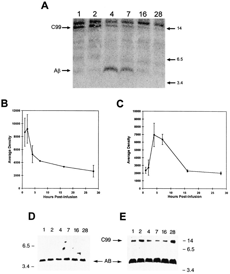
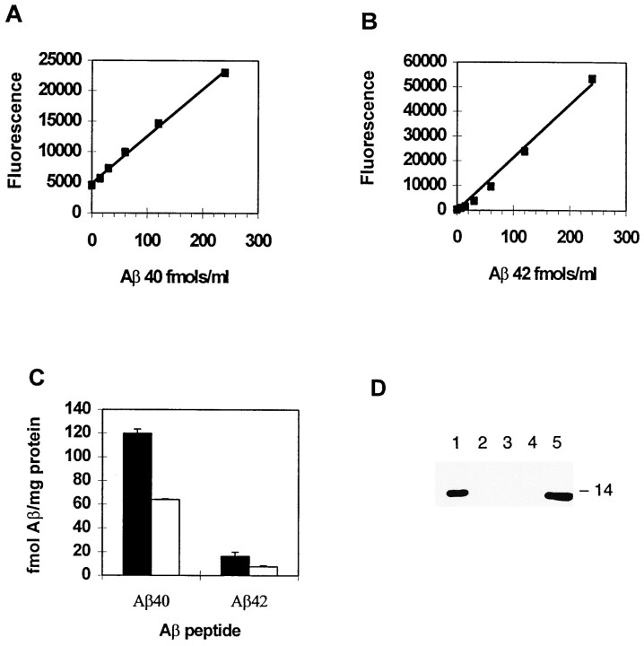
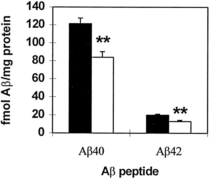
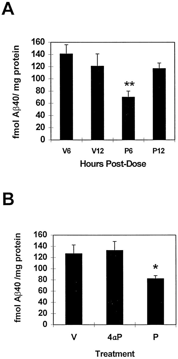
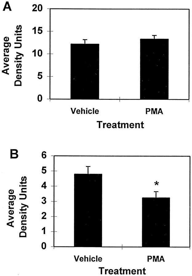
Fibrillar amyloid deposits are defining pathological lesions in Alzheimer's disease brain and are thought to mediate neuronal death. Amyloid is composed primarily of a 39-42 amino acid protein fragment of the amyloid precursor protein (APP), called amyloid beta-protein (Abeta). Because deposition of fibrillar amyloid in vitro has been shown to be highly dependent on Abeta concentration, reducing the proteolytic release of Abeta is an attractive, potentially therapeutic target. Here, the turnover rate of brain Abeta has been determined to define treatment intervals over which a change in steady-state concentration of Abeta could be measured. Mice producing elevated levels of human Abeta were used to determine approximate turnover rates for Abeta and two of its precursors, C99 and APP. The t1/2 for brain Abeta was between 1.0 and 2.5 hr, whereas for C99, immature, and fully glycosylated forms of APP695 the approximate t1/2 values were 3, 3, and 7 hr, respectively. Given the rapid Abeta turnover rate, acute studies were designed using phorbol 12-myristate 13-acetate (PMA), which had been demonstrated previously to reduce Abeta secretion from cells in vitro via induction of protein kinase C (PKC) activity. Six hours after intracortical injection of PMA, Abeta levels were significantly reduced, as measured by both Abeta40- and Abeta42-selective ELISAs, returning to normal by 12 hr. An inactive structural analog of PMA, 4alpha-PMA, had no effect on brain Abeta levels. Among the secreted N-terminal APP fragments, APPbeta levels were significantly reduced by PMA treatment, whereas APPalpha levels were unchanged, in contrast to most cell culture studies. These results indicate that Abeta is rapidly turned over under normal conditions and support the therapeutic potential of elevating PKC activity for reduction of brain Abeta.


Figures

Fig. 1.
Fig. 1.
Time-dependent changes in specific activity of mouse brain Aβ and C99. [35S]Methionine (500 μCi) was infused into the femoral vein of gene-targeted mice. At indicated hourly time points after midpoint of infusion, mice were perfused with Ringer’s solution, and brain APP fragments were isolated by immunoprecipitation and visualized using electrophoresis and exposure of resolved proteins to phosphorimage screens.A, Representative phosphorimage showing radiolabeled Aβ and C99. Graphs illustrating change in density of C99 (B) or Aβ (C) with time are shown. n = 3 at each time point. D, E, Representative immunoblots confirming equivalent absolute levels of Aβ and C99 during these experiments. Immobilized proteins used to obtain the phosphorimage in A were detected using antibody 6E10. D, Aβ; E, C99 and Aβ (from a longer exposure of D).
Fig. 2.
Fig. 2.
Change in specific activity of mouse brain APP with time. Mice were treated as reported in the legend to Figure 1 and Materials and Methods. A, Representative phosphorimage showing radiolabeled immature (i) and fully glycosylated (m) APP695. B, C, Time-dependent change in specific activity of immature (B) and fully glycosylated APP695 (C). n = 3 at each time point. D, Representative Western blot showing relatively constant absolute levels of APP, although specific activity was changing. E, Immunoblot confirming the predominant forms of APP synthesized by gene-targeted mouse. The most prominent band in the gene-targeted mouse brain sample (lane 2) co-electrophoreses with immature human APP695 from transgenic rat brain (lane 1) and lowest APP form isolated from human cortex (lane 3). The top band in the full-length APP complex (lane 2) co-electrophoreses with fully glycosylated human APP 695 from transgenic rat (lane 1).
Fig. 3.
Fig. 3.
ELISAs selective for Aβ40 and Aβ42 detect levels of endogenous Aβ, correctly reflecting gene dosage.A, Standard curve of Aβ1–40 generated using ELISA with a polyclonal antibody selective for Aβ40. B,Standard curve of Aβ1–42 using 42-selective ELISA. C,Soluble Aβ extracted from brains of gene-targeted mice having one copy (white bars) or two copies (black bars) of the targeted allele. n = 3 in each group. D, Immunoblot showing that the DEA extraction method does not release detectable levels of C99, an abundant, membrane-spanning form of APP. Lanes 2–4, Extracts immunoprecipitated with antibody 97, which recognizes the C terminus of C99; lane 1, recombinant C100 added into DEA extract before immunoprecipitation; lane 5, 0.5 ng of C100 loaded directly on to the gel.
Fig. 4.
Fig. 4.
Effect of PMA on levels of Aβ40 and Aβ42. Aβ proteins were significantly reduced 6 hr after treatment. PMA was injected into the parietal cortex of the gene-targeted mouse. Parietal cortex was removed, and Aβ was extracted using DEA–NaCl buffer, neutralized to pH 8.0, and analyzed by ELISA; vehicle values are inblack, and PMA values are in white. Aβ40 and Aβ42 were reduced by 31 (p < 0.0001) and 35% (p < 0.0001), respectively. Number of animals per group: 40 assay, vehicle, 30; PMA, 39; 42 assay, vehicle, 24; PMA, 33.
Fig. 5.
Fig. 5.
Time-dependent reduction in Aβ levels after PMA treatment. Aβ levels were unaffected by 4αPMA. A,Significant reductions in Aβ40 at 6 hr (P6,p < 0.001) were gone by 12 hr (P12). V, Vehicle; P, PMA. Number of animals per group: V6, 6; V12, 6; P6, 11; P12, 12. B,4αPMA (4αP) did not lower cortical Aβ40 levels after 6 hr compared with the active analog PMA (P, p < 0.03). Number of animals per group: V, 6; 4αP, 7;P, 6.
Fig. 6.
Fig. 6.
Effect of PMA on levels of APPα and APPβ in parietal cortex 6 hr after treatment. Cortical APPβ levels were significantly reduced by PMA treatment, whereas APPα levels are unchanged. These APP fragments were assayed from the DEA extracts used to measure Aβ. Equivalent amounts of protein were MeOH-precipitated and immunoblotted with either 6E10 (A) to visualize APPα or 54 (B) to visualize APPβ. The reduction of APPβ is significant to p < 0.02. Number of animals per group: vehicle, 19; PMA, 23.

References

    1. Baranyi A, Szente MB, Woody CD. Intracellular injection of phorbol ester increases the excitability of neurons of the motor cortex of awake cats. Brain Res. 1987;424:396–401. - PubMed
    1. Busciglio J, Gabuzda DH, Matsudaira P, Yankner BA. Generation of β-amyloid in the secretory pathway in neuronal and nonneuronal cells. Proc Natl Acad Sci USA. 1993;90:2092–2096. - PMC - PubMed
    1. Buxbaum JD, Oishi M, Chen HI, Pinkas-Kramarski R, Jaffe EA, Gandy SE, Greengard P. Cholinergic agonists and interleukin 1 regulate processing and secretion of the Alzheimer β/A4 amyloid protein precursor. Proc Natl Acad Sci USA. 1992;89:10075–10078. - PMC - PubMed
    1. Buxbaum JD, Koo EH, Greengard P. Protein phosphorylation inhibits production of Alzheimer amyloid β/A4 peptide. Proc Natl Acad Sci USA. 1993;90:9195–9198. - PMC - PubMed
    1. Cai XD, Golde TE, Younkin SG. Release of excess amyloid β protein from a mutant amyloid β protein precursor. Science. 1993;259:514–516. - PubMed

LinkOut - more resources