. 1998 Mar 1;18(5):1806-17.
doi: 10.1523/JNEUROSCI.18-05-01806.1998.

Intrastriatal mesencephalic grafts affect neuronal activity in basal ganglia nuclei and their target structures in a rat model of Parkinson's disease

Affiliations

Intrastriatal mesencephalic grafts affect neuronal activity in basal ganglia nuclei and their target structures in a rat model of Parkinson's disease

N Nakao et al. J Neurosci. .

Abstract

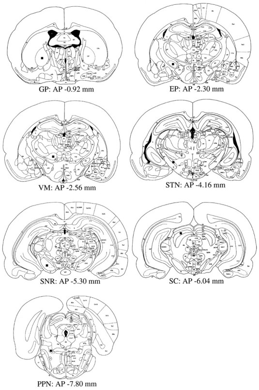
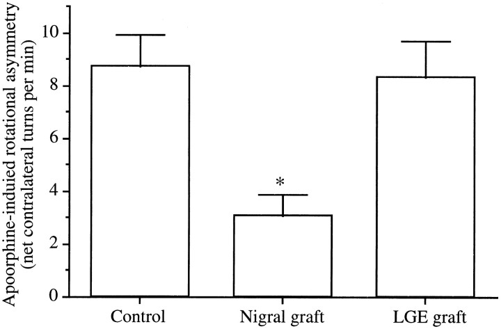
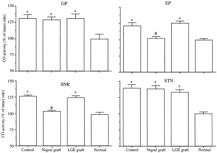
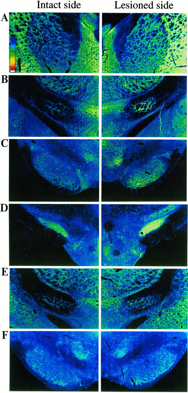
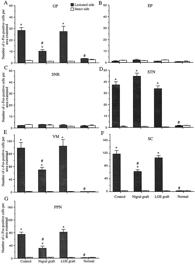
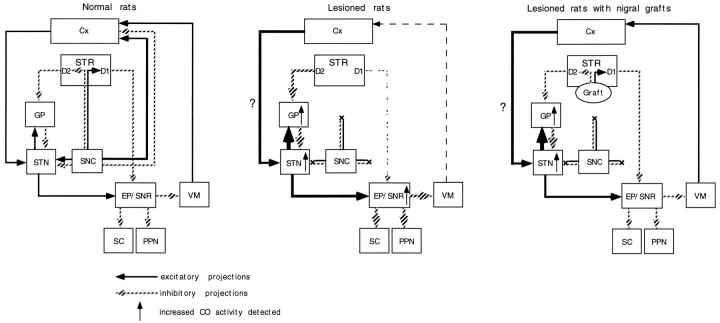
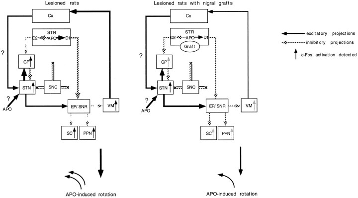
Nigrostriatal dopamine (DA) lesions lead to changes of neuronal activity in basal ganglia nuclei such as the globus pallidus (GP, the rodent homolog of lateral globus pallidus), entopeduncular nucleus (EP, the rodent homolog of medial globus pallidus), substantia nigra pars reticulata (SNR), and subthalamic nucleus (STN). We investigated in rats whether embryonic mesencephalic DA neurons grafted in the striatum may affect the lesion-induced alterations of neuronal activity in these structures. Regional neuronal activity was determined by use of quantitative cytochrome oxidase histochemistry. It was also examined in lesioned rats whether the grafts may regulate the expression of c-Fos after systemic administration of apomorphine in the basal ganglia nuclei as well as their target structures, including the ventromedial thalamic nucleus (VM), superior colliculus (SC), and pedunculopontine nucleus (PPN). Lesioned rats exhibited an increased activity of CO in the GP, EP, SNR, and STN ipsilateral to the lesion. Intrastriatal nigral grafts reversed the increases in the CO activity in the EP and SNR, whereas the grafts failed to affect the enzyme activity in the GP or STN. Apomorphine induced an increased expression of c-Fos in the GP, STN, VM, SC, and PPN on the lesioned side. The enhanced expression of this protein in all the structures except for the STN was attenuated by nigral grafts. The present results indicate that intrastriatal DA neuron grafts can normalize the lesion-induced changes of neuronal activity in the output nuclei of the basal ganglia as well as their target structures.


Figures

Fig. 1.
Fig. 1.
Schematic representation of anatomical regions in which c-Fos-positive cell numbers were evaluated (drawn on atlas of Paxinos and Watson, 1982). Closed boxes represent the regions at which a sampling grid for the determination of c-Fos-positive cell density was placed.
Fig. 2.
Fig. 2.
Net contralateral apomorphine-induced rotation asymmetry (full turns per minute) over the 60 min test session. Data represent the mean ± SEM. *p < 0.0001; significant difference from controls (one-way ANOVA with post hoc Scheffé’s test).
Fig. 3.
Fig. 3.
Cytochrome oxidase (CO) activity ipsilateral to nigrostriatal lesion in basal ganglia nuclei. CO activity on the lesioned side is expressed as a percentage of that on the intact side. Data are the mean ± SEM. *p < 0.0001; significant difference from normal animals (one-way ANOVA withpost hoc Scheffé’s test). #p< 0.0001; significant difference from controls (one-way ANOVA withpost hoc Scheffé’s test).
Fig. 4.
Fig. 4.
Digitized images of cytochrome oxidase (CO) activity in basal ganglia nuclei. In rats with unilateral nigrostriatal lesions, increased optical densities are noted in the GP (A), EP (B), SNR (C), and STN (D) ipsilateral to the lesion. In the lesioned animals with nigral grafts, the increases in CO activity are attenuated in the EP (E) and SNR (F).
Fig. 5.
Fig. 5.
Apomorphine-induced c-Fos activation in basal ganglia nuclei and their target structures. Values are the mean ± SEM of numbers of c-Fos-immunopositive cells per analyzed area. *p < 0.0001; significant difference from the intact side (paired two-tailed Student’s t test). #p < 0.0001; significant difference from controls (one-way ANOVA with post hoc Scheffé’s test).
Fig. 6.
Fig. 6.
Photomicrographs demonstrating c-Fos-immunopositive cells in the GP (A, B), EP (C), SNR (D), and STN (E) ipsilateral to nigrostriatal lesions after systemic injections of apomorphine. In animals with unilateral nigrostriatal lesions, the numbers of c-Fos-positive cells are increased in the GP (A) and STN (E) ipsilateral to the lesion after systemic apomorphine. No differences in the c-Fos cell numbers between the lesioned and intact sides are seen in the EP (C) or SNR (D). The apomorphine-induced enhancement of c-Fos expression in the GP (B) is attenuated in the lesioned rats with nigral grafts. Scale bar, 100 μm.
Fig. 7.
Fig. 7.
Photomicrographs showing c-Fos-immunopositive cells in the VM (A, B), SC (C, D), and PPN (E, F) on the lesioned side after systemic administrations of apomorphine. In the lesioned animals, the numbers of c-Fos-positive cells are increased in the VM (A), SC (C), and PPN (E) on the lesioned side after systemic apomorphine challenge. The apomorphine-induced enhancement of c-Fos expression in these structures is attenuated in the lesioned rats with nigral grafts (B, D, F). Scale bar, 100 μm.
Fig. 8.
Fig. 8.
Schematic representation of a hypothetical model of the functional organization of the basal ganglia based on the results of cytochrome oxidase histochemistry (modified from Albin et al., 1989). Lesions of the nigrostriatal bundle can deprive DA innervation of not only the striatum (STR) but also the the subthalamic nucleus (STN) and cortex (Cx). The hyperactivity of the entopeduncular nucleus (EP) and substantia nigra pars reticulata (SNR) is attributable to the lesion-induced suppression of direct inhibitory inputs from the striatum (direct pathway). In this model, the striatopallido–subthalamic pathway (indirect pathway) does not play a major role in the increased activity of the STN. The DA denervation of the STN and cortex could contribute to the overactivity of the STN through direct and indirect mechanisms, respectively. The hyperactivity of the STN may result in the increased activity of the globus pallidus (GP). The graft-derived restoration of DA levels in the striatum can attenuate the increased activity of the EP and SNR by a trans-synaptic mechanism. On the other hand, the intrastriatal grafts fail to affect the increased activity in the GP or STN.
Fig. 9.
Fig. 9.
Diagrammatic representation of a hypothetical model of a cascade of changes of neuronal activity in the basal ganglia after systemic injections of apomorphine (modified from Albin et al., 1989). After apomorphine (APO) challenge, the activity of striatopallidal GABAergic neurons with hypersensitive D2 receptors is suppressed, with consequent disinhibition of the globus pallidus (GP). Apomorphine could directly induce c-Fos activation in the DA-denervated subthalamic nucleus (STN). The lesion-induced DA denervation of the neocortex (Cx) could also affect the excitability of the STN to cause the c-Fos activation in the STN after injections of apomorphine. Stimulation of striatal D2 receptors with supersensitive state may suppress the activity of the entopeduncular nucleus (EP) and substantia nigra pars reticulata (SNR) through an inhibition of the activity of glutamatergic STN neurons (indirect pathway). The suppression of the activity of the EP and SNR can lead to disinhibition of the activity in their target structures, such as the ventromedial thalamic nucleus (VM), superior colliculus (SC), and pedunculopontine nucleus (PPN). Stimulation of striatal D1 receptors, which directly suppresses the neuronal activity in the EP and SNR (direct pathway), also leads to bursting activity in the VM, SC, and PPN by a disinhibitory mechanism. The graft-mediated reversal of the increased levels of striatal D1 and D2 receptors can partly reverse the altered activity of basal ganglia circuitry to attenuate the apomorphine-induced bursting activity of the GP as well as the basal ganglia target structures.

Similar articles

Cited by

References

    1. Abercrombie M. Estimation of nuclear population from microtome section. Anat Rec. 1946;94:239–247. - PubMed
    1. Afsharpour S. Topographical projections of the cerebral cortex to the subthalamic nucleus. J Comp Neurol. 1985;236:14–28. - PubMed
    1. Albin RL, Young AB, Penney JB. The functional anatomy of basal ganglia disorders. Trends Neurosci. 1989;12:366–375. - PubMed
    1. Berger B, Thierry AM, Tassin JP, Moyne MA. Dopaminergic innervation of the rat prefrontal cortex: a fluorescence histochemical study. Brain Res. 1976;106:133–145. - PubMed
    1. Björklund A. Dopaminergic transplants in experimental parkinsonism: cellular mechanisms of graft-induced functional recovery. Curr Opin Neurobiol. 1992;2:683–689. - PubMed

Publication types

MeSH terms