. 1998 Feb 17;95(4):1387-91.
doi: 10.1073/pnas.95.4.1387.

Parathyroid hormone activation of the 25-hydroxyvitamin D3-1alpha-hydroxylase gene promoter

Affiliations

Parathyroid hormone activation of the 25-hydroxyvitamin D3-1alpha-hydroxylase gene promoter

H L Brenza et al. Proc Natl Acad Sci U S A. .

Abstract

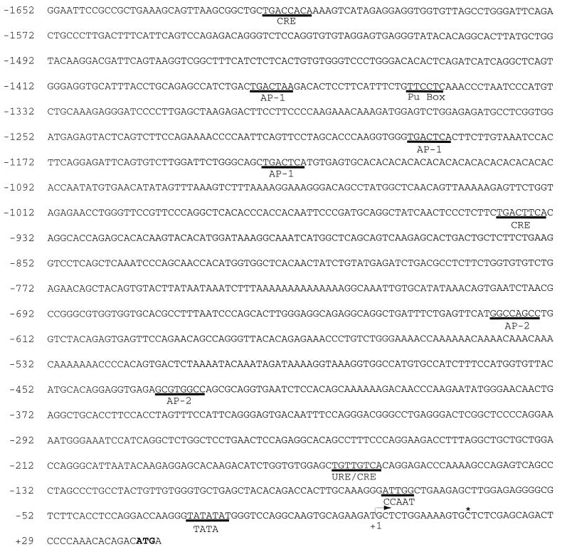
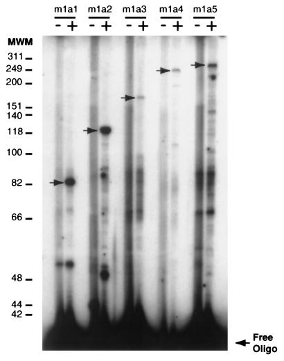
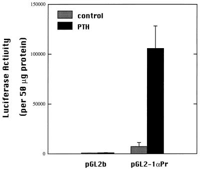
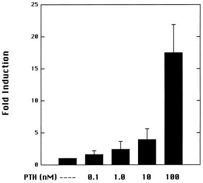
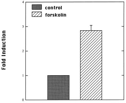
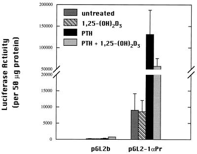
The DNA flanking the 5' sequence of the mouse 1alpha-hydroxylase gene has been cloned and sequenced. A TATA box has been located at -30 bp and aCCAAT box has been located at -79 bp. The gene's promoter activity has been demonstrated by using a luciferase reporter gene construct transfected into a modified pig kidney cell line, AOK-B50. Parathyroid hormone stimulates this promoter-directed synthesis of luciferase by 17-fold, whereas forskolin stimulates it by 3-fold. The action of parathyroid hormone is concentration-dependent. 1,25-Dihydroxyvitamin D3 does not suppress basal promoter activity and marginally suppresses parathyroid hormone-driven luciferase reporter activity. The promoter has three potential cAMP-responsive element sites, and two perfect and one imperfect AP-1 sites, while no DR-3 was detected. These results indicate that parathyroid hormone stimulates 25-hydroxyvitamin D3-1alpha-hydroxylase by acting on the promoter of the 1alpha-hydroxylase gene.


Figures

Figure 1
Figure 1
Nucleotide sequence of the 5′ flanking region of the mouse 1α-hydroxylase gene. The transcriptional start site is designated as +1. Putative cis regulatory elements are underlined. The translational start codon is indicated in bold. An asterisk (∗) indicates the end of the published mouse 1α-hydroxylase cDNA.
Figure 2
Figure 2
Primer extension analysis of mouse kidney 1α-hydroxylase mRNA population. Primer extension has been performed with op/op mouse kidney mRNA (+ lanes) by using five different labeled primers: m1a1, m1a2, m1a3, m1a4, and m1a5 (see Materials and Methods section for specific sequences). Control lanes (−) represent primer extensions performed with the primer alone. Primer extension products have been fractionated on a sequencing gel. Arrows represent specific 1α-hydroxylase cDNAs. Molecular weight markers (MWM) are indicated in nucleotide bases.
Figure 3
Figure 3
Transcriptional activity of the mouse 1α-hydroxylase 1.7-kb 5′ flanking sequence. AOK-B50 cells were transiently transfected with the promoter luciferase reporter construct, pGL2–1αPr, or empty pGL2b by using lipofectin. Transfected cells were treated for 24 h with 100 nM PTH or vehicle (water). Luciferase activity was normalized to protein content for each sample. Experiments were repeated three times in duplicate. Values are mean ± SD.
Figure 4
Figure 4
Dose-dependent stimulation of mouse 1α-hydroxylase gene promoter by PTH. AOK-B50 cells were transfected with pGL2–1αPr. Transfected cells were treated for 24 h with the indicated concentrations of PTH. The induction is relative to luciferase activity detected in untreated transfected cells. Experiments were repeated three times in duplicate.
Figure 5
Figure 5
Activation of mouse 1α-hydroxylase promoter by forskolin. AOK-B50 cells were transfected with pGL2–1αPr. Transfected cells were treated for 24 h with 10 μM forskolin or vehicle (ethanol). Data are presented as fold activation relative to vehicle-treated transfected cells. Values were obtained from three independent experiments performed in duplicate.
Figure 6
Figure 6
1,25-(OH)2D3 suppression of PTH-stimulated luciferase activity in AOK-B50 cells. Cells were transfected with pGL2–1αPr. Transfected cells were treated with 10 nM 1,25-(OH)2D3, 100 nM PTH, and both 1,25-(OH)2D3 and PTH or vehicle (water and/or ethanol) for 24 h. Luciferase activity is corrected for protein. Experiments were performed at least two times in duplicate. Values are mean ± SD.

Similar articles

Cited by

References

    1. DeLuca H F. The Harvey Lectures. New York: Academic; 1981. , Series 75, pp. 333–379.
    1. Ross T K, Darwish H M, DeLuca H F. Vitam Horm. 1994;49:281–326. - PubMed
    1. Prader A, Illig R, Heierli E. Helv Paediatr Acta. 1961;16:452–468. - PubMed
    1. Scriver C R. N Engl J Med. 1978;299:976–979. - PubMed
    1. Fraser D, Kooh S W, Kind H P, Holick M F, Tanaka Y, DeLuca H F. N Engl J Med. 1973;289:817–822. - PubMed

Publication types

Associated data

LinkOut - more resources