. 1998 Feb 17;95(4):1432-7.
doi: 10.1073/pnas.95.4.1432.

RAFT1 phosphorylation of the translational regulators p70 S6 kinase and 4E-BP1

Affiliations

RAFT1 phosphorylation of the translational regulators p70 S6 kinase and 4E-BP1

P E Burnett et al. Proc Natl Acad Sci U S A. .

Abstract

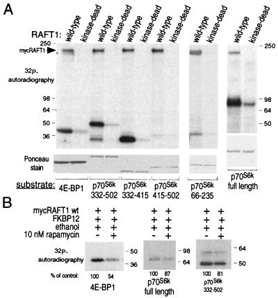
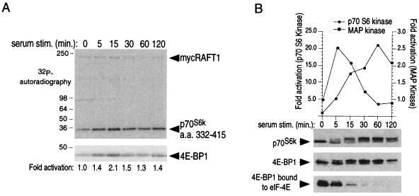
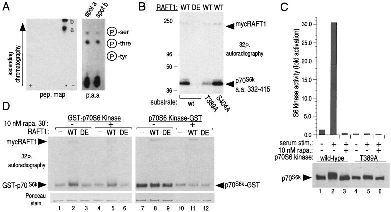
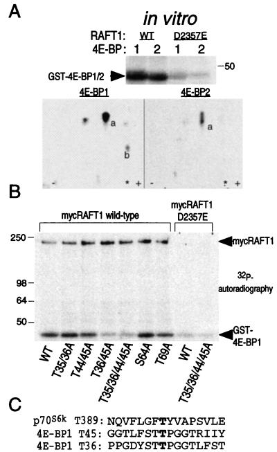
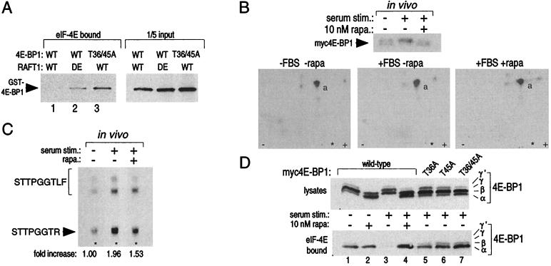
The complex of rapamycin with its intracellular receptor, FKBP12, interacts with RAFT1/FRAP/mTOR, the in vivo rapamycin-sensitive target and a member of the ataxia telangiectasia mutated (ATM)-related family of kinases that share homology with the catalytic domain of phosphatidylinositol 3-kinase. The function of RAFT1 in the rapamycin-sensitive pathway and its connection to downstream components of the pathway, such as p70 S6 kinase and 4E-BP1, are poorly understood. Here, we show that RAFT1 directly phosphorylates p70(S6k), 4E-BP1, and 4E-BP2 and that serum stimulates RAFT1 kinase activity with kinetics similar to those of p70(S6k) and 4E-BP1 phosphorylation. RAFT1 phosphorylates p70(S6k) on Thr-389, a residue whose phosphorylation is rapamycin-sensitive in vivo and necessary for S6 kinase activity. RAFT1 phosphorylation of 4E-BP1 on Thr-36 and Thr-45 blocks its association with the cap-binding protein, eIF-4E, in vitro, and phosphorylation of Thr-45 seems to be the major regulator of the 4E-BP1-eIF-4E interaction in vivo. RAFT1 phosphorylates p70(S6k) much more effectively than 4E-BP1, and the phosphorylation sites on the two proteins show little homology. This raises the possibility that, in vivo, an unidentified kinase analogous to p70(S6k) is activated by RAFT1 phosphorylation and acts at the rapamycin-sensitive phosphorylation sites of 4E-BP1.


Figures

Figure 1
Figure 1
RAFT1 phosphorylates 4E-BP1 and the C-terminal half of p70S6k in vitro. (A) Wild-type and kinase-dead RAFT1 expressed in HEK293 cells were purified by immunoprecipitation and incubated in a kinase assay with [γ-32P]ATP and the indicated fusion proteins of 4E-BP1 or p70S6k. The phosphorylated proteins are revealed by autoradiography (Upper), and a Ponceau stain reveals the relative amounts of each fusion protein in the assays (Lower). (B) FKBP12-rapamycin partially blocks RAFT1 phosphorylation of 4E-BP1 and p70S6k. Kinase assays using the indicated substrates were performed as in A except that the immunoprecipitates were first incubated on ice for 1 h with 100 nM FKBP12 with or without 10 nM rapamycin. These results are representative of five similar experiments.
Figure 2
Figure 2
Serum stimulates RAFT1 kinase activity toward p70S6k and 4E-BP1. (A) Wild-type RAFT1 purified from quiescent HEK293 cells stimulated with 10% serum for the indicated times and purified by immunoprecipitation was incubated in a kinase assay with indicated fusion proteins and [γ-32P]ATP. After SDS-PAGE, transfer to poly(vinylidene difluoride) and Ponceau staining, the bands corresponding to p70S6k 332–415 and 4E-BP1 were excised, and the amount of radioactivity incorporated into each was determined by scintillation counting. The increases in 32P incorporation were similar for p70S6k 332–415 and 4E-BP1, and the fold activation for p70S6k is shown below the autoradiograph. The results shown are representative of three similar experiments. (B) Time course of p70S6k phosphorylation (Top) and activation (graph) and 4E-BP1 phosphorylation and release from eIF-4E (Middle and Bottom). p70S6k and 4E-BP1 were detected with immunoblotting. Kinetics of endogenous MAP kinase activation is shown for comparison (graph).
Figure 3
Figure 3
RAFT1 phosphorylates p70S6k on Thr-389. (A) Chymotryptic peptide map of RAFT1-phosphorylated p70S6k 332–415 and phosphoamino acid analysis of the purified peptides. Peptides “a” is an incomplete digest of peptide “b,” and tryptic peptide maps reveal only one spot (data not shown). (B) Substitution of a T389A mutant for wild-type p70S6k 332–415 eliminates the majority of RAFT1 phosphorylation. (C) T389A p70S6k expressed in HEK293 cells is not activated by serum stimulation (graph) and displays decreased laddering on SDS-PAGE. (D) Wild-type (WT) but not kinase-dead (DE) RAFT1 phosphorylates HEK293-purified p70S6k fused to GST at the N terminus (Left) but not at the C terminus (Right). All the p70S6k fusion proteins are capable of autophosphorylating (−). The amount of added p70S6k is roughly equal except for those with p70S6k-GST from rapamycin-treated cells, which contain about one-third of the amount of the others (Lower).
Figure 4
Figure 4
RAFT1 phosphorylates 4E-BP1 on Thr-36 and Thr-45. (A) Phosphoamino acid analysis reveals that RAFT1 phosphorylates 4E-BP1 on threonine (data not shown). Tryptic peptide maps (Lower) of RAFT-phosphorylated 4E-BP1 and 4E-BP2 (Upper) share a major peptide (marked “a” in Lower) that migrates as a single spot in the 4E-BP1 map but as three closely spaced spots in the 4E-BP2 map. The asterisk indicates where the sample was applied. Spot “b” is seen in maps of 4E-BP1 phosphorylated by both wild-type and kinase-dead RAFT1 and represents the site of phosphorylation of a contaminating kinase. Spots visible to the left and right of spot “a” in the 4E-BP1 map are partial digestion products of the peptide in spot “a” (data not shown). (B) Thr-36 and Thr-45 are the major sites of phosphorylation by RAFT1 on 4E-BP1. The indicated GST-4E-BP1 mutants were phosphorylated in vitro by RAFT1 as in Fig. 1A. Phosphorylation of mutants S64A and T69A are shown for comparison. (C) Comparison of RAFT1 phosphorylation sites on p70S6k and 4E-BP1.
Figure 5
Figure 5
Thr-36 and Thr-45 are phosphorylated in vivo, and mutations of these residues to alanine create a 4E-BP1 variant that binds constitutively to eIF-4E in vitro and in vivo. (A) 4E-BP1 phosphorylated in vitro by wild-type (WT) RAFT1, but not by kinase-dead (DE) RAFT, cannot bind to eIF-4E (Left, lanes 1 and 2). Even after an incubation with wild-type RAFT1 in a kinase assay, the T36/45A mutant still binds to eIF-4E (Left, lane 3). Equal amounts of GST-4E-BP1 were offered for binding in each condition (Right). Kinase assays of 30-min duration were performed as in Fig. 1A except that only 100 ng of fusion protein was used. The reaction products were then offered for binding to recombinant eIF-4E prebound to 7-methyl-GTP Sepharose. Bound 4E-BP1 was detected by immunoblotting with an anti-GST antibody. (B) Tryptic peptide maps (Lower) of 4E-BP1 isolated form cells labeled in vivo with 32P (Upper) and treated with the indicated conditions. Peptide “a” is eliminated in maps prepared with a T36/45A mutant (data not shown). (C) Serum stimulation increases phosphate content at Thr-45 2-fold, and rapamycin blocks 50% of the increase. Peptide “a” was purified from the TLC plates of B and analyzed as described in Materials and Methods. The spots were quantitated with densitometry. (D) Alanine mutations at Thr-36 and Thr-45 prevent serum-induced release of 4E-BP1 from eIF-4E and mimic the effects of rapamycin. HEK293 cells were transfected with plasmids encoding wild-type or the indicated mutant 4E-BP1s, treated with rapamycin and serum, and the migration pattern of 4E-BP1 (Upper) or the amount of 4E-BP1 bound to eIF-4E (Lower) was determined by immunoblotting.

References

    1. Nielsen F C, Ostergaard L, Nielsen J, Christiansen J. Nature (London) 1995;377:358–362. - PubMed
    1. Brown E J, Schreiber S L. Cell. 1996;86:517–520. - PubMed
    1. Sonenberg N. In: Translational Control. Hershey J W B, Mathews M B, Sonenberg N, editors. Plainview, NY: Cold Spring Harbor Lab. Press; 1996. pp. 245–269.
    1. Martel R R, Klicius J, Galet S. Can J Physiol Pharmacol. 1977;55:48–51. - PubMed
    1. Barbet N C, Schneider U, Helliwell S B, Stansfield I, Tuite M F, Hall M N. Mol Biol Cell. 1996;7:25–42. - PMC - PubMed

Publication types

MeSH terms