. 1998 Feb 17;95(4):1511-6.
doi: 10.1073/pnas.95.4.1511.

A unique glucose-dependent apoptotic pathway induced by c-Myc

Affiliations

A unique glucose-dependent apoptotic pathway induced by c-Myc

H Shim et al. Proc Natl Acad Sci U S A. .

Abstract

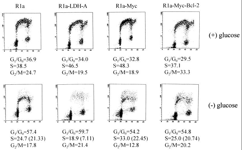
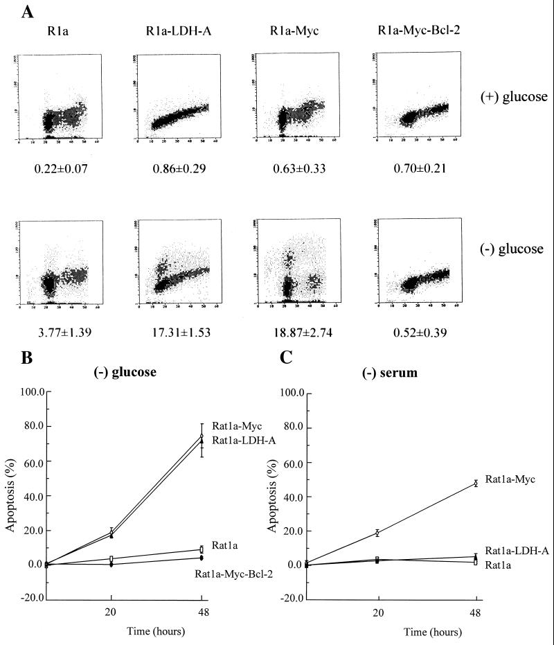
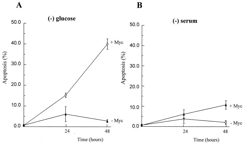
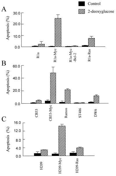
The lactate dehydrogenase A (LDH-A) gene, whose product participates in normal anaerobic glycolysis and is frequently increased in human cancers, has been identified as a c-Myc-responsive gene. It was of interest, therefore, to compare the effect of glucose deprivation in c-Myc-transformed and nontransformed cells. We observed that glucose deprivation or treatment with the glucose antimetabolite 2-deoxyglucose caused nontransformed cells to arrest in the G0/G1 phase of the cell cycle. In contrast, c-Myc-transformed fibroblasts, lymphoblastoid, or lung carcinoma cells underwent extensive apoptosis. Ectopic expression of LDH-A alone in Rat1a fibroblasts was sufficient to induce apoptosis with glucose deprivation but not with serum withdrawal, suggesting that LDH-A mediates the unique apoptotic effect of c-Myc when glycolysis is blocked. The apoptosis caused by glucose deprivation was blocked by Bcl-2 expression but appeared to be independent of wild-type p53 activity. These studies provide insights on the coupling of glucose metabolism and the cell cycle in c-Myc-transformed cells and may in the future be exploited for cancer therapeutics.


Figures

Figure 1
Figure 1
Cell cycle distribution and BrdUrd incorporation characteristics of Rat1a, Rat1a-LDH-A, Rat1a-Myc, and Rat1a-Myc-Bcl-2 cells. Two-dimensional flow cytometric distributions of DNA content stained by propidium iodide (abscissa) and BrdUrd labeling (ordinate) are shown for each cell line cultured for 20 h in the presence (Upper) or absence (Lower) of glucose. Distributions of nuclei in each compartment (G0/G1, S, or G2/M phases) were estimated by curve fitting and are shown below. (Lower) Numbers in parentheses for the S phase indicate the percentage of BrdUrd-labeled cells. (Upper) The percentage of cells in S phase and those labeled with BrdUrd were equal.
Figure 2
Figure 2
(A) Glucose-deprivatio-induced apoptotic cell death of c-Myc- or LDH-A-overexpressing cells. Rat1a, Rat1a-LDH-A, Rat1a-Myc, and Rat1a-Myc-Bcl-2 cells were cultured for 20 h in the presence (Upper) or absence (Lower) of glucose. Cells were harvested, and DNA strand breaks were labeled with biotin-dUTP by using TdT and then stained with propidium iodide. DNA content determined by propidium iodide staining is shown on the abscissa and DNA strand breaks content is shown on the ordinate. The numbers below indicate the percentage of cells that were apoptotic and labeled with biotin-dUTP. (B) Time course of glucose-deprivation-induced apoptotic death of Rat1a-Myc and Rat1a-LDH-A cells as compared with Rat1a and Rat1a-Myc-Bcl-2 cells. (C) Time course of serum-deprivation-induced apoptotic death of Rat1a-Myc cells as compared with Rat1a-LDH-A and Rat1a cells.
Figure 3
Figure 3
Myc-mediated apoptosis caused by glucose deprivation appears to be independent of wild-type p53 activity. (A) A mouse embryo fibroblast cell line, (10.1)Val5-Myc (+Myc) that overexpresses c-Myc and lacks the wild-type p53 function at 39°C, underwent more extensive apoptosis caused by glucose deprivation when compared with (10.1)Val5 cells (−Myc) that do not overexpress murine c-Myc. (B) Mouse fibroblast cells were grown in DMEM with 10% FBS and deprived of serum (0.1% FBS) for 24 or 48 h before flow cytometric analysis for apoptosis.
Figure 4
Figure 4
The glycolysis inhibitor 2-DG induces apoptosis in c-Myc-overexpressing cells. (A) Rat fibroblasts exposed to 10 mM 2-DG for 1 day were harvested for flow cytometric analysis of apoptosis. (B) Various lymphoid cells were exposed to 10 mM 2-DG for 1 day before they were fixed for analysis of apoptosis. (C) Human small cell lung cancer cells were treated with 10 mM 2-DG for 1 day before subjected to the apoptosis assay.

References

    1. Cole M D. Annu Rev Genet. 1986;20:361–384. - PubMed
    1. Dalla-Favera R, Bregni M, Erikson J, Patherson D, Gallo R C, Croce C M. Proc Natl Acad Sci USA. 1982;79:7824–7827. - PMC - PubMed
    1. Dang C V, Lee L A. c-Myc Function in Neoplasia. Austin, TX: Landes; 1995.
    1. Eilers M, Picard D, Yamamoto K R, Bishop J M. Nature (London) 1989;340:66–68. - PubMed
    1. Eilers M, Schirm S, Bishop J M. EMBO J. 1991;10:133–141. - PMC - PubMed

Publication types

LinkOut - more resources