. 1998 Feb 17;95(4):1630-4.
doi: 10.1073/pnas.95.4.1630.

Menin, the product of the MEN1 gene, is a nuclear protein

Affiliations

Menin, the product of the MEN1 gene, is a nuclear protein

S C Guru et al. Proc Natl Acad Sci U S A. .

Abstract

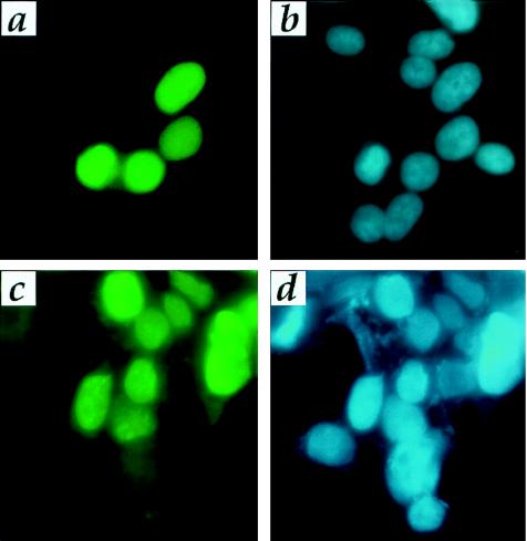
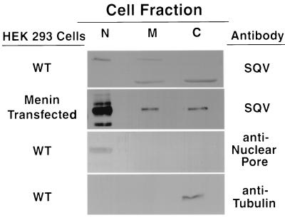
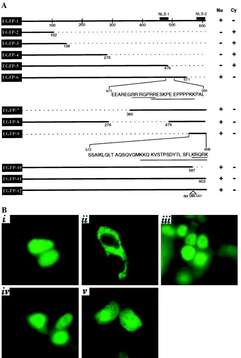
The MEN1 gene, mutations in which are responsible for multiple endocrine neoplasia type 1 (MEN1), encodes a 610-amino acid protein, denoted menin. The amino acid sequence of this putative tumor suppressor offers no clue to the function or subcellular location of the protein. We report herein, based on immunofluorescence, Western blotting of subcellular fractions, and epitope tagging with enhanced green fluorescent protein, that menin is located primarily in the nucleus. Enhanced green fluorescent protein-tagged menin deletion constructs identify at least two independent nuclear localization signals (NLS), both located in the C-terminal fourth of the protein. Among the 68 known independent disease-associated mutations, none of the 22 missense and 3 in-frame deletions affect either of the putative NLS sequences. However, if expressed, none of the truncated menin proteins resulting from the 43 known frameshift/nonsense mutations would retain both the NLSs. The precise role(s) of menin in the nucleus remain to be understood.


Figures

Figure 1
Figure 1
Immunofluorescence of menin-transfected HEK-293T cells with menin and myc epitope antibodies. Twenty-four hours after transfection with pcDNA3.1-menin, cells were processed for immunofluorescence with menin antibody (KC27) or with myc antibody followed by fluorescein isothiocyanate-conjugated secondary antibody detection. Immunofluorescence pattern with menin antibody (a) and the DAPI staining (b) showing the nuclei from the same cells. Immunofluorescence with myc antibody (c) and the DAPI staining (d) showing the nuclei from the same cells. Note that not all cells are positive, because this is a transient transfection. Endogenous levels of menin are not detectable above background.
Figure 2
Figure 2
Immunoblot of representative nuclear (N), membrane (M), and cytoplasmic (C) fractions of HEK-293T cells transfected with vector only (WT) or with menin. Fifty micrograms of protein was loaded for each fraction for the menin blot in WT cells, 5 μg of protein was used for the menin blot in menin-transfected cells, 25 μg of protein is present in each lane for the nuclear pore protein blot, and 12.5 μg of protein was used per lane for the tubulin blot. The blots shown for tubulin and for nuclear pore protein were from WT cells, but blots of fractions from menin-transfected cells gave comparable results.
Figure 3
Figure 3
(A) Schematic of deletion constructs of menin fused to EGFP and localization of green fluorescence observed in the transfected cells. All constructs (EGFP1–EGFP12) were generated with EGFP (boxed) at the N terminus of the menin coding region. The extent of the menin coding region retained in each construct is represented by a solid thick line and the deleted region is represented by a dotted thin line. The position of the starting and the terminating amino acid in each deletion construct is also indicated, except for amino acids 1 and 610. The presence (+) or absence (−) of green fluorescence observed in the nucleus (Nu) or cytoplasm (Cy) for each construct is shown. The sequence around the two putative independent NLSs mapped in this study (NLS-1 and NLS-2) are shown for constructs EGFP6 and EGFP9, respectively. Underlined sequences represent potential single basic type and bipartite sequences in both NLS-1 and NLS-2. (B) Representative examples of green fluorescence in HEK-293T cells transfected with EGFP-menin fusion constructs. The presence of green fluorescence in the nucleus or cytoplasm of cells transfected with EGFP1 (i), EGFP5 (ii), EGFP6 (iii), and EGFP9 (iv) is shown. The diffuse pattern of fluorescence observed in cells transfected with EGFP vector alone (v) is also shown

References

    1. Metz D C, Jensen R T, Bale A E, Skarulis M C, Eastman R C, Nieman L, Norton J A, Friedman E, Larsson C, Amorossi A, et al. In: The Parathyroids. Bilezekian J P, Levine M A, Marcus R, editors. New York: Raven; 1994. pp. 591–646.
    1. Chandrasekharappa S C, Guru S C, Manickam P, Olufemi S E, Collins F S, Emmert-Buck M R, Debelenko L V, Zhuang Z, Lubensky I A, Liotta L A, et al. Science. 1997;276:404–407. - PubMed
    1. Agarwal S K, Kester M B, Debelenko L V, Heppner C, Emmert-Buck M R, Skarulis M C, Doppman J L, Kim Y S, Lubensky I A, Zhuang Z, et al. Hum Mol Genet. 1997;6:1169–1175. - PubMed
    1. The European Consortium on MEN1. Hum Mol Genet. 1997;6:1177–1183. - PubMed
    1. Heppner C, Kester M B, Agarwal S K, Debelenko L V, Emmert-Buck M R, Guru S C, Manickam P, Olufemi S E, Skarulis M C, Doppman J L, et al. Nat Genet. 1997;16:375–378. - PubMed

LinkOut - more resources