. 1998 Feb 17;95(4):1730-4.
doi: 10.1073/pnas.95.4.1730.

A role for B cells in the development of T cell helper function in a malaria infection in mice

Affiliations
Free PMC article

A role for B cells in the development of T cell helper function in a malaria infection in mice

J Langhorne et al. Proc Natl Acad Sci U S A. .
Free PMC article

Abstract

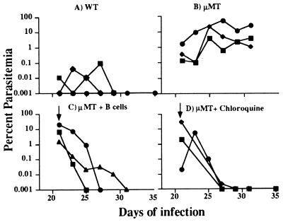
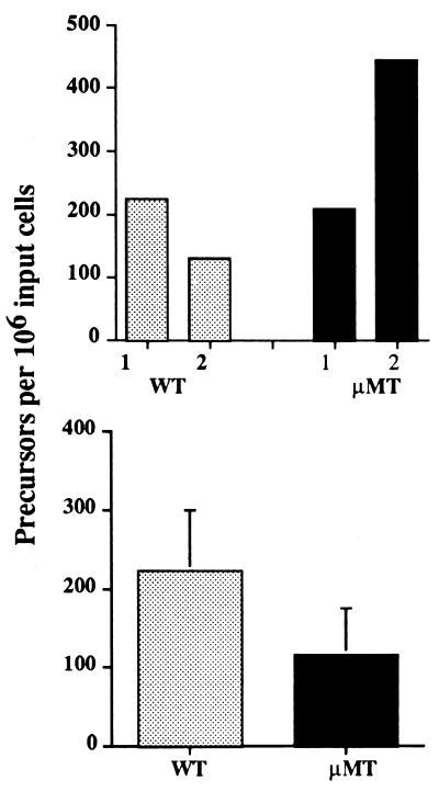
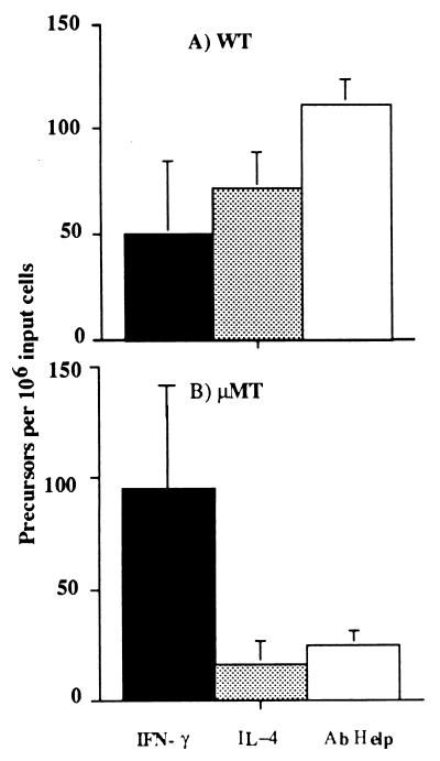
B cell knockout mice are unable to clear a primary erythrocytic infection of Plasmodium chabaudi chabaudi. However, the early acute infection is controlled to some extent, giving rise to a chronic relapsing parasitemia that can be reduced either by drug treatment or by adoptive transfer of B cells. Similar to mice rendered B-cell deficient by lifelong treatment with anti-mu antibodies, B cell knockout mice (muMT) retain a predominant CD4+ Th1-like response to malarial antigens throughout a primary infection. This contrasts with the response seen in control C57BL/6 mice in which the CD4+ T-cell response has switched to that characteristic of Th2 cells at the later stages of infection, manifesting efficient help for specific antibodies in vitro and interleukin 4 production. Both chloroquine and adoptive transfer of immune B cells reduced parasite load. However, the adoptive transfer of B cells resulted in a Th2 response in recipient muMT mice, as indicated by a relative increase in the precursor frequency of helper cells for antibody production. These data support the idea that B cells play a role in the regulation of CD4+ T subset responses.


Figures

Figure 1
Figure 1
Course of a primary P. chabaudi infection in female WT (A) and μMT mice (B) and in μMT receiving immune B cells (C) or chloroquine and pyrimethamine (D). Each line represents the infection in an individual mouse. The arrow indicates the time of adminstration of immune B cells (2 × 10⁷ given intravenously) or chloroquine and pyrimethamine (as described in text).
Figure 2
Figure 2
Precursor frequency analysis of CD4+ T cells from P. chabaudi-infected WT (□) and μMT (▪) mice able to proliferate in response to malarial Ags in vitro. The data are expressed as precursor frequencies, calculated as described in text. (Upper) At 8 days of infection. The bars show the responses in 2 experiments, each performed with a pool of cells from 2 mice. (Lower) At 30 days of infection. The bars represent the mean precursor frequencies obtained in 6 independent experiments. The standard errors of the mean are shown.
Figure 3
Figure 3
Comparison of precursor frequencies of CD4+ T cells producing IFN-γ (▪) or Il-4 (░⃞) or providing help for B cells (□) from WT (A) and μMT (B) mice obtained 40 days after infection with P. chabaudi. The histograms represent the mean precursor frequencies derived from the frequencies calculated from five, six, and four separate experiments for IFN-γ, Ab Help, and IL-4, respectively. The standard errors of the means are shown.
Figure 4
Figure 4
Comparison of precursor frequencies of CD4+ T cells taken from individual WT and μMT mice after treatment with chloroquine or after adoptive transfer of immune B cells. CD4+ T cells were taken at 50 days of infection from WT and μMT mice (a and c, respectively), WT and μMT mice treated with Chloroquine at 30 days of infection (b and d, respectively), and μMT mice that received 2 × 10⁷ immune B cells i.v. at 30 days of infection (e). The percentages of B220+ cells in the spleens of the reconstituted mice were: (1) 6.75%; (2) 6.95%; (3) 6.36%, and (4) 1%. Precursor frequencies of IFN-γ (▪) and T helper cells for Ab production (□) are shown for each mouse.

References

    1. Hsieh C-S, Heimberger A B, Gold J S, O’Garra A, Murphy K M. Proc Natl Acad Sci USA. 1992;89:6065–6060. - PMC - PubMed
    1. Macatonia S E, Hsieh C-S, Murphy K M, O’Garra A. Int Immunol. 1993;5:1119–1128. - PubMed
    1. Constant S L, Bottomly K. Annu Rev Immunol. 1997;15:297–322. - PubMed
    1. Seder R A, Paul W E. Annu Rev Immunol. 1994;12:635–637. - PubMed
    1. Langhorne J, Gillard S, Simon B, Slade S, Eichmann K. Int Immunol. 1989;1:416–424. - PubMed

Publication types