. 1998 Feb 17;95(4):1770-5.
doi: 10.1073/pnas.95.4.1770.

From differentiation to proliferation: the secretory amyloid precursor protein as a local mediator of growth in thyroid epithelial cells

Affiliations

From differentiation to proliferation: the secretory amyloid precursor protein as a local mediator of growth in thyroid epithelial cells

C U Pietrzik et al. Proc Natl Acad Sci U S A. .

Abstract

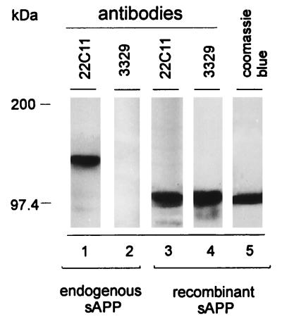
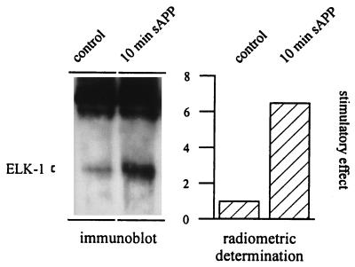
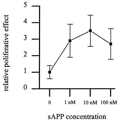
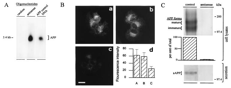
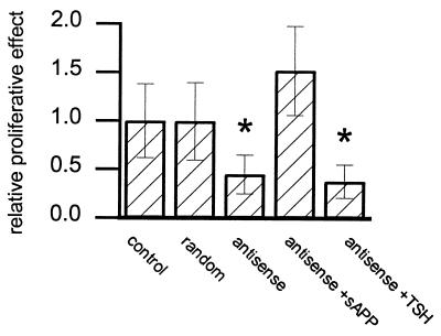
In various species, thyrotropin (TSH) is known to stimulate both differentiation and proliferation of thyroid follicle cells. This cell type has also been shown to express members of the Alzheimer amyloid precursor (APP) protein family and to release the secretory N-terminal domain of APP (sAPP) in a TSH-dependent fashion. In this study on binding to the cell surfaces, exogenously added recombinant sAPP stimulated phosphorylation mediated by mitogen-activated protein kinase and effectively evoked proliferation in the rat thyroid epithelial cell line FRTL-5. To see whether this proliverative effect of sAPP is of physiological relevance, we used antisense techniques to selectively inhibit the expression of APP and the proteolytic release of sAPP by cells grown in the presence of TSH. The antisense-induced inhibition was detected by immunoblot, immunoprecipitation, and immunocytochemical analyses. After the reduced APP expression and sAPP secretion, we observed a strong suppression of the TSH-induced cell proliferation down to 35%. Recombinant sAPP but not TSH was able to overcome this antisense effect and to completely restore cell proliferation, indicating that sAPP acts downstream of TSH, in that it is released from thyroid epithelial cells during TSH-induced differentiation. We propose that sAPP operates as an autocrine growth factor mediating the proliferative effect of TSH on neighboring thyroid epithelial cells.


Figures

Figure 1
Figure 1
Immunocytochemical localization of endogenous APP in FRTL-5 cells (A) and of recombinant sAPP (B and D). Visualization of endogenous APP with an antibody directed against the cytoplasmic C-terminal domain (antiserum 2189) resulted in a characteristic crescent perinuclear staining indicative of a Golgi localization of APP (A). For visualization of recombinant sAPP, cells were incubated with 100 nM recombinant His-tagged sAPP for 20 min at 4°C, i.e., under conditions that halted membrane flow. The recombinant sAPP was immunolabeled with specific antibodies recognizing only recombinant sAPP, but not endogenous APP (antibody 3329; see Fig. 2), and detected with either fluorescently labeled (B) or gold-conjugated antibodies followed by silver enhancement (D). The phase-contrast image corresponding to B is shown in C. [Bars = 10 μm (A and B) and 2 μm (D).]
Figure 2
Figure 2
Antibody 3329 selectively recognizes recombinant sAPP. In immunoblots monoclonal antibody 22C11 reacted with both the secreted sAPP of FRTL-5 cells (lane 1) and the purified recombinant sAPP expressed in E. coli (lane 3). Coomassie blue staining of the purified recombinant sAPP is shown in lane 5. The polyclonal rabbit antibody 3329 reacted exclusively with the recombinant sAPP (lane 4) and showed no reaction with endogenous sAPP (lane 2). This antibody was therefore used for binding studies with recombinant sAPP on FRTL-5 cells.
Figure 3
Figure 3
Effect of 10 nM recombinant sAPP on the stimulation of MAP kinase activity in the absence of TSH. Detection of ELK-1 fusion protein by immunoblot analysis using a phospho-ELK-1-specific antibody showed a significant increase in Elk-1 phosphorylation after the addition of 10 nM sAPP for 10 min as compared with untreated control cells. The stimulatory effect of 10 nM sAPP was determined radiometrically by the use of [γ-32P]ATP and radiolabeled ELK-1 fusion protein. The control value was set as 1.
Figure 4
Figure 4
Effect of recombinant sAPP on BrdUrd incorporation in TSH-stimulated FRTL-5 cells. To investigate the contribution of sAPP to the proliferative effect, FRTL-5 cells were incubated with the indicated concentrations of sAPP. Note that addition of 10 nM sAPP resulted in a 3.5-fold proliferative increase compared with controls. Higher concentrations (100 nM) of sAPP resulted in a decreased stimulatory effect.
Figure 5
Figure 5
(A) Binding and effect of antisense oligonucleotides on the expression of APP and the release of sAPP. Northern blot analysis of total RNA preparations from FRTL-5 cells using [γ-32P]ATP-radiolabeled oligonucleotides. The antisense oligonucleotides bound to the RNA (lane antisense), whereas the random oligonucleotides did not (lane random). A PCR fragment from APP (lane APP control DNA) was used as a standard, indicating the size of APP mRNA (3.4 kb) in the total RNA preparation. (B) Immunofluorescence localization of endogenous APP using an antibody directed against the C terminus of APP (a). Effect of 25 μM random oligonucleotide-treated cells (b) and 25 μM antisense oligonucleotide-treated cells (c) on the expression of APP. (Bar = 10 μm.) The total fluorescence intensity of cells was measured by the use of a laser scanning microscope (d). Total protein synthesis in FRTL-5 cells remained unaltered during treatment with oligonucleotides (data not shown), whereas the expression of APP was selectively reduced after antisense treatment (c). (C) Effect of antisense oligonucleotides on the synthesis of APP and the secretory release of sAPP. Cells grown in the presence or absence of oligonucleotides for 48 hr were biosynthetically radiolabeled with [35S]methionine/[35S]cysteine (3.7 MBq/ml) for 3 hr. APP was immunoprecipitated from cell lysates with an antibody directed against the C terminus of APP, analyzed by SDS/PAGE, and quantified radiometrically (Top). Immunoblot analysis showed the effect of antisense oligonucleotides on the secretion of sAPP by using an antibody directed against the N terminus of APP (Bottom). Note that the expression of APP and the release of sAPP in antisense-treated cells was significantly decreased as compared with control cells.
Figure 6
Figure 6
Effect of antisense and random oligonucleotides on the proliferation of FRTL-5 cells. Note that the proliferative effect was reduced to 35% (∗, P < 0.05) when cells were cultured in medium containing 25 μM antisense oligonucleotides (bar antisense) compared with the proliferative effect of cells cultured in the presence or absence of 25 μM random oligonucleotides (bars control or random). The inhibitory effect of antisense oligonucleotides was overcome by the addition of 10 nM sAPP (bar antisense + sAPP). Note that increased concentrations of TSH (50 milliunits/ml) to FRTL-5 cells did not compensate for the inhibitory effect of 25 μM antisense oligonucleotides (bar antisense + TSH).

References

    1. Robakis N K, Ramakrishna N, Wolfe G, Wisniewski H M. Proc Natl Acad Sci USA. 1987;84:4190–4194. - PMC - PubMed
    1. Sisodia S S, Price D L. FASEB J. 1995;9:366–370. - PubMed
    1. Selkoe D J. Annu Rev Cell Biol. 1994;10:373–403. - PubMed
    1. Sandbrink R, Masters C L, Beyreuther K. J Biol Chem. 1994;269:14227–14234. - PubMed
    1. Saitoh T, Roch J M, Jin L W, Ninomiya H, Otero D A C, Yamamoto K, Masliah E. In: Amyloid Precursor Protein in Development, Aging and Alzheimer’s Disease. Masters C L, Beyreuther K, Trillet M, Christen Y, editors. Heidelberg: Springer; 1994. pp. 90–99.

Publication types

MeSH terms

Associated data