. 1998 Feb 17;95(4):1782-7.
doi: 10.1073/pnas.95.4.1782.

Genes in the pX region of human T cell leukemia virus I influence Vav phosphorylation in T cells

Affiliations

Genes in the pX region of human T cell leukemia virus I influence Vav phosphorylation in T cells

W Mahana et al. Proc Natl Acad Sci U S A. .

Abstract

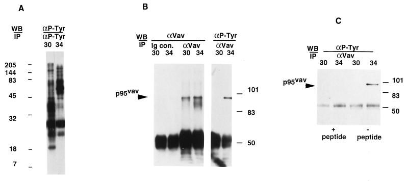
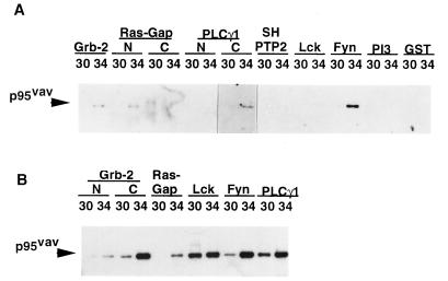
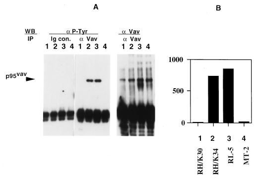
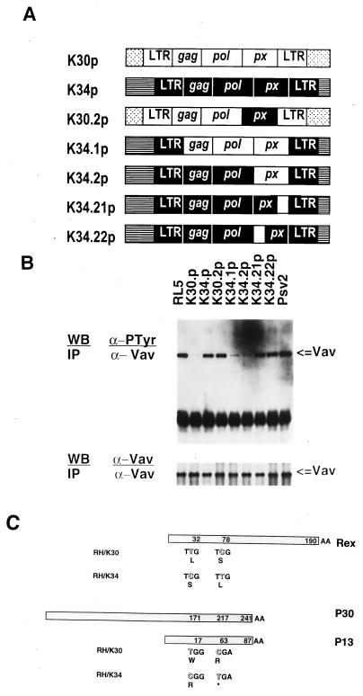
Human T cell leukemia virus I (HTLV-I) causes acute leukemic disease in a low percentage of infected individuals through obscure mechanisms. Our studies compare two rabbit HTLV-I-infected T cell lines: one, RH/K34, causes lethal experimental leukemia and the other, RH/K30, mediates asymptomatic infection. We show herein that the product of the protooncogene vav is constitutively Tyr-phosphorylated in RH/K34 but not in RH/K30. A role for the retrovirus in phosphorylation of Vav was assigned by transfection experiments with molecular clones of HTLV-I derived from the two lines. The HTLV-I molecular clone from RH/K30, but not that from RH/K34, down-regulates Vav phosphorylation in a Herpesvirus ateles-transformed T cell line. Use of recombinant virus clones revealed that a pX region sequence differing by two nucleotides between the two clones mediates this down-regulation. Because Vav is involved in T cell signaling and Vav phosphorylation occurs upon activation of T cells, control of the activation state of Vav by viral proteins may relate to the leukemogenic potential of certain HTLV-I-infected cells.


Figures

Figure 1
Figure 1
Analysis of Tyr phosphorylation profiles of HTLV-I-transformed cell lines RH/K30 and RH/K34 and identification of phosphorylated Vav in RH/K34. (A) Rabbit HTLV-I-transformed T cell lines RH/K30 (3) and RH/K34 (9) (107 cells) were washed in PBS and treated with lysis buffer. Lysates were precleared with normal mouse serum and immunoprecipitated with agarose conjugated anti-Tyr(P) 4G10 antibody. Samples were mixed with 100 μl of Laemmli sample buffer, 10-μl aliquots were separated by SDS/PAGE on a 14% gel and transferred onto Immobilon P membrane, and blots were developed with peroxidase-labeled 4G10 antibody, using the enhanced chemiluminescence substrate. Numbers to the left of the blot indicate migration of molecular size standards from a Kaleidoscope (Bio-Rad) standard. (B) RH/K30 and RH/K34 cells (107 cells) were washed then lysed, precleared with normal rabbit serum, immunoprecipitated with normal rabbit Ig or with rabbit anti-Vav antibody, separated by SDS/PAGE (8% gel), and transferred, and blots were developed with anti-Vav antibody or with anti-Tyr(P) 4G10 antibody and corresponding peroxidase-labeled antibody. (C) Precleared RH/K30 and RH/K34 cell lysates (107 cells) were immunoprecipitated with anti-Vav antibody in presence or in absence of specific peptide (20 μg/ml) corresponding to amino acid residues 523–547 of Vav, separated by SDS/PAGE (10% gel), transferred, and blotted with 4G10 antibody.
Figure 2
Figure 2
In vitro association of Vav with SH2 and SH3 domains from different molecules involved in signal transduction. (A) RH/K30 and RH/K34 cell lysates (107 cells) were precleared with GST-glutathione-agarose conjugate, then precipitated with agarose-immobilized GST containing different SH2 domains corresponding to Grb2, Ras-Gap, PLCg1, SH-PTP2, LCK, Fyn, and PI3, separated by SDS/PAGE (10% gel), transferred onto an Immobilon P membrane, and developed with rabbit anti-Vav antibody and peroxidase-labeled goat anti-rabbit Ig. (B) Precleared cell lysates were precipitated with agarose-immobilized GST containing different SH3 domains corresponding to Grb2, N and C Ras-Gap, Lck, Fyn, and PLCg1 and processed as in A.
Figure 3
Figure 3
Vav phosphorylation in different T cell lines. (A) Precleared cell lysates (107 cells) from RH/K30, RH/K34, RL-5, and MT-2 cell lines (designated as lanes 1–4, respectively) were immunoprecipitated with normal rabbit Ig (Ig con) or anti-Vav, transferred onto Immobilon P membranes, and immunoblotted with anti-Tyr(P) or with rabbit anti-Vav antibodies. (B) Densitometric analysis of the blot obtained with anti-Tyr(P) in A by using arbitrary densitometry units. Identity of cell line is shown.
Figure 4
Figure 4
Modulation of Vav phosphorylation in RL-5 cells by transfection with HTLV-I DNA clones. (A) Schematic representation of the K30p and K34p HTLV-I molecular clones and chimeric clones used to transfect the RL-5 cell line. Open and solid boxes correspond to the genome segments of HTLV-I clones K30 and K34, respectively. For example, the clones designated K34.21p and K34.22p correspond to the K34 with single pX region substitutions characteristic of K30. (B Upper) Precleared cell lysates (107 cells) from RL-5 cells and RL-5 transfected with molecular clone K30p or K34p, with chimeric clones, or with control pSv2 plasmid were immunoprecipitated with anti-Vav antibody, transferred onto Immobilon P membrane, and immunoblotted with anti-Tyr(P) antibody. (Lower) Duplicate samples immunoblotted with anti-Vav. (C) Schematic representation of HTLV-I molecular clones K30p and K34p showing predicted amino acid sequence differences in their pX region for the proteins Rex, p13, and p30.

References

    1. Cann A J, Chen I S Y. In: Virology. Fields B N, editor. New York: Raven; 1990. pp. 1520–1527.
    1. de Thé G, Bomford R. AIDS Res Hum Retroviruses. 1993;9:381–386. - PubMed
    1. Sawasdikosol S, Kindt T J. In: AIDS Research Review. Koff W, Wong-Staal F, Kennedy R C, editors. Vol. 2. New York: Mercel Dekker; 1992. pp. 211–233.
    1. Seto A, Kawanishi M, Matsuda S, Ogawa K, Eguchi T, Miyoshi I. Jpn J Cancer Res. 1987;78:1150. - PubMed
    1. Simpson M R, Leno M, Hubbard B S, Kindt T J. J Infect Dis. 1996;173:722–726. - PubMed

MeSH terms

Associated data

LinkOut - more resources