. 1998 Feb 17;95(4):1812-7.
doi: 10.1073/pnas.95.4.1812.

Negative regulation of granulocytic differentiation in the myeloid precursor cell line 32Dcl3 by ear-2, a mammalian homolog of Drosophila seven-up, and a chimeric leukemogenic gene, AML1/ETO

Affiliations

Negative regulation of granulocytic differentiation in the myeloid precursor cell line 32Dcl3 by ear-2, a mammalian homolog of Drosophila seven-up, and a chimeric leukemogenic gene, AML1/ETO

M Y Ahn et al. Proc Natl Acad Sci U S A. .

Abstract

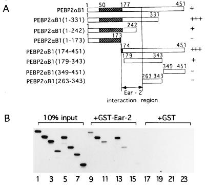
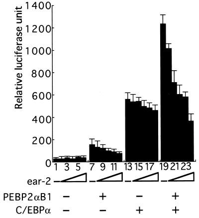
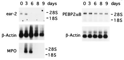
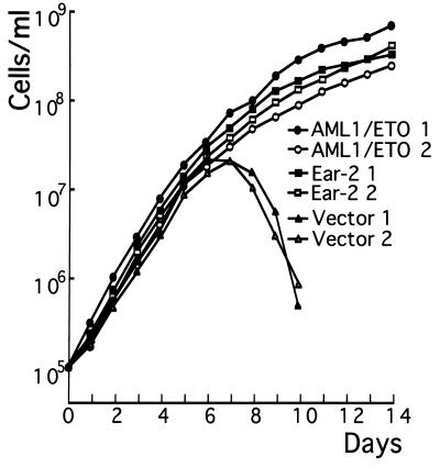
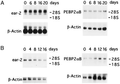
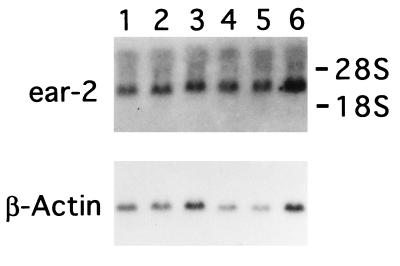
The polyomavirus enhancer binding protein 2alphaB (AML1/PEBP2alphaB/Cbfa2) plays a pivotal role in granulocyte colony-stimulating factor (G-CSF)-mediated differentiation of a myeloid progenitor cell line, 32Dc13. In this article, we report the identification of a PEBP2alphaB interacting protein, Ear-2, an orphan member of the nuclear hormone receptor superfamily that directly binds to and can inhibit the function of PEBP2alphaB. Ear-2 is expressed in proliferating 32Dc13 cells in presence of interleukin 3 but is down-regulated during differentiation induced by G-CSF. Interestingly, AML1/ETO(MTG8), a leukemogenic chimeric protein can block the differentiation of 32Dc13 cells, which is accompanied by the sustained expression of ear-2. Overexpression of Ear-2 can prevent G-CSF-induced differentiation, strongly suggesting that ear-2 is a key negative regulator of granulocytic differentiation. Our results indicate that a dynamic balance existing between PEBP2alphaB and Ear-2 appears to determine the choice between growth or differentiation for myeloid cells.


Figures

Figure 1
Figure 1
Physical interaction between PEBPαB1 and Ear2. (A) Full-length PEBPαB1 and its deletion derivatives are schematically shown. Stippled boxes represent the Runt domain. Degrees of interaction shown in B are summarized as −, +, and +++ on the right. (B) PEBPαB1 and its deletion derivatives were translated by programmed reticulocyte lysate in the presence of [35S]methionine. Ten percent of the material used in the GST pull down experiment is shown (lanes 1–8). In vitro-translated proteins were mixed with GST–Ear-2-bound beads (lanes 9–16) or the GST-bound beads (lanes 17–24), and a GST pull down assay was performed. Lanes: 1, 9, and 17, PEBP2αB1; 2, 10, and 18, PEBP2αB1(1–331); 3, 11, and 19, PEBP2αB1(1–242); 4, 12, and 20, PEBP2αB1(1–173); 5, 13, and 21, PEBP2αB1(174–451); 6, 14, and 22, PEBP2αB1(179–343); 7, 15, and 23, PEBP2αB1(349–451); 8, 16, and 24, PEBP2αB1(263–343). The proteins bound to the matrix were then analyzed by SDS/PAGE on 15% gels and autoradiography.
Figure 2
Figure 2
Ear-2 represses transactivation mediated by PEBP2αB1. A Jurkat T cell line was transfected with 1 μg of MCSF-R-luc, 2 μg of pEF-PEBP2αB1, 2 μg of MSV-C/EBP, 1 μg of pEF-PEBP2β2, 0.5 μg of RSV-β-gal, and increasing amounts of pCMX-ear-2. Lanes: 1, 7, 13, and 19, 0 μg; 2, 8, 14, and 20, 1 μg; 3, 9, 15, and 21, 2 μg; 4, 10, 16, and 22, 4 μg; 5, 11, 17, and 23, 8 μg; 6, 12, 18, and 24, 12 μg. Luciferase activities were normalized to the β-galactosidase activities of each extract. Each value represents the mean of three independent experiments. Standard deviations are indicated by error bars.
Figure 3
Figure 3
Northern blot analysis of endogenous PEBP2αB1 and ear-2 mRNA in 32Dcl3 cells during granulocytic differentiation. Poly(A)+ RNAs were prepared from 32Dcl3 stably transfected with the pBabeneo vector alone at days 0, 3, 6, 8, and 9 after the addition of G-CSF. Five micrograms of poly(A)+ RNA was analyzed by Northern blotting using 32P-labeled ear-2 cDNA as a probe. To detect an early differentiation maker, MPO, its cDNA was used as a probe for the same blot after it was stripped. A different membrane containing 2.5 μg of poly(A)+ RNA was hybridized with 32P-labeled PEBP2αB1 cDNA as a probe. As an internal control, β-actin mRNA expression was analyzed for each membrane after striping. Positions of 18S and 28S rRNAs are indicated.
Figure 4
Figure 4
Western blot analysis of Ear-2 or AML1/ETO(MTG8) synthesized in 32Dcl3 cells infected with retrovirus expressing pBabeneo-ear-2 or AML1/ETO(MTG8), respectively. (A) Twenty micrograms of whole cell extracts from pBabeneo vector transfected clone (lane 1) and pBabeneo-ear-2 transfected clones (lanes 3–5) were subjected to Western blot analysis with anti-Xpress antibody (CLONTECH). An arrow indicates the Ear-2 band. In vitro-translated Ear-2 was used as a marker (lane 2). (B) Twenty micrograms of whole cell extracts of 32Dcl3 cells transfected with pBabeneo-AML1/ETO(MTG8) was analyzed by using anti-PEBP2αB1 (lanes 1–3). An arrow indicates the AML1/ETO(MTG8) band. A 32Dcl3 clone transfected with pBabeneo vector alone was used as a control (lanes 4).
Figure 5
Figure 5
Growth curve of 32Dcl3 cells in response to G-CSF. Approximately 1 × 10⁶ cells of ear-2 or AML1/ETO(MTG8)-expressing clones or those received only the vector growing in the presence of IL-3 were washed twice with PBS and cultured in the medium containing recombinant human G-CSF (500 units/ml). At each time point, viable cells excluding trypan blue were counted by using a hemocytometer. Two independent experiments were performed. ▪ and □, Ear-2 1 and Ear-2 2, respectively; • and ○, AML1/ETO(MTG8) 1 and AML1/ETO(MTG8) 2, respectively; ▴ and ▵, Vector 1 and Vector 2, respectively.
Figure 6
Figure 6
Northern blot analysis of PEBP2αB1 and ear-2 mRNA. Poly(A)+ RNAs from 32Dcl3 cells stably transfected with pBabeneo-ear-2 (A) or pBabeneo-AML1/ETO(MTG8) (B) were analyzed by Northern blotting using 5 μg or 2.5 μg of poly(A)+ RNA for the expression of ear-2 or PEBP2αB, respectively. Numbers indicate days in the medium containing G-CSF. Of the three bands representing the ear-2 mRNA in A, top two correspond to the exogenous and the bottom one to the endogenous mRNA. As an internal control, β-actin mRNA expression was analyzed for each membrane after stripping. The positions of 18S and 28S rRNA are indicated. AML1/ETO(MTG8) RNA was not detected by the labeled PEBP2αB1 probe in B Right. Expression of AML1/ETO(MTG8) was confirmed, however, by using the AML1/ETO(MTG8) cDNA as a probe (data not shown).
Figure 7
Figure 7
Northern blot analysis of endogenous ear-2 mRNA in the cell clones expressing AML1/ETO(MTG8). Poly(A)+ RNAs from parental 32Dcl3 cells (lane 1), 32Dcl3 cell clone stably transfected with pBabeneo vector alone (lane 2), or 32Dcl3 cell clones stably transfected with pBabeneo-AML1/ETO(MTG8) (lanes 3–6), which were cultured in the medium containing IL-3, were analyzed by using 5 μg of poly(A)+ RNA and ear-2 cDNA as a probe. β-actin mRNA is shown as an internal control. The positions of 18S and 28S rRNA are indicated.

References

    1. Avalos B R. Blood. 1996;88:761–777. - PubMed
    1. Murakami H, Nagata S. In: The Cytokine Handbook. 3rd Ed. Thomson A W, editor. London: Academic; 1997. , in press.
    1. Valtieri M, Tweardy D J, Caracciolo D, Johnson K, Mavilio F, Altmann S, Santoli D, Rovera G. J Immunol. 1987;138:3829–3835. - PubMed
    1. Tanaka T, Tanaka K, Ogawa S, Kurokawa M, Mitani K, Nishida J, Shibata Y, Yazaki Y, Hirai H. EMBO J. 1995;14:341–350. - PMC - PubMed
    1. Zhang Y W, Bae S C, Huang G, Fu Y X, Lu J, Ahn M Y, Kanno Y, Kanno T, Ito Y. Mol Cell Biol. 1997;17:4133–4145. - PMC - PubMed

Publication types

MeSH terms

Substances

LinkOut - more resources