. 1998 Feb 17;95(4):1899-902.
doi: 10.1073/pnas.95.4.1899.

Molecular cloning and expression of the human delta7-sterol reductase

Affiliations

Molecular cloning and expression of the human delta7-sterol reductase

F F Moebius et al. Proc Natl Acad Sci U S A. .

Abstract

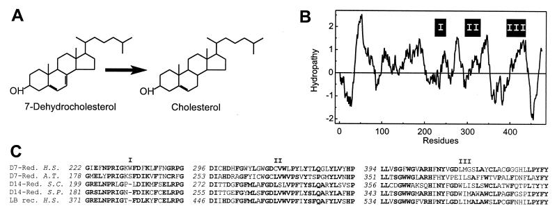
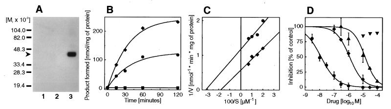
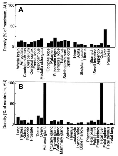
Inhibitors of the last steps of cholesterol biosynthesis such as AY9944 and BM15766 severely impair brain development. Their molecular target is the Delta7-sterol reductase (EC 1.3.1.21), suspected to be defective in the Smith-Lemli-Opitz syndrome, a frequent inborn disorder of sterol metabolism. Molecular cloning of the cDNA revealed that the human enzyme is a membrane-bound protein with a predicted molecular mass of 55 kDa and six to nine putative transmembrane segments. The protein is structurally related to plant and yeast sterol reductases. In adults the ubiquitously transcribed mRNA is most abundant in adrenal gland, liver, testis, and brain. The Delta7-sterol reductase is the ultimate enzyme of cholesterol biosynthesis in vertebrates and is absent from yeast. Microsomes from Saccharomyces cerevisiae strains heterologously expressing the human cDNA remove the C7-8 double bond in 7-dehydrocholesterol. The conversion to cholesterol depends on NADPH and is potently inhibited by AY9944 (IC50 0.013 microM), BM15766 (IC50 1.2 microM), and triparanol (IC50 14 microM). Our work paves the way to clarify whether a defect in the delta7-sterol reductase gene underlies the Smith-Lemli-Opitz syndrome.


Figures

Figure 1
Figure 1
(A) NADPH-dependent reduction of 7-dehydrocholesterol. (B) Hydropathy analysis with an average window size of 19 amino acid residues plotted at one residue intervals. On the ordinate, hydrophobicity is indicated by positive numbers. The positions of segments highly conserved throughout sterol reductases and related proteins are shown (I, II, and III). (C) Partial alignment of highly conserved domains (I, II, and III) in the amino acid sequences of the Δ7-sterol reductases from man (D7-Red., H.S.) and Arabidopsis thaliana (D7-Red., A.T., GenBank accession no. U49398), the Δ14-sterol reductases from Saccharomyces cerevisiae (D14-Red., S.C., no. S69420) and Saccharomyces pombe (D14-Red., S.P., no. L36039), and the human lamin B receptor (LB rec., H.S., no. L25931). Gaps are indicated (–). Numbers in italics refer to the position in the amino acid sequence. Residues identical in the majority of sequences are shown in bold.
Figure 2
Figure 2
Heterologous expression of the 5′ truncated Δ7-sterol reductase cDNA (D7-ORF) and a myc-tagged derivative (mycD7-ORF) in S. cerevisiae strain JB811 (ade2–1 leu2–3,112 pep4–3 trp1–289 ura3–52). (A) For Western blotting, 5 μg of microsomal protein was separated on a 12% (wt/vol) SDS gel under reducing conditions (sample buffer containing 10 mM DTT), immunostained with 20 ng/ml 9E10 c-myc antibody as in ref. , and developed with CDP-Star chemiluminescent reagent. The arrow indicates the migration of a c-myc immunoreactive band with a migration corresponding to 40 kDa (lane 1, mock; lane 2, D7-ORF; lane 3, mycD7-ORF). (B) Time course of Δ7-sterol reductase activity. Microsomes (0.5 mg) prepared from strains transformed with the vector without cDNA (mock, ▪), D7-ORF (•), and mycD7-ORF (⧫) were incubated anaerobically for the indicated times with 7-dehydrocholesterol in the presence of 2 mM NADPH. Mean values from duplicate determinations are shown. (C) Lineweaver Burk plot from yeast microsomes expressing D7-ORF (•) and mycD7-ORF (⧫). From this experiment Km values of 30 μM and 50 μM, respectively, were determined for D7-ORF and mycD7-ORF (Vmax 0.85 and 1.65 nmol/min per mg of protein, respectively). (D) Inhibition of the Δ7-sterol reductase (D7-ORF) by AY9944 (•, IC50 = 0.013 μM), BM15766 (⧫, IC50 = 1.2 μM), triparanol (▴, IC50 14.2 μM), and tamoxifen (▾, IC50 > 100 μM). Data shown are mean ± SD (n = 3).
Figure 3
Figure 3
Tissue distribution of the two Δ7-sterol reductase transcripts. Northern Blots with 2 μg of poly(A)+ RNA (Multiple tissue Northern blot, CLONTECH) were probed with the 32P-labeled human Δ7-sterol reductase (A) and human sterol Δ8-Δ7 isomerase cDNAs (B), respectively, and exposed for 16 hr. The arrows indicate the migration of the 2.9-kb and 2.3-kb Δ7-sterol reductase (A) and the 1.3-kb sterol isomerase (B) mRNAs, respectively.
Figure 4
Figure 4
Ubiquitous expression of the Δ7-sterol reductase mRNA. Northern dot blots with 100–500 ng poly(A)+ RNA normalized to ubiquitin (Human RNA master blot, CLONTECH) were probed with the 32P-labeled human Δ7-sterol reductase cDNA and exposed for 4 hr. Hybridization signals were quantified with a phosphorimager and normalized to adult adrenal gland and fetal liver (100%), respectively. Data shown are the mean from two different blots. Variability was less than 10%.

Similar articles

Cited by

References

    1. Smith D W, Lemli L, Opitz J M. J Pediatr. 1964;64:210–217. - PubMed
    1. Tint G S, Irons M, Elias E R, Batta A K, Frieden R, Chen T S, Salen G. N Engl J Med. 1994;330:107–113. - PubMed
    1. Batta A K, Tint G S, Shefer S, Abuelo D, Salen G. J Lipid Res. 1995;36:705–713. - PubMed
    1. Shefer S, Salen G, Batta A K, Honda A, Tint G S, Irons M, Elias E R, Chen T C, Holick M F. J Clin Invest. 1995;96:1779–1785. - PMC - PubMed
    1. Llirbat B, Wolf C, Chevy F, Citadelle D, Bereziat G, Roux C. J Lipid Res. 1997;38:22–34. - PubMed

Publication types

Substances

Associated data