. 1998 Feb 17;95(4):1921-6.
doi: 10.1073/pnas.95.4.1921.

Identification and characterization of a cell membrane nucleic acid channel

Affiliations

Identification and characterization of a cell membrane nucleic acid channel

B Hanss et al. Proc Natl Acad Sci U S A. .

Abstract

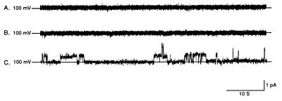
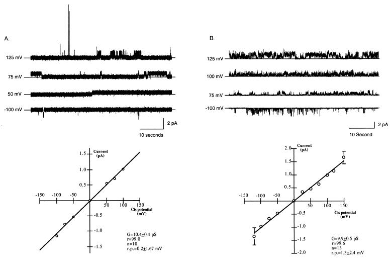
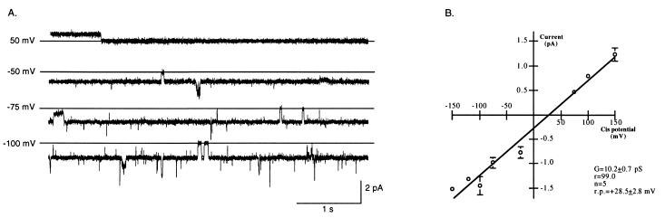
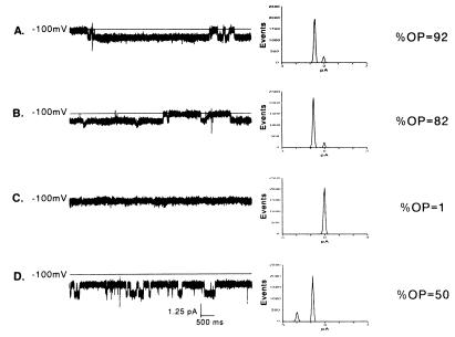
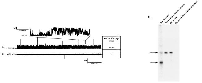
We have identified a 45-kDa protein purified from rat renal brush border membrane that binds short single-stranded nucleic acid sequences. This activity was purified, reconstituted in proteoliposomes, and then fused with model planar lipid bilayers. In voltage-clamp experiments, the reconstituted 45-kDa protein functioned as a gated channel that allows the passage of nucleic acids. Channel activity was observed immediately after addition of oligonucleotide. Channel activity was not observed in the absence of purified protein or of oligonucleotide or when protein was heat-inactivated prior to forming proteoliposomes. In the presence of symmetrical buffered solution and oligonucleotide, current passed linearly over the range of holding potentials tested. Conductance was 10.4 +/- 0.4 picosiemens (pS) and reversal potential was 0.2 +/- 1.7 mV. There was no difference in channel conductance or reversal potential between phosphodiester and phosphorothioate oligonucleotides. Ion-substitution experiments documented a shift in reversal potential only when a concentration gradient for oligonucleotide was established, indicating that movement of oligonucleotide alone was responsible for current. Movement of oligonucleotide across the bilayer was confirmed by using 32P-labeled oligonucleotides. Channel open probability decreased significantly in the presence of heparan sulfate. These studies provide evidence for a cell surface channel that conducts nucleic acids.


Figures

Figure 1
Figure 1
Representative 1-min current traces from a lipid bilayer experiment in the presence or absence of purified protein and/or oligonucleotide. Purified protein functioned as a gated channel when reconstituted in lipid bilayers and channel activity was observed only when both protein and oligonucleotide were present in the bilayer system. (A) Current trace, obtained at a holding potential of +100 mV, after formation of a lipid bilayer in the absence of both purified protein and oligonucleotide. The lipid bilayer alone was electrically stable and without channel activity. (B) Current trace, obtained at a holding potential of +100 mV, after the addition of NAC-PLs but not oligonucleotide. After the addition of NAC-PLs alone the bilayer remained stable without evidence of channel activity. The bilayer also remained stable after the addition of oligonucleotide in the absence of NAC-PLs (data not shown). (C) Current trace, obtained at a holding potential of +100 mV, after addition of both NAC-PLs and 5 μM Oligo. In the presence of both purified protein and oligonucleotide, channel activity was observed with clear transitions between open and closed states.
Figure 2
Figure 2
Summary of channel activity in buffered solutions identical in the concentration of salt and either Oligo or S-Oligo on both sides of the membrane (symmetrical buffered solution). Experiments were performed at different holding potentials after addition of identical buffered solution (200 mM CsCl/20 mM Tris⋅HCl, pH 7.4) and either Oligo or S-Oligo (5 μM) to both bilayer chambers. (A) Characterization of the channel in the presence of phosphodiester oligonucleotides (Oligo). (Upper) Representative 1-min traces from a typical experiment with both NAC-PLs and Oligo. For each trace, the holding potential is indicated and the solid horizontal line indicates zero current. (Lower) Current–voltage relationship of the channel in symmetrical Oligo and electrolyte solution. The data are the mean ± SEM of 10 experiments; error bars that are not visible are included within the symbol. The line represents a best fit linear regression analysis. The change in current was linear over the range of holding potentials tested with no sign of rectification. Reversal potential in symmetrical solutions was 0.2 ± 1.67 mV and conductance was 10.4 ± 0.4 pS. G, conductance; r.p., reversal potential; n, number. (B) Characterization of the channel in the presence of phosphorothioate oligonucleotides (S-Oligo). (Upper) Representative 1-min traces from a typical experiment with S-Oligo. For each trace, the holding potential is indicated and the solid horizontal line indicates zero current. (Lower) Current–voltage relationship of the channel in symmetrical S-Oligo and electrolyte solution. The data are the mean ± SEM of 13 experiments; error bars not visible are included within the symbol. Reversal potential in symmetrical solutions was 1.3 ± 2.4 mV and conductance was 9.9 ± 0.5 pS. There was no difference in channel conductance or reversal potential between S-Oligo- and Oligo-stimulated channel activity.
Figure 3
Figure 3
Summary of channel activity in the presence of a 1,000-fold concentration gradient for S-Oligo. (A) Representative 5-s traces from an experiment obtained at different holding potentials in the presence of a 1,000-fold S-Oligo gradient (500 μM S-Oligo cis and 0.5 μM S-Oligo trans). The solid horizontal line in each trace indicates the closed state. (B) Current–voltage relationship summarizing five experiments in which the presence of an oligonucleotide current was measured after establishing a 1,000-fold concentration gradient for S-Oligo. Conductance of the channel was 10.2 ± 0.7 pS and there was a substantial shift in reversal potential to +28.5 ± 2.8 mV. This shift in reversal potential is consistent with movement of oligonucleotide through the channel.
Figure 4
Figure 4
Blockade of channel activity with the polyanion heparan sulfate. Continuous 10-s traces (Left), histograms of the distribution of current (Middle), and open probability (Right) in which two doses of heparan sulfate were used to block channel activity. Traces were obtained at a holding potential of −100 mV in an experimental buffer containing 200 mM CsCl and 5 μM S-Oligo (pH 7.4). Heparan sulfate produced a dose-dependent decrease in open probability. Addition of heparan sulfate at 10 μg/ml to both solution chambers decreased open probability from 0.92 (A) to 0.82 (B). Channel activity was completely blocked when heparan sulfate was increased in both chambers to 20 μg/ml (C). Channel activity was restored when heparan sulfate was washed from both chambers (D).
Figure 5
Figure 5
Direct measurement of oligonucleotide movement across the lipid bilayer. Movement of oligonucleotide across the bilayer was confirmed by tracking the movement of [32P]S-Oligo. [32P]S-Oligo was added to the trans buffer chamber (input chamber) and 20–40 min later samples were collect from both buffer chambers. Radioactivity in the samples was measured by liquid scintillation and analyzed by gel electrophoresis. (A) Representative 30-min current trace obtained at a holding potential of +150 mV. The channel was highly active with clear transition between open and closed states (Inset). During the 30-min collection period approximately 32 fmol of [32P]S-Oligo moved across the lipid membrane. (B) A 30-min current trace obtained from an experiment in which NAC was heat-denatured before forming PLs. Channel activity was not observed and [32P]S-Oligo was not detected in the collection chamber, indicating that oligonucleotide did not move across the bilayer when NAC was rendered inactive. (C) A representative autoradiogram from an experiment to confirm that full-length oligonucleotide moved across the membrane. Samples from the trans (input) and cis (collection) chambers were loaded on a 15% polyacrylamide/7 M urea gel. Before loading, radioactivity in the samples was measured and appropriately diluted to load approximately the same number of counts in each well. Lanes: 1, radiolabeled size markers; 2, sample from the input chamber (diluted 1:10,000 with deionized H2O); 3, sample from collection chamber after 35 min of channel activity; 4, sample from collection chamber obtained 1 h after adding heat-inactivated NAC. In the presence of active NAC, a single band comigrated with the 20-base size marker and the input chamber aliquot. There was no degradation of oligonucleotide as it crossed the bilayer and there was no free 32P detected in the collection chamber solution. Identical results were observed when experiments were performed with radiolabeled Oligo (data not shown).

Similar articles

Cited by

References

    1. Gilkeson G S, Bernstein K, Pippen A M, Clarke S H, Marion T, Pisetsky D S, Ruiz P, Lefkowith J B. Clin Immunol Immunopathol. 1995;76:59–67. - PubMed
    1. Messina J P, Gilkeson G S, Pisetsky D S. Cell Immunol. 1993;147:148–157. - PubMed
    1. Agrawal S, Temsamani J, Tang J Y. Proc Natl Acad Sci USA. 1991;88:7595–7599. - PMC - PubMed
    1. Goodarzi G, Watabe M, Watabe K. Biopharm Drug Dispos. 1992;13:221–227. - PubMed
    1. Cossum P A, Sasmor H, Dellinger D, Truong L, Cummins L, Owens S R, Markham P M, Shea J P, Crooke S. J Pharmacol Exp Ther. 1993;267:1181–1190. - PubMed

Publication types

LinkOut - more resources