. 1998 Feb 15;12(4):462-72.
doi: 10.1101/gad.12.4.462.

High mobility group protein-1 (HMG-1) is a unique activator of p53

Affiliations

High mobility group protein-1 (HMG-1) is a unique activator of p53

L Jayaraman et al. Genes Dev. .

Abstract

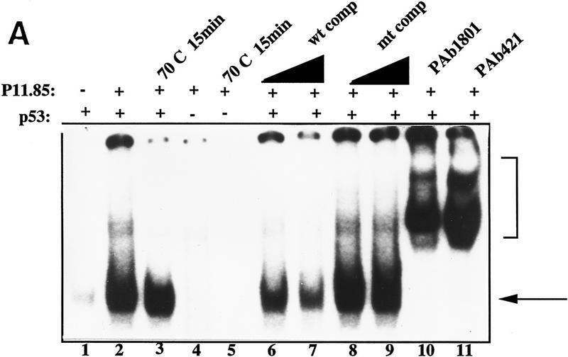
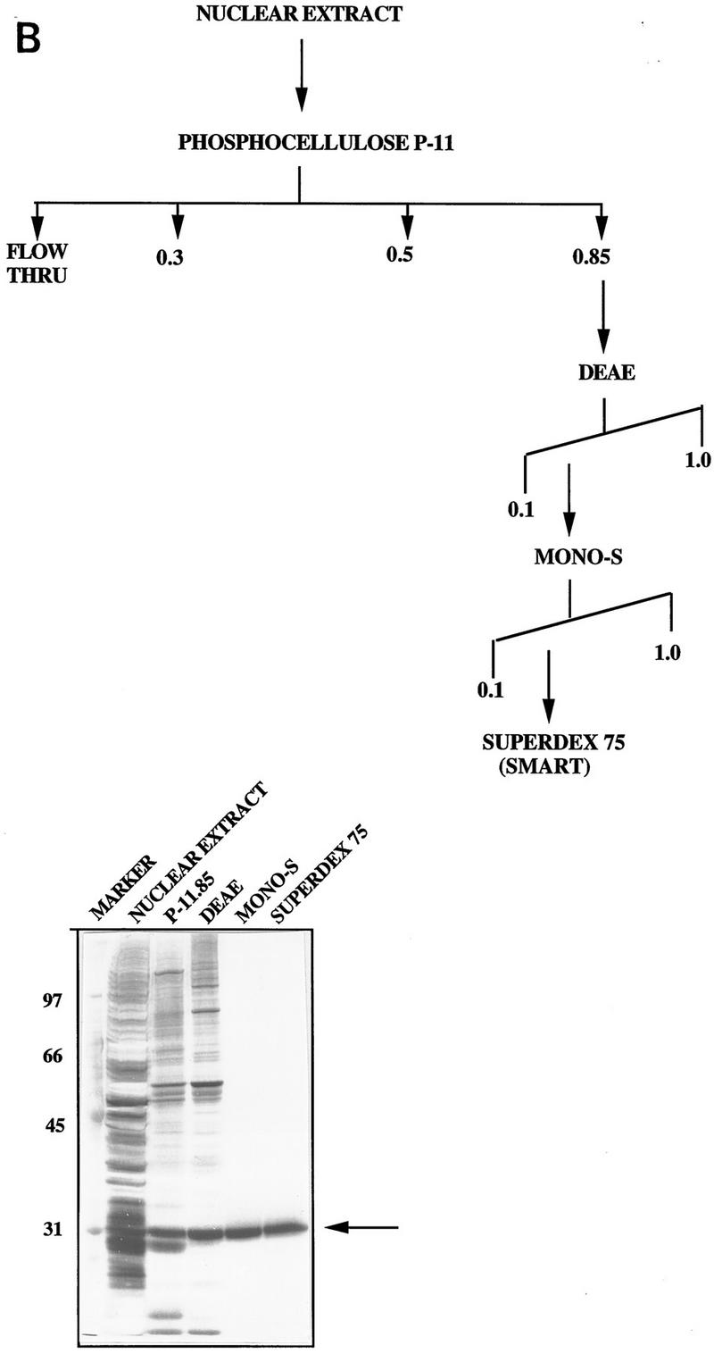
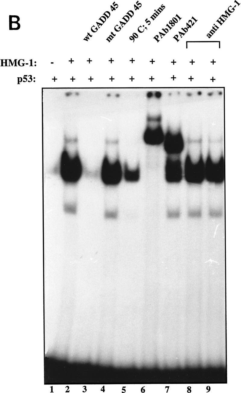
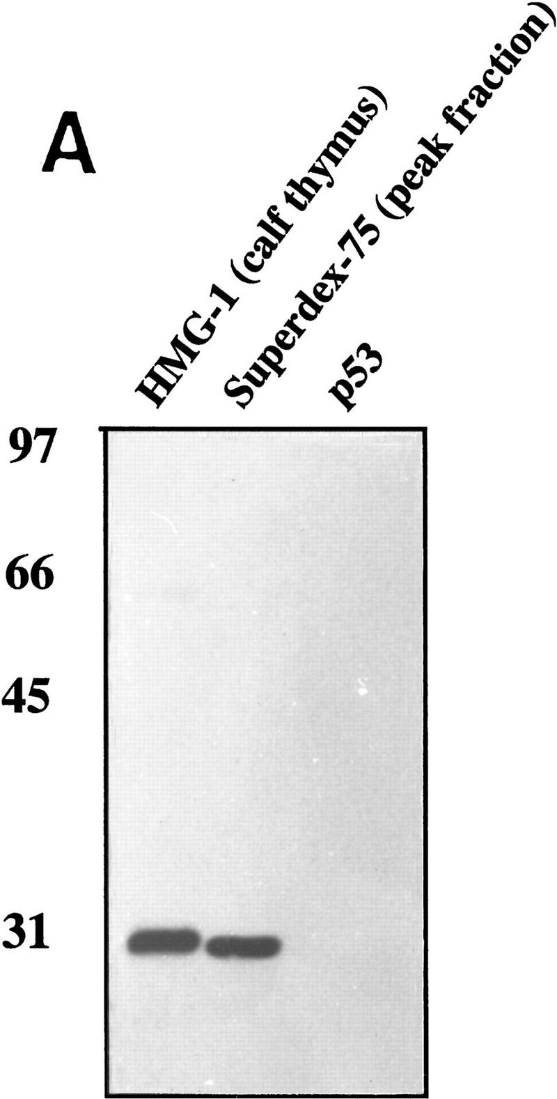
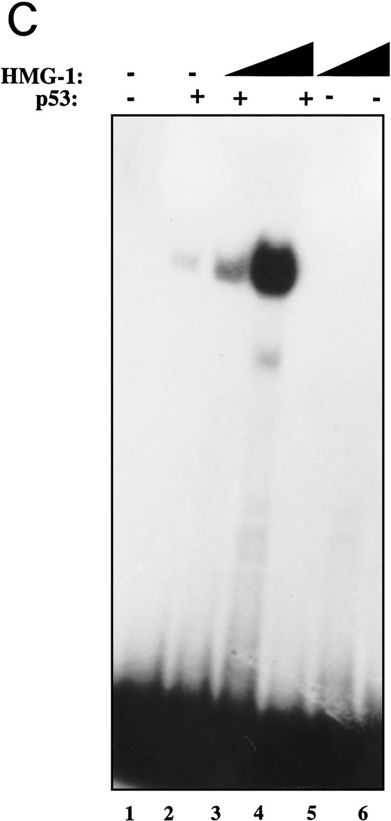
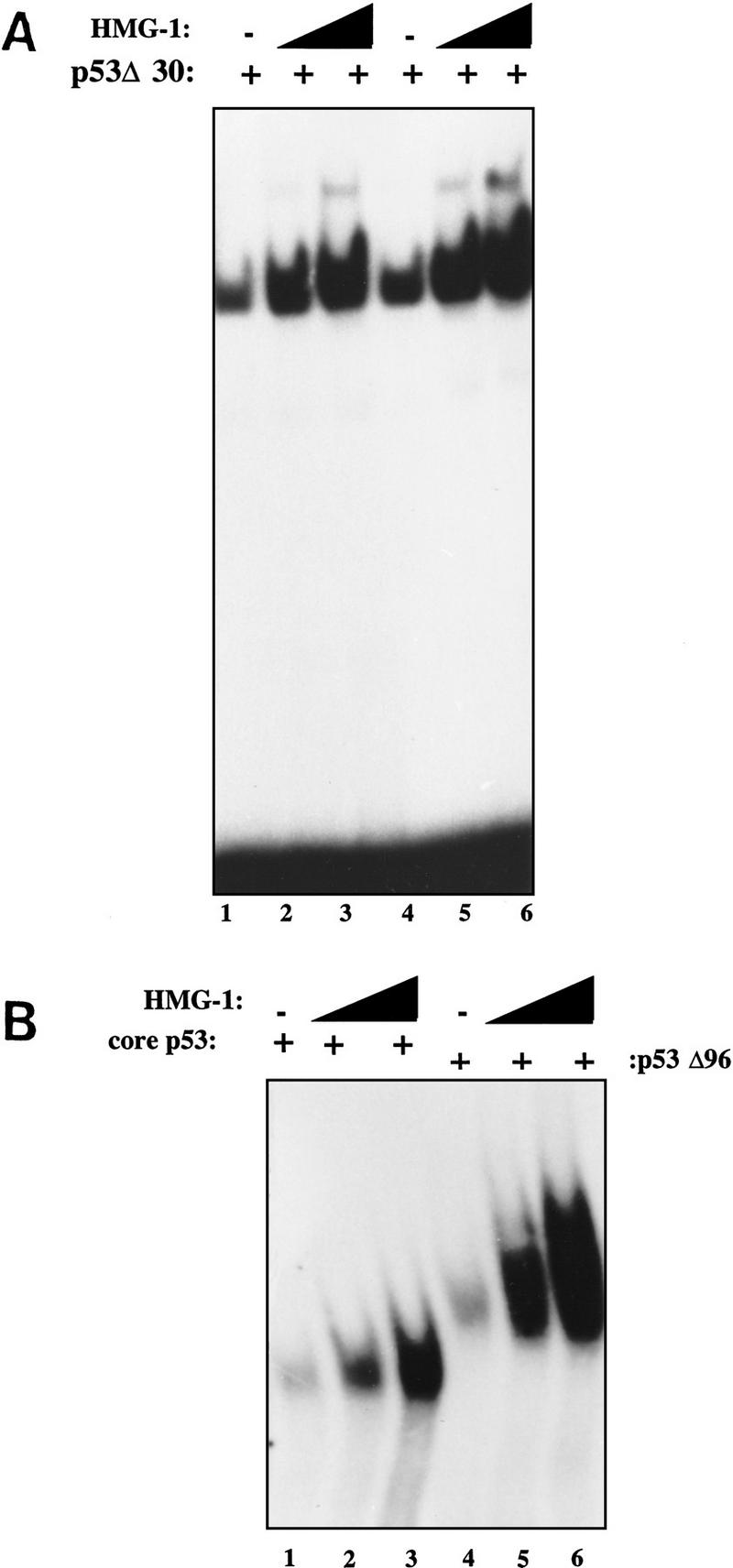
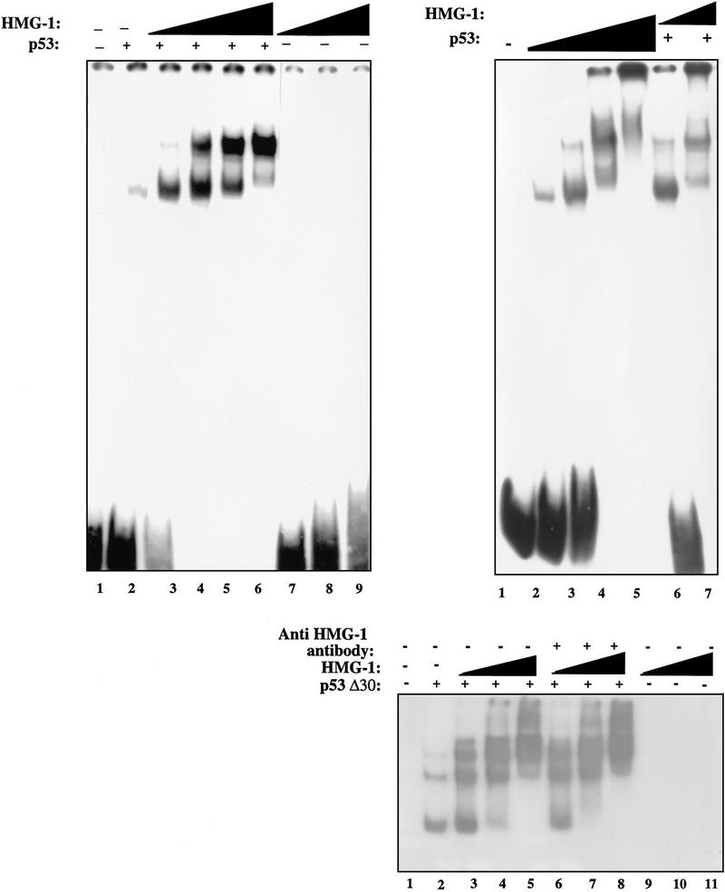
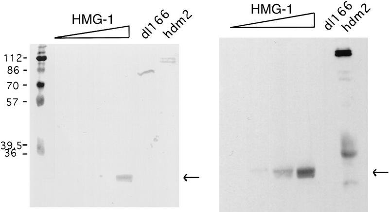
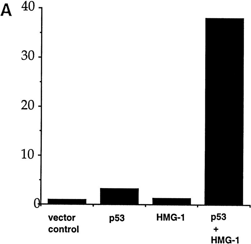
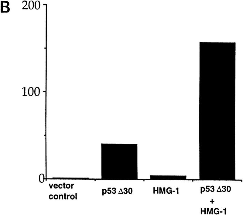
The binding of p53 protein to DNA is stimulated by its interaction with covalent as well as noncovalent modifiers. We report the identification of a factor from HeLa nuclear extracts that activates p53 DNA binding. This factor was purified to homogeneity and identified as the high mobility group protein, HMG-1. HMG-1 belongs to a family of highly conserved chromatin-associated nucleoproteins that bend DNA and facilitate the binding of various transcription factors to their cognate DNA sequences. We demonstrate that recombinant His-tagged HMG-1 enhances p53 DNA binding in vitro and also that HMG-1 and p53 can interact directly in vitro. Unexpectedly, HMG-1 also stimulates DNA binding by p53Delta30, a carboxy-terminally deleted form of the protein that is considered to be constitutively active, suggesting that HMG-1 stimulates p53 by a mechanism that is distinct from other known activators of p53. Finally, using transient transfection assays we show that HMG-1 can increase p53 and p53Delta30-mediated transactivation in vivo. HMG-1 promotes the assembly of higher order p53 nucleoprotein structures, and these data, along with the fact that HMG-1 is capable of bending DNA, suggest that HMG-1 may activate p53 DNA binding by a novel mechanism involving a structural change in the target DNA.


Figures

Figure 1
Figure 1
(A) HeLa nuclear fraction P-11.85 stimulates p53 DNA binding. p53 protein (90 ng) was bound to 32P-labeled GADD45 oligonucleotides either alone (lane 1) or in the presence of the P-11.85 fraction and p53 monoclonal antibodies pAb 421 and pAb 1801 or competitor DNA as indicated and analyzed by EMSA and autoradiography. Lanes 3 and 5 contain the P-11.85 fraction heat-treated for 5 min at 70°C. Reaction mixtures in lanes 4 and 5 contain no p53 protein. Wild-type (wt) or mutant (mt) consensus site-containing oligonucleotides were added in 10-fold (lanes 6,8) or 20-fold (lanes 7,9) molar excess over probe. The arrow indicates the p53–DNA complex; the bracket indicates p53–DNA complexes supershifted by antibodies. Not shown is free unbound probe at bottom of gel. (B) Purification of the p53 activator protein from HeLa nuclear fraction P-11.85. (Top) Protocol for purification of p53 DNA-binding stimulatory activity from HeLa nuclear extract. (Bottom) Aliquots of peak fraction from each step in A were analyzed by SDS-PAGE and silver staining. The arrow indicates purified protein.
Figure 1
Figure 1
(A) HeLa nuclear fraction P-11.85 stimulates p53 DNA binding. p53 protein (90 ng) was bound to 32P-labeled GADD45 oligonucleotides either alone (lane 1) or in the presence of the P-11.85 fraction and p53 monoclonal antibodies pAb 421 and pAb 1801 or competitor DNA as indicated and analyzed by EMSA and autoradiography. Lanes 3 and 5 contain the P-11.85 fraction heat-treated for 5 min at 70°C. Reaction mixtures in lanes 4 and 5 contain no p53 protein. Wild-type (wt) or mutant (mt) consensus site-containing oligonucleotides were added in 10-fold (lanes 6,8) or 20-fold (lanes 7,9) molar excess over probe. The arrow indicates the p53–DNA complex; the bracket indicates p53–DNA complexes supershifted by antibodies. Not shown is free unbound probe at bottom of gel. (B) Purification of the p53 activator protein from HeLa nuclear fraction P-11.85. (Top) Protocol for purification of p53 DNA-binding stimulatory activity from HeLa nuclear extract. (Bottom) Aliquots of peak fraction from each step in A were analyzed by SDS-PAGE and silver staining. The arrow indicates purified protein.
Figure 2
Figure 2
Identification of HMG-1 as the p53 stimulating protein from HeLa P-11.85 fraction. (A) HMG-1 purified from calf thymus or HeLa P-11.85 Superdex-75 peak fraction or immunopurified p53 protein was subjected to SDS-PAGE and transferred to nitrocellulose, and the resulting blots were probed with affinity-purified polyclonal anti-HMG-1 antibody. (B) DNA binding by p53 (90 ng) in the absence (lane 1) or presence of calf thymus-purified HMG-1 (100 ng) (lanes 28) was analyzed by EMSA as in Fig. 1. Antibodies and 20-fold excess competitor DNA were added as indicated. Lane 8 contains mixtures in which antibody and proteins were added simultaneously, whereas in lane 9 HMG-1 was preincubated with anti-HMG-1 antibody for 20 min prior to addition to reaction mixture. (C) Recombinant HMG-1 enhances DNA binding by p53. DNA-binding mixtures contained recombinant His–HMG-1 (50 or 100 ng), either in the absence (lanes 5,6) or presence of p53 (90 ng) (lanes 24). Lane 1 contains probe alone. (D) DNA-binding reaction mixtures were as above but in the absence of poly[d(I-C)]. Binding mixtures contained either 8 (lanes 24,12,13) or 4 ng (lanes 79,10,11) of p53 in the absence (lanes 2,7,10,11) or presence (lanes 3,4, 8,9,12,13) of His–HMG-1 (60 or 120 ng). HMG-1 (60 ng) was present in mixtures shown in lanes 12 and 13. Monoclonal antibody pAb 421 was added as indicated. Only the DNA–protein complexes are shown as in Fig 1A.
Figure 2
Figure 2
Identification of HMG-1 as the p53 stimulating protein from HeLa P-11.85 fraction. (A) HMG-1 purified from calf thymus or HeLa P-11.85 Superdex-75 peak fraction or immunopurified p53 protein was subjected to SDS-PAGE and transferred to nitrocellulose, and the resulting blots were probed with affinity-purified polyclonal anti-HMG-1 antibody. (B) DNA binding by p53 (90 ng) in the absence (lane 1) or presence of calf thymus-purified HMG-1 (100 ng) (lanes 28) was analyzed by EMSA as in Fig. 1. Antibodies and 20-fold excess competitor DNA were added as indicated. Lane 8 contains mixtures in which antibody and proteins were added simultaneously, whereas in lane 9 HMG-1 was preincubated with anti-HMG-1 antibody for 20 min prior to addition to reaction mixture. (C) Recombinant HMG-1 enhances DNA binding by p53. DNA-binding mixtures contained recombinant His–HMG-1 (50 or 100 ng), either in the absence (lanes 5,6) or presence of p53 (90 ng) (lanes 24). Lane 1 contains probe alone. (D) DNA-binding reaction mixtures were as above but in the absence of poly[d(I-C)]. Binding mixtures contained either 8 (lanes 24,12,13) or 4 ng (lanes 79,10,11) of p53 in the absence (lanes 2,7,10,11) or presence (lanes 3,4, 8,9,12,13) of His–HMG-1 (60 or 120 ng). HMG-1 (60 ng) was present in mixtures shown in lanes 12 and 13. Monoclonal antibody pAb 421 was added as indicated. Only the DNA–protein complexes are shown as in Fig 1A.
Figure 3
Figure 3
HMG-1 can stimulate DNA binding by p53Δ30 as well as by core p53. (A) Ten nanograms (lanes 13) or 20 ng (lanes 46) of p53Δ30 was bound to 32P-labeled GADD45 oligonucleotides and analyzed by EMSA in the absence (lanes 1,4) or presence (lanes 2,3,5,6) of HMG-1 (50 or 100 ng). (B) DNA-binding mixtures contained either core domain p53 (lanes 13) or p53Δ96 (lanes 46) in the presence (lanes 2,3,5,6) or absence (lanes 1,4) of HMG-1 (50 or 100 ng). Shown are p53–DNA complexes only as in Fig. 1A.
Figure 4
Figure 4
HMG-1 promotes co-operative p53 interactions on DNA. (A, Left) Reaction mixtures contained increasing amounts of HMG-1 (50, 75, 100, or 150 ng) in the absence (lanes 79) or presence (lanes 36) of full-length p53 (50 ng) using a 120-bp 32P-labeled DNA fragment containing the GADD45 p53 binding site. Lane 1 contains no protein. Mixtures in lane 2 contained 50 ng of p53 alone. (Right) Reaction mixtures contain increasing amounts (50, 100, 200, or 400 ng) of p53 (lanes 25) or increasing amounts of HMG-1 (50 or 125 ng) with 50 ng of p53 (lanes 6,7). Lane 1 contains no protein. (B) Reaction mixtures contained p53Δ30 (10 ng), alone (lane 2) or with 75 (lanes 3,6), 100 (lanes 4,7), or 150 (lanes 5,8) ng of HMG-1 with or without excess anti-HMG-1 antibody as indicated. Mixtures in lanes 911 contained HMG-1 protein at the above concentrations without p53.
Figure 5
Figure 5
p53 binds HMG-1 in vitro. Calf-thymus-purified HMG-1 (0.25, 0.5, 1.0, and 2.0 μg), truncated dl166 HDM2 (1 μg) or full-length HDM2 (1 μg) as indicated, and molecular mass marker polypeptides were resolved by SDS-PAGE and transferred to nitrocellulose. (Right) Filter was incubated with human p53 protein (1 μg in 5 ml) followed by detection with pAb 421 as described in Materials and Methods. (Left) Ponceau S staining of filter. The lower amounts of HMG-1 were below the level of detection by Ponceau S. Arrows indicate position of HMG-1 protein.
Figure 6
Figure 6
HMG-1 stimulates transactivation by wild-type p53 as well as p53Δ30 in transfected cells. (A,B) H1299 cells were transiently transfected with wild-type p53 (300 ng) (A) or p53Δ30 (35 ng) (B) or HMG-1 expression plasmid (2.5 μg) or both p53 and HMG-1 expression plasmids as indicated. In all cases, total DNA transfected was normalized with equivalent amounts of control parental vectors. The reporter construct used was cyclin G–luc (1 μg). (Y-axis) Luciferase activity is represented as fold transactivation relative to samples transfected with reporter constructs and parental control plasmids. The fold activation represents an average of triplicate samples.
Figure 6
Figure 6
HMG-1 stimulates transactivation by wild-type p53 as well as p53Δ30 in transfected cells. (A,B) H1299 cells were transiently transfected with wild-type p53 (300 ng) (A) or p53Δ30 (35 ng) (B) or HMG-1 expression plasmid (2.5 μg) or both p53 and HMG-1 expression plasmids as indicated. In all cases, total DNA transfected was normalized with equivalent amounts of control parental vectors. The reporter construct used was cyclin G–luc (1 μg). (Y-axis) Luciferase activity is represented as fold transactivation relative to samples transfected with reporter constructs and parental control plasmids. The fold activation represents an average of triplicate samples.

Similar articles

Cited by

References

    1. Agarwal A, Schatz DG. RAG1 and RAG2 form a stable postcleavage synaptic complex with DNA containing signal ends in V(D)J recombination. Cell. 1997;89:43–53. - PubMed
    1. Balagurumoorthy P, Sakamoto H, Lewis MS, Zambrano N, Clore GM, Groneborn AM, Appella E, Harrington RE. Four p53 DNA-binding domain peptides bind natural p53 response elements and bend the DNA. Proc Natl Acad Sci. 1995;92:8591–8595. - PMC - PubMed
    1. Barak Y, Juven T, Haffner R, Oren M. mdm2 expression is induced by wild type p53 activity. EMBO J. 1993;12:461–468. - PMC - PubMed
    1. Baxevanis AD, Landsman D. The HMG-1 box protein family: Classification and functional relationships. Nucleic Acids Res. 1995;23:1604–1613. - PMC - PubMed
    1. Bazett-Jones DP, LeBlanc B, Herfort M, Moss T. Short-range DNA looping by the Xenopus HMG-box transcription factor, xUBF. Science. 1994;264:1134–1137. - PubMed

Publication types

MeSH terms

LinkOut - more resources