. 1998 Feb 15;12(4):538-46.
doi: 10.1101/gad.12.4.538.

Vaccinia NPH-I, a DExH-box ATPase, is the energy coupling factor for mRNA transcription termination

Affiliations

Vaccinia NPH-I, a DExH-box ATPase, is the energy coupling factor for mRNA transcription termination

L Deng et al. Genes Dev. .

Abstract

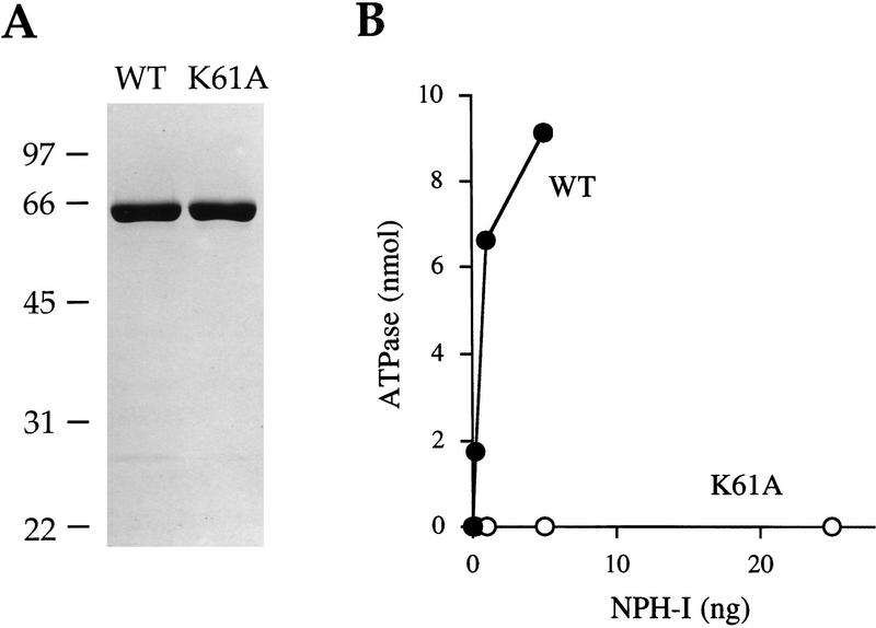
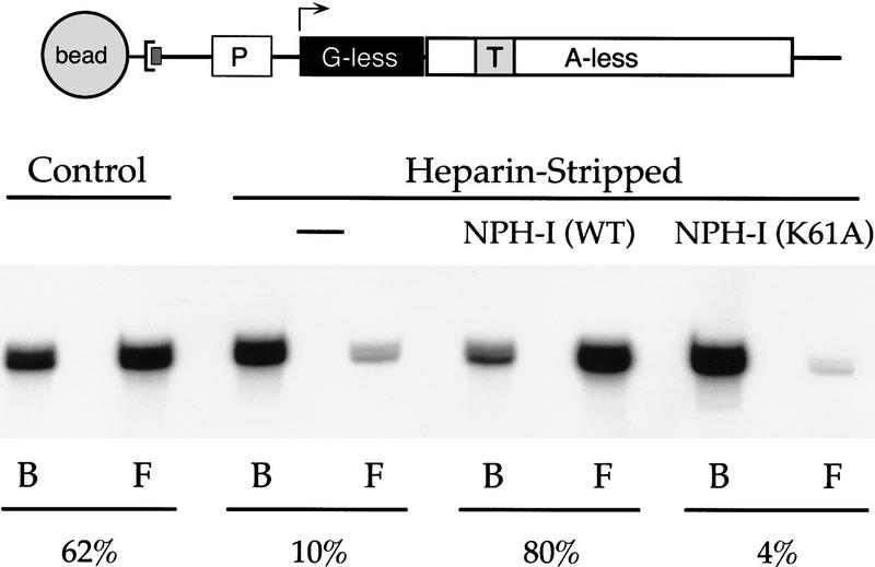
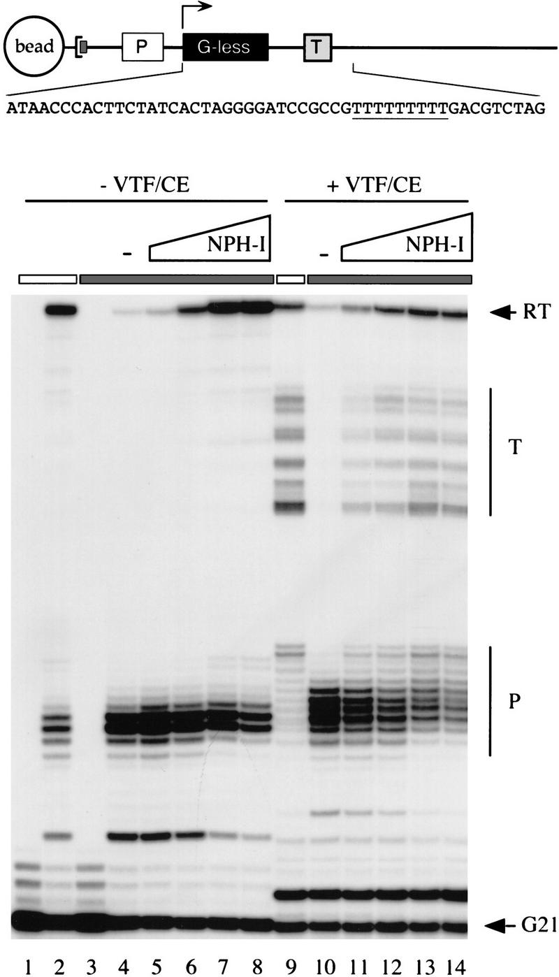
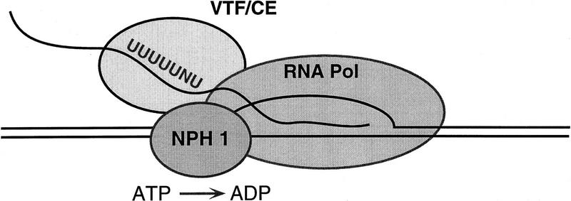
Vaccinia virus RNA polymerase terminates transcription in response to a specific signal UUUUUNU in the nascent RNA. Transduction of this signal to the elongating polymerase requires a trans-acting viral termination factor (VTF/capping enzyme), and is coupled to the hydrolysis of ATP. Recent studies suggest that ATP hydrolysis is catalyzed by a novel termination protein (factor X), which is tightly associated with the elongation complex. Here, we identify factor X as NPH-I (nucleoside triphosphate phosphohydrolase-I), a virus-encoded DNA-dependent ATPase of the DExH-box family. We report that NPH-I serves two roles in transcription (1) it acts in concert with VTF/CE to catalyze release of UUUUUNU-containing nascent RNA from the elongation complex, and (2) it acts by itself as a polymerase elongation factor to facilitate readthrough of intrinsic pause sites. A mutation (K61A) in the GxGKT motif of NPH-I abolishes ATP hydrolysis and eliminates the termination and elongation factor activities. Related DExH proteins may have similar roles at postinitiation steps during cellular mRNA synthesis.


Figures

Figure 1
Figure 1
Amino acid sequence of vaccinia NPH-I. The GxGKT and DExH motifs are demarcated by boxes.
Figure 2
Figure 2
Purification and ATPase activity of recombinant NPH-I. (A) Aliquots (3 μg) of the phosphocellulose preparations of wild-type NPH-I (WT) and the K61A mutant protein were electrophoresed through a 10% polyacrylamide gel containing 0.1% SDS. Polypeptides were visualized by staining with Coomassie brilliant blue dye. The positions and sizes (in kD) of coelectrophoresed marker proteins are indicated on the left. (B) ATPase activity. Assays were performed as described in Materials and Methods. Activity (nmoles Pi release/30 min) is plotted as a function of the input wild-type and K61A phosphocellulose enzyme preparations.
Figure 3
Figure 3
Sedimentation analysis and DNA binding by recombinant NPH-I. (A) Glycerol gradient sedimentation was performed as described in Materials and Methods. Aliquots (2 μl) of the indicated gradient fractions were assayed for DNA-dependent ATPase activity. The activity profile is shown. The peak positions of marker proteins sedimented in a parallel gradient are indicated by the arrows. (B) DNA-binding assay. Reaction mixtures (20 μl) containing 20 mm Tris-HCl (pH 8.0), 2 mm DTT, 25 fmoles of a 5′ 32P-labeled 40-mer DNA oligonucleotide (5′-CCATGTTTCTGTACTAATCTAGAACGAGTCTATATATTCC), and 25, 5, or 1 ng of wild-type NPH-I of K61A (proceeding from left to right within each titration series) were incubated for 30 min at 30°C. NPH-I was omitted from control reactions (lanes−). The samples were adjusted to 5% glycerol and then applied to a native 8% polyacrylamide gel in 0.25× TBE. Electrophoresis was at 20 mA for 45 min. Free DNA and a protein–DNA complex or retarded electrophoretic mobility (asterisk) were visualized by autoradiographic exposure of the dried gel.
Figure 4
Figure 4
NPH-I restores VTF-responsiveness to heparin-stripped elongation complexes. A schema of the G21(TER29)A78 DNA template is shown. The DNA contains a biotinylated nucleotide incorporated uniquely at the 3′ end of the template DNA strand, which anchors the DNA to a streptavidin-coated magnetic bead. The transcription unit consists of a vaccinia early promoter (P) fused to a 20-nucleotide G-less cassette, which is flanked by a run of three G residues at positions +21 to +23. Situated downstream of the G-less cassette is a 57-nucleotide A-less cassette flanked at its 3′ end by a run of four A residues at positions +78 to + 81. Placed within the A-less cassette is a termination signal, TTTTTTTTT, spanning positions +29 to +37. Ternary complexes containing pulse-labeled OMeG21 RNA were assembled as described in Materials and Methods, then treated for 2 min with 625 μg/ml of heparin (Heparin-Stripped). Control complexes were mock-incubated in the absence of heparin. After bead purification and washing, the ternary complexes were incubated for 30 min on ice in 20 mm Tris-HCl (pH 8.0), 2 mm DTT, with no additional protein, or with 25 ng of the phosphocellulose fraction of wild-type NPH-I or K61A. The G21 complexes were then walked to A78 during a 5-min chase at 30°C in the presence of 6 mm MgCl2 and 1 mm each of 3′ dATP, GTP, CTP, and UTP. The mixtures were then supplemented with 200 fmoles of recombinant VTF/CE (Luo et al. 1995) and incubated for an additional 5 min at 30°C. The bead-bound A78 RNA (lane B) was separated from released A78 RNA (lane F; free) by microcentrifugation of the reaction mixtures. Nucleic acid was then recovered from the pellet and supernatant fractions by phenol extraction and ethanol precipitation. The transcription products were analyzed by electrophoresis through a 17% polyacrylamide gel containing 7 m urea in TBE (90 mm Tris, 90 mm borate, and 2.5 mm EDTA). The labeled A78 transcript was visualized by autoradiography. The percent of RNA released (indicated below the autoradiograph) was quantitated by scanning the gel with a PhosphorImager.
Figure 5
Figure 5
Effects of DExH box mutations on ATPase and transcription release. NPH-I mutants D141A and E142A were expressed in bacteria and purified from soluble lysates by Ni-agarose and phosphocellulose chromatography. The purity of the phosphocellulose preparations was equivalent to that of wild-type and K61A (data not shown). (A) ATPase assays were performed as described in Materials and Methods. Activity (nmoles Pi release/30 min) is plotted as a function of the input D141A (○) and E142A (□) phosphocellulose enzyme preparations. The K61A protein (▵) was assayed in parallel. (B) Transcript release. Assays were performed as described in the legend to Fig. 4. Purified heparin-stripped G21 complexes were incubated with 5 ng of the phosphocellulose preparations of wild-type NPH-I, D141A, or E142A, then walked to A78 and challenged with 200 fmoles of recombinant VTF/CE. Bead-bound and released RNAs were separated by centrifugation. The bound and free samples were deproteinized, ethanol-precipitated, and analyzed by denaturing gel electrophoresis. The percent of A78 RNA released is plotted in bar graph format.
Figure 6
Figure 6
NPH-I suppresses intrinsic pausing and restores termination competence to heparin-stripped complexes. The architecture of the G21(TER) DNA template is shown in cartoon form. The DNA contains a biotinylated nucleotide incorporated uniquely at the 3′ end of the template DNA strand, which anchors the DNA to a streptavidin-coated magnetic bead. A vaccinia virus early promoter element specifies the initiation of transcription at position +1 of a 20-nucleotide G-less cassette. A termination signal (TTTTTTTTT) is situated downstream of the G-less cassette. The sequence of the first 50 nucleotides of the transcribed region (non-template strand) is shown. Pulse-labeled G21 ternary complexes assembled on the G21(TER) template were treated with 625 μg/ml of heparin (lanes 38 and 1014, denoted by the shaded horizontal bar over the lanes) or with buffer (lanes 1, 2, and 9, denoted by an open bar over the lanes) before bead purification and washing. The purified complexes were incubated for 30 min on ice with increasing amounts of the peak glycerol gradient fraction (no. 20) of wild-type NPH-I as follows: 0.4 U (lanes 5,11); 1.6 U (lanes 6,12); 6.3 U (lanes 7,13); 25 U (lanes 8,14). NPH-I was omitted from control reactions (lanes 4,10, indicated by −). Pulse-labeled RNAs were isolated from samples of heparin-stripped (lane 3) and untreated (lane 1) ternary complexes. The remaining samples were chased in 20-μl elongation reaction mixtures containing 20 mm Tris-HCl (pH 8.0), 6 mm MgCl2, 2 mm DTT, and 1 mm ATP, 1 mm GTP, 1 mm CTP, and 10 μm UTP. Where indicated (lanes 914), the mixtures were supplemented with 500 fmoles of recombinant VTF/CE just before the addition of NTPs. After incubation for 10 min at 30°C, the elongation reactions were quenched by addition of buffer containing SDS and urea. The samples were extracted with phenol:chloroform and labeled RNA was recovered by ethanol precipitation. Transcription products were analyzed by electrophoresis through a 17% denaturing polyacrylamide gel and radiolabeled transcripts were visualized by autoradiography. The positions of pulse-labeled G21 RNA and the runoff transcript (RT) are indicated by arrows. The terminated (T) and paused (P) RNAs are indicated by vertical bars.
Figure 7
Figure 7
A model of the pretermination complex.

Similar articles

Cited by

References

    1. Brennan CA, Dombroski AJ, Platt T. Transcription termination factor Rho is an RNA-DNA helicase. Cell. 1987;48:945–952. - PubMed
    1. Broyles SS, Moss B. Identification of the vaccinia virus gene encoding nucleoside triphosphate phosphohydrolase I, a DNA-dependent ATPase. J Virol. 1987;61:1738–1742. - PMC - PubMed
    1. Broyles SS, Yuen L, Shuman S, Moss B. Purification of a factor required for transcription of vaccinia virus early genes. J Biol Chem. 1988;263:10754–10760. - PubMed
    1. Burns CM, Richardson JP. NusG is required to overcome a kinetic limitation to Rho function as an intragenic terminator. Proc Natl Acad Sci. 1995;92:4738–4742. - PMC - PubMed
    1. Burova E, Hung SC, Sagitov V, Stitt BL, Gottesman ME. Escherichia coli NusG protein stimulates transcription elongation rates in vivo and in vitro. J Bacteriol. 1995;177:1388–1392. - PMC - PubMed

Publication types

MeSH terms

LinkOut - more resources