. 1998 Feb 15;12(4):547-56.
doi: 10.1101/gad.12.4.547.

Different core promoters possess distinct regulatory activities in the Drosophila embryo

Affiliations

Different core promoters possess distinct regulatory activities in the Drosophila embryo

S Ohtsuki et al. Genes Dev. .

Abstract

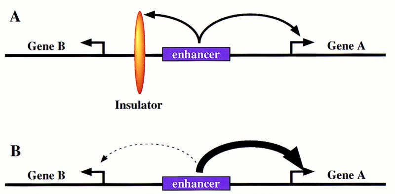
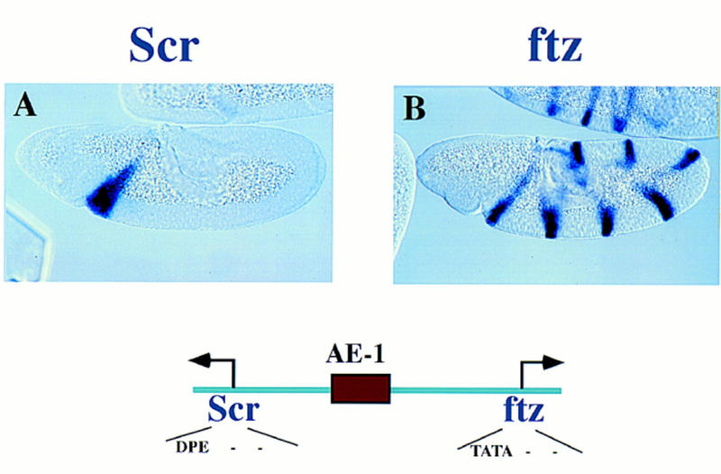
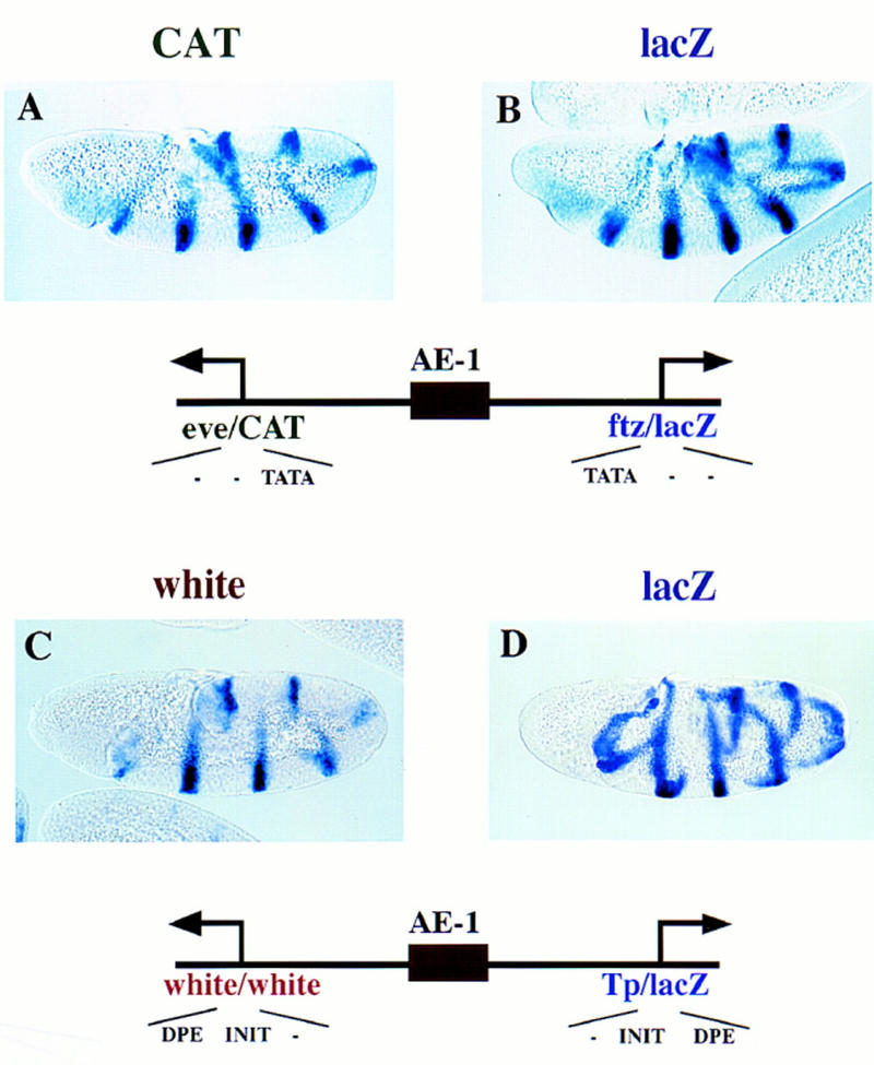
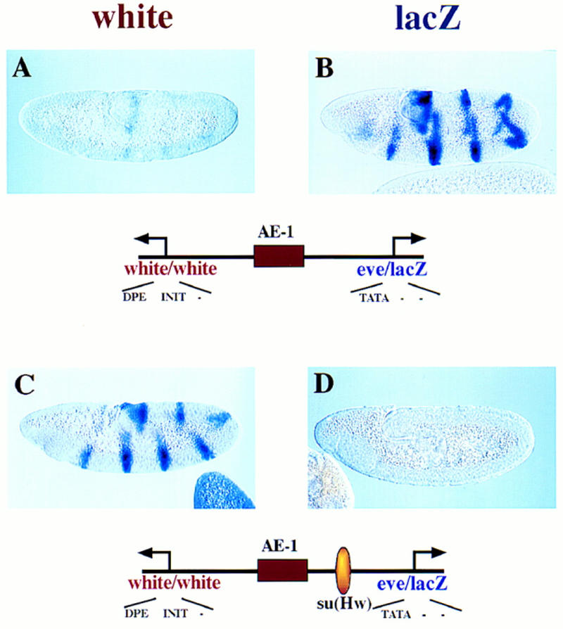
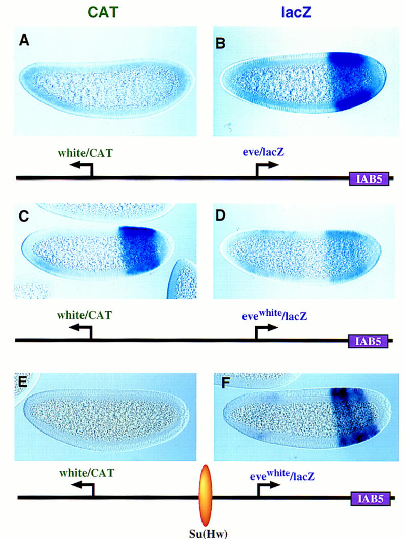
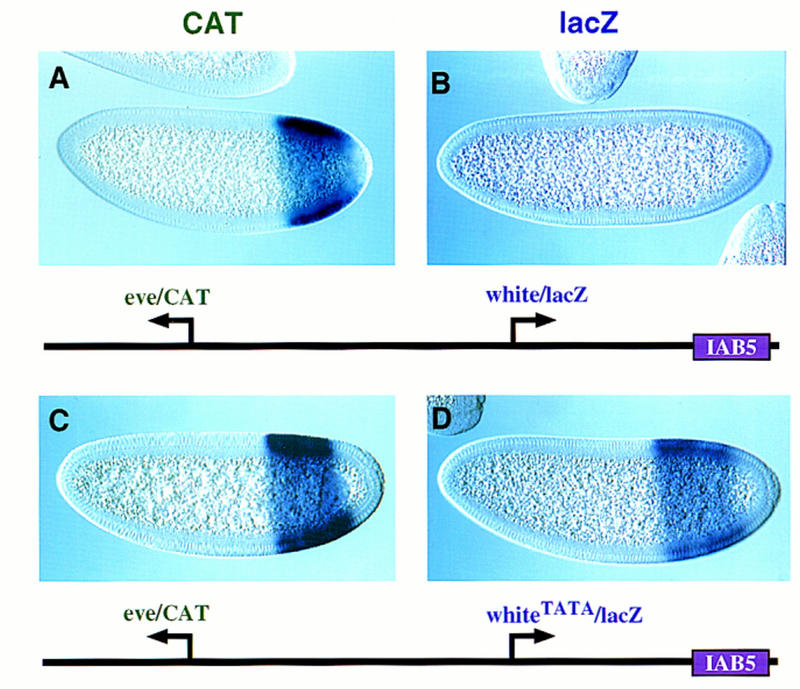
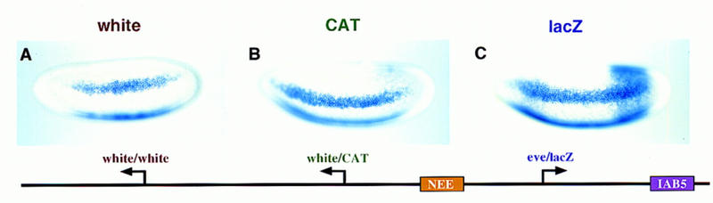
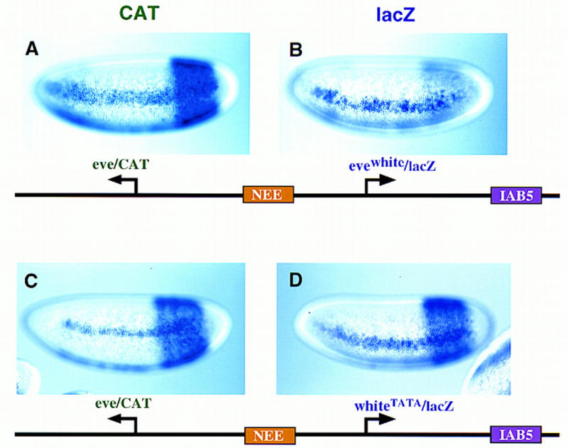
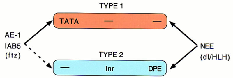
There are numerous examples of shared enhancers interacting with just a subset of target promoters. In some cases, specific enhancer-promoter interactions depend on promoter competition, whereby the activation of a preferred target promoter precludes expression of linked genes. Here, we employ a transgenic embryo assay to obtain evidence that promoter selection is influenced by the TATA element. Both the AE1 enhancer from the Drosophila Antennapedia gene complex (ANT-C) and the IAB5 enhancer from the Bithorax complex (BX-C) preferentially activate TATA-containing promoters when challenged with linked TATA-less promoters. In contrast, the rho neuroectoderm enhancer (NEE) does not discriminate between these two classes of promoters. Thus, certain upstream activators, such as Ftz, prefer TATA-containing promoters, whereas other activators, including Dorsal, work equally well on both classes of promoters. These results provide in vivo evidence that different core promoters possess distinct regulatory activities. We discuss the possibility that an invariant TFIID complex can adopt different conformations on the core promoter.


Figures

Figure 1
Figure 1
Regulation of enhancer–promoter interactions. The diagrams depict two divergently transcribed genes, A and B, with a common enhancer located in the intergenic region. (A) An insulator DNA is located between gene B and the enhancer. In principle, this blocks interactions of the enhancer with gene B, without altering the activation of gene A. (B) Promoter competition. In principle, the enhancer can activate both gene A and gene B, but prefers the promoter region of gene A. Enhancer–gene A interactions preclude activation of gene B.
Figure 2
Figure 2
Regulatory specficity in the Scr–ftz interval of the ANT-C. Embryos were hybridized with either a dioxigenin-labeled Scr or ftz antisense RNA probe and visualized via histochemical staining. The embryos are undergoing the rapid phase of germ-band elongation (4–5 hr postfertilization) and are oriented with anterior to the left and dorsal up. Scr is expressed within the primordia of parasegment 2 (PS 2), which gives rise to regions of the labial and prothoracic segments. ftz is expressed in a series of pair–rule stripes. The diagram below the embryos shows the location of the AE1 enhancer within the Scr–ftz interval. AE1 specifically interacts with the ftz promoter to maintain the seven-stripe pattern. It does not activate the linked Scr gene.
Figure 3
Figure 3
AE1 can coactivate linked reporter genes. Transgenic embryos carry fusion genes containing the 430-bp AE1 enhancer placed between divergently transcribed reporter genes that can be independently assayed. Embryos are undergoing the rapid phase of germ-band elongation. (A,B) Transgenic embryos carry a fusion gene with linked CAT and lacZ reporter genes. The arrows indicate the location and orientation of the transcription start sites. The leftward CAT gene was linked to the eve promoter, whereas the rightward lacZ reporter gene was attached to the ftz promoter. A was hybridized with a CAT antisense RNA probe; B was hybridized with a lacZ probe. Both reporter genes are expressed in a series of seven stripes, indicating that AE1 activates both the ftz and eve promoters. The diagrams indicate that the promoters contain TATA sequences, but lack optimal Inr (INIT) and Dpe (DPE) sequences. (C,D) Transgenic embryos carry a fusion gene with linked white and lacZ reporter genes. The white gene contains a mini-white promoter sequence, whereas lacZ was placed under the control of the core promoter sequence from the transposase gene (Tp) located within the P-element vector. C was hybridized with a white antisense RNA probe; D was hybridized with a lacZ probe. Both reporter genes are activated by AE1 and expressed in a series of stripes. The diagrams indicate that the promoters lack TATA sequences, but contain INIT and DPE elements.
Figure 4
Figure 4
Promoter competition influences AE1 activity. Transgenic embryos contain a P transposon with divergently transcribed white and lacZ reporter genes that are under the control of different core promoter sequences. The AE1 enhancer was placed between the linked genes, as summarized in the diagrams below the stained embryos. The embryos were hybridized with digoxigenin-labeled white or lacZ antisense RNA probes. (A,B) The white and lacZ reporter genes are driven by minimal white and eve promoter sequences, respectively. The eve/lacZ gene is expressed in a series of seven stripes, but the white gene exhibits just residual staining. It would appear that AE1-eve interactions preclude activation of the linked white gene, because AE1 can activate white in the absence of eve (e.g., see Fig. 3C). (C,D) Same as A and B except that the 340-bp su(Hw) insulator DNA from the gypsy retrotransposon was placed between the AE1 enhancer and eve/lacZ fusion gene. This silences lacZ staining and results in the activation of white.
Figure 5
Figure 5
The IAB5 enhancer prefers TATA-containing promoters. Transgenic embryos were stained and oriented as described in the previous figure legends, except that these are younger embryos (between cellularization and the onset of gastrulation). The IAB5 enhancer was placed downstream of the rightward lacZ reporter gene. The distal CAT gene is under the control of the white promoter. The proximal lacZ gene is driven by eve (B) or an evewhite chimeric promoter (D,F) whereby the eve TATA region was replaced with the corresponding sequences in white. (A,B) CAT (A) and lacZ (B) staining patterns obtained with linked white/CAT and eve/lacZ genes. The IAB5 enhancer selects eve over white, so that the eve/lacZ reporter gene exhibits strong expression whereas white/CAT is silent. (C,D) Same as A and B except that the proximal lacZ gene is under the control of the evewhite chimeric promoter (D). There is only weak expression of the lacZ reporter in the presumptive abdomen (D). IAB5 now mediates strong expression of the distal white/CAT fusion gene (C). The residual lacZ staining observed in anterior regions (D) may be a position effect resulting from the site of P insertion. (E,F) Same as C and D except that the 340-bp gypsy insulator DNA [su(Hw)] was placed between the leftward CAT gene and rightward lacZ reporter. The insulator blocks IAB5–white interactions, so that CAT is not expressed above backround levels. Instead, IAB5 directs strong expression of the evewhite/lacZ in the presumptive abdomen, indicating that the chimeric promoter is not defective. The weak staining seen in head regions is caused by sequences contained within the P-transformation vector (Small et al. 1992).
Figure 6
Figure 6
TATA is an important determinant of IAB5–eve interactions. Transgenic embryos carry the indicated P transposons and are oriented as described in the legend to Fig. 5. (A,B) Nuclear cleavage 14 embryos that carry a P transposon with the distal CAT gene driven by the core eve promoter and the rightward lacZ gene driven by mini-white. The IAB5 enhancer selectively interacts with the eve promoter, and directs intense expression of the CAT reporter in the presumptive abdomen (A). In contrast, the white/lacZ reporter gene is not expressed above background levels (B). (C,D) Cellularizing embryos carrying the same P transposon as A and B except that a synthetic TATA sequence was inserted into the mini-white promoter. IAB5 activates the lacZ reporter gene in the presumptive abdomen (D). The eve/CAT fusion gene is also activated by IAB5 (C). These results suggest that the whiteTATA promoter is almost as active as eve.
Figure 7
Figure 7
Independent activities of the IAB5 and NEE enhancers. Transgenic embryos carry the P-transformation vector shown in the diagram and are oriented as described in the previous legends to the figures. This synthetic gene complex contains three different reporter genes, white, CAT, and lacZ. The white and CAT genes are driven by the mini-white promoter, whereas lacZ contains the eve promoter. All three reporter genes exhibit robust expression in the lateral neurogenic ectoderm, indicating that the NEE enhancer interacts equally well with the mini-white and eve promoters. In contrast, lacZ is strongly activated in the presumptive abdomen (C), whereas white and CAT exhibit little or no expression in this region (A,B).
Figure 8
Figure 8
Independent activities of NEE and IAB5 on chimeric promoters. Transgenic embryos express the indicated P-transformation vectors, and are oriented as described previously. The rho NEE was placed in the intergenic region between the reporters, whereas IAB5 is located 3′ of the lacZ gene. The leftward CAT gene contains the eve promoter, whereas lacZ is driven by different chimeric promoters. (A,B) CAT and lacZ staining patterns obtained with the evewhite promoter, which contains an Inr element but lacks TATA and Dpe sequences. CAT transcripts are detected in lateral stripes and the presumptive abdomen (A), indicating activation of the eve/CAT gene by both the NEE and IAB5 enhancers. In contrast, lacZ is expressed primarily in lateral stripes; there is only residual staining in the abdomen (B). This staining pattern indicates that the evewhite promoter is strongly activated by the NEE enhancer, but only weakly interacts with IAB5. (C,D) CAT and lacZ staining patterns obtained with a synthetic white promoter (whiteTATA) that contains a TATA sequence. Both reporter genes exhibit robust expression in lateral stripes and the abdomen, indicating that the NEE and IAB5 enhancers work equally well on the eve and whiteTATA promoters.
Figure 9
Figure 9
Different core promoters possess distinct regulatory activities. The IAB5 and AE1 enhancers preferentially activate TATA-containing promoters (type I) when given a choice between linked TATA and Inr/Dpe (type II) promoters. In contrast, the NEE activates both classes of promoters. These results suggest that the IAB5 and AE1 activators, particularly ftz, prefer type I promoters. NEE activators, including Dorsal (dl) and bHLH proteins, appear to be promiscuous and work equally well on both classes of core promoters. We propose that the TFIID complex adopts different conformations on type I and type II promoters. Basal targets for the Ftz activator may be displayed in a more accessible conformation when TFIID binds TATA. In contrast, basal targets for the Dorsal and bHLH activators may be equally accessible whether TFIID binds TATA or Inr/Dpe elements.

Similar articles

Cited by

References

    1. Arnosti DN, Barolo S, Levine M, Small S. The eve stripe 2 enhancer employs multiple modes of transcriptional synergy. Development. 1996;122:205–214. - PubMed
    1. Barolo S, Levine M. Hairy mediates dominant repression in the Drosophila embryo. EMBO J. 1997;16:2883–2891. - PMC - PubMed
    1. Burke TW, Kadonaga JT. Drosophila TFIID binds to a conserved downstream basal promoter element that is present in many TATA-box-deficient promoters. Genes & Dev. 1996;10:711–724. - PubMed
    1. ————— The downstream core promoter element, DPE, is conserved from Drosophila to humans and is recognized by TAFII60 of Drosophila. Genes & Dev. 1997;11:3020–3031. - PMC - PubMed
    1. Burley SK, Roeder RG. Biochemistry and structural biology of transcription factor IID (TFIID) Annu Rev Biochem. 1996;65:769–799. - PubMed

Publication types

MeSH terms

LinkOut - more resources