. 1998 Feb 23;140(4):751-65.
doi: 10.1083/jcb.140.4.751.

gp25L/emp24/p24 protein family members of the cis-Golgi network bind both COP I and II coatomer

Affiliations

gp25L/emp24/p24 protein family members of the cis-Golgi network bind both COP I and II coatomer

M Dominguez et al. J Cell Biol. .

Abstract

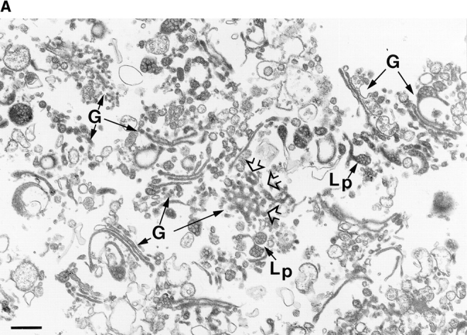
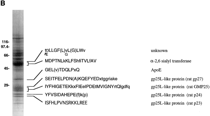
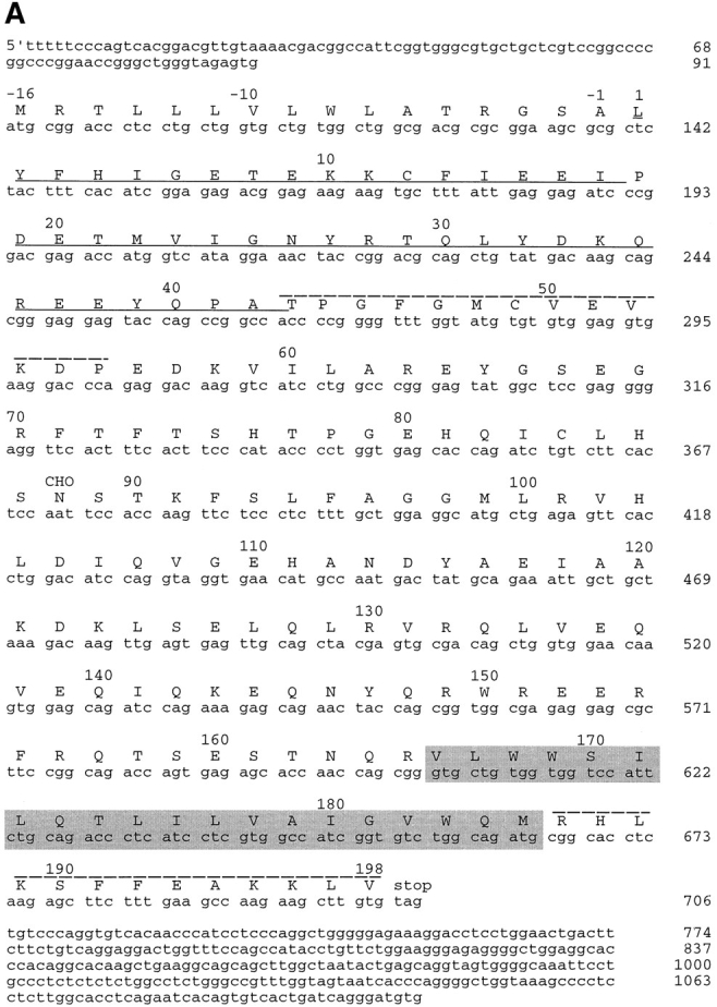
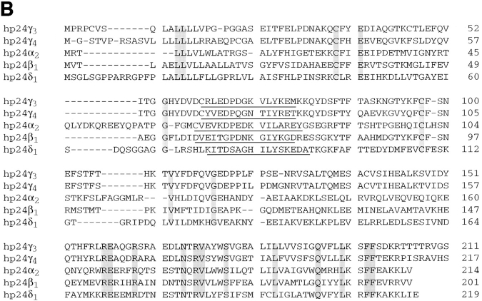
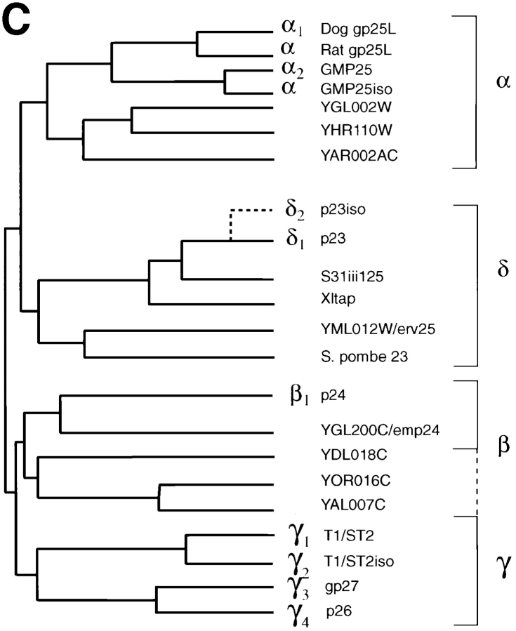
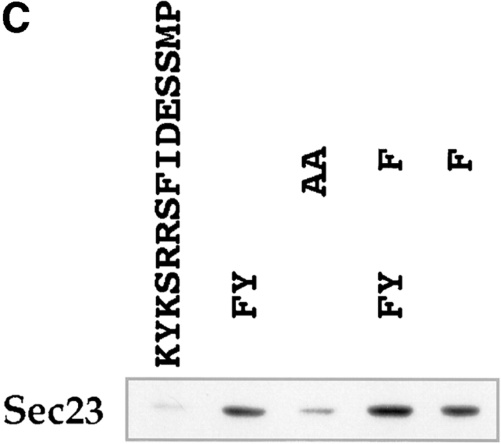
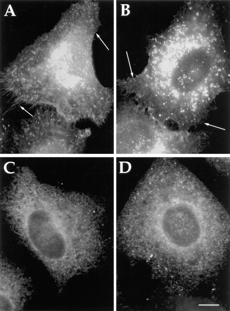
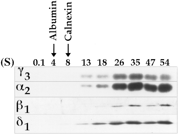
Abstract. Five mammalian members of the gp25L/ emp24/p24 family have been identified as major constituents of the cis-Golgi network of rat liver and HeLa cells. Two of these were also found in membranes of higher density (corresponding to the ER), and this correlated with their ability to bind COP I in vitro. This binding was mediated by a K(X)KXX-like retrieval motif present in the cytoplasmic domain of these two members. A second motif, double phenylalanine (FF), present in the cytoplasmic domain of all five members, was shown to participate in the binding of Sec23 (COP II). This motif is part of a larger one, similar to the F/YXXXXF/Y strong endocytosis and putative AP2 binding motif. In vivo mutational analysis confirmed the roles of both motifs so that when COP I binding was expected to be impaired, cell surface expression was observed, whereas mutation of the Sec23 binding motif resulted in a redistribution to the ER. Surprisingly, upon expression of mutated members, steady-state distribution of unmutated ones shifted as well, presumably as a consequence of their observed oligomeric properties.


Figures

Figure 1
Figure 1
(A) Random view of the purified hepatic Golgi fraction. Random sampling of the fraction (160-fold purification over the homogenate for the Golgi marker GalT) visualized after cross sectioning of filtered fraction reveals an abundance of stacked Golgi saccules (G) with a mottled content (Lp), especially in saccular distensions identified previously in Golgi apparatus in situ as apoE-containing lipoprotein particles (Dahan et al., 1994). Fenestrated structures (open arrowheads) are also evident. Bar, 400 nm. (B) Identification and NH2-terminal sequence of Golgi integral membrane proteins. The membrane proteins of the Golgi fraction were extracted with Triton X-114, subjected to SDS-PAGE, transferred to PVDF membranes, stained by Coomassie brilliant blue, and then processed for NH2-terminal sequencing. Upper and lower case residues represent, respectively, certain and less certain amino acids. Repeated sequencing at five different regions of the broad band, indicated by the square bracket, has revealed only the indicated sequence. Four proteins of the p24 family were identified: Rat p23, the homologue to human p23; rat p24, the rat homologue of CHOp24; rat GMP25, the rat homologue to human GMP25, and a fourth previously uncharacterized family member, p26.
Figure 1
Figure 1
(A) Random view of the purified hepatic Golgi fraction. Random sampling of the fraction (160-fold purification over the homogenate for the Golgi marker GalT) visualized after cross sectioning of filtered fraction reveals an abundance of stacked Golgi saccules (G) with a mottled content (Lp), especially in saccular distensions identified previously in Golgi apparatus in situ as apoE-containing lipoprotein particles (Dahan et al., 1994). Fenestrated structures (open arrowheads) are also evident. Bar, 400 nm. (B) Identification and NH2-terminal sequence of Golgi integral membrane proteins. The membrane proteins of the Golgi fraction were extracted with Triton X-114, subjected to SDS-PAGE, transferred to PVDF membranes, stained by Coomassie brilliant blue, and then processed for NH2-terminal sequencing. Upper and lower case residues represent, respectively, certain and less certain amino acids. Repeated sequencing at five different regions of the broad band, indicated by the square bracket, has revealed only the indicated sequence. Four proteins of the p24 family were identified: Rat p23, the homologue to human p23; rat p24, the rat homologue of CHOp24; rat GMP25, the rat homologue to human GMP25, and a fourth previously uncharacterized family member, p26.
Figure 2
Figure 2
cDNA cloning and comparison with other family members. (A) cDNA sequence and the deduced amino acid sequence of GMP25. Residue +1 represents the first amino acid as determined from the NH2-terminal sequence analysis with the preceding amino acids representing the signal sequence. The underlined sequence corresponds to the sequence amplified by RT-PCR of rat liver total RNA that was then used as a probe to screen the human brain library. The putative transmembrane sequence as deduced from the algorithm of Rost et al. (1995) is highlighted by shading. CHO indicates the single predicted site of N-glycosylation. Peptide sequences to generate antibodies used for subcellular fractionation (residues 43–55) and immunolocalization on cryosections (residues 186–189) are indicated. (B) Comparison of coding sequences. Alignment of obtained sequences was performed using the Multialin Program. Amino acids found to be conserved are highlighted. Sequences to generate further peptide antibodies for immunofluorescence and subcellular fractionation studies in the family of membrane proteins are underlined. (C) Cluster tree. Sequence relationships among the deduced protein sequences was performed using the multiclusteral option of PIMA multisequence alignment based on the amino acid classification scheme described by Smith and Smith (1990). Branches of the deduced tree represented by the four major membrane proteins found in hepatic Golgi fractions are indicated on the right.
Figure 2
Figure 2
cDNA cloning and comparison with other family members. (A) cDNA sequence and the deduced amino acid sequence of GMP25. Residue +1 represents the first amino acid as determined from the NH2-terminal sequence analysis with the preceding amino acids representing the signal sequence. The underlined sequence corresponds to the sequence amplified by RT-PCR of rat liver total RNA that was then used as a probe to screen the human brain library. The putative transmembrane sequence as deduced from the algorithm of Rost et al. (1995) is highlighted by shading. CHO indicates the single predicted site of N-glycosylation. Peptide sequences to generate antibodies used for subcellular fractionation (residues 43–55) and immunolocalization on cryosections (residues 186–189) are indicated. (B) Comparison of coding sequences. Alignment of obtained sequences was performed using the Multialin Program. Amino acids found to be conserved are highlighted. Sequences to generate further peptide antibodies for immunofluorescence and subcellular fractionation studies in the family of membrane proteins are underlined. (C) Cluster tree. Sequence relationships among the deduced protein sequences was performed using the multiclusteral option of PIMA multisequence alignment based on the amino acid classification scheme described by Smith and Smith (1990). Branches of the deduced tree represented by the four major membrane proteins found in hepatic Golgi fractions are indicated on the right.
Figure 3
Figure 3
Identification and orientation of the α2 sequence as a type I integral membrane glycoprotein. (A) Orientation of α2 by pronase digestion of hepatic Golgi fractions. 50 μg of Golgi fraction protein were subjected to proteolytic digestion. At a pronase concentration of 25 μg/ml, α2 was completely digested when 0.1% Triton X-100 was added, (lane 4) but unaffected in absence of detergent (lane 3). (B) Glycanase digestion of α2. 50 μg of Golgi fraction protein was untreated (lane 1, Control), treated with neuraminidase (lane 2, NA), neuraminidase and O-glycosidase (lane 3, NA+O+gly), or GpaseF (lane 4, GPase F). Treatment without (lane 5) or with endo H (lane 6) did not affect the mobility of the α2 protein. All immunoblots were with affinity-purified antibody to residues 43–55 of the α2 sequence (refer to Fig. 2 A).
Figure 3
Figure 3
Identification and orientation of the α2 sequence as a type I integral membrane glycoprotein. (A) Orientation of α2 by pronase digestion of hepatic Golgi fractions. 50 μg of Golgi fraction protein were subjected to proteolytic digestion. At a pronase concentration of 25 μg/ml, α2 was completely digested when 0.1% Triton X-100 was added, (lane 4) but unaffected in absence of detergent (lane 3). (B) Glycanase digestion of α2. 50 μg of Golgi fraction protein was untreated (lane 1, Control), treated with neuraminidase (lane 2, NA), neuraminidase and O-glycosidase (lane 3, NA+O+gly), or GpaseF (lane 4, GPase F). Treatment without (lane 5) or with endo H (lane 6) did not affect the mobility of the α2 protein. All immunoblots were with affinity-purified antibody to residues 43–55 of the α2 sequence (refer to Fig. 2 A).
Figure 4
Figure 4
Immunofluorescence localization of p24 family members after transient transfection in HeLa cells. The different cDNAs of all hp24 family members were cotransfected into HeLa cells as described in Materials and Methods. At 72 h after transfection, cells were fixed and then processed for indirect immunofluorescence using corresponding and specific primary antibodies. Their localization was compared to the Golgi resident protein NAGT I (red), and, as can be seen, α2 (A, green), β1 (B, green), γ3/4 (C, green), and β1 (D, green) proteins colocalized (yellow) with NAGT I to the Golgi apparatus. An additional localization of α2 to the nuclear envelope is indicated by an arrow in A. Bar, 5 μm.
Figure 5
Figure 5
In situ distribution of the α2 protein in rat liver hepatocyte ultrathin cryosections. Cryosections of rat liver hepatocytes were immunolabeled with the rabbit anti-C–terminus antibody against the α2 sequence followed by goat anti– rabbit IgG 10-nm-gold conjugates. Representative profiles of labeling within secretory compartments in liver hepatocytes are shown. Profiles of flattened cisternae of the rough ER are labeled along their length (A and B, arrows); note the gold particles lining mostly the cytosolic surface of the rough ER cisternae where the COOH-terminal domain of the α2 sequence is expected. α2 labeling is distributed throughout tubular smooth membranous profiles (arrowheads) in the Golgi/bile canalicular region of hepatocytes; prominent gold particle labeling can be seen in tubulovesicular membranous profiles closely approaching one side of a given Golgi apparatus (B–D, brackets). Labeling within the Golgi apparatus for the α2 protein is similarly restricted to one of the saccules on one side of a Golgi stack, whereas most other saccules reveal negligible labeling. Mitochondria (M), peroxisomes (P), and bile canaliculus (bc) were largely devoid of labeling. Bars, 400 nm.
Figure 6
Figure 6
Analytical fractionation of the α2 protein in rat liver homogenates. The MLP fraction from rat liver homogenates was centrifuged on linear sucrose gradients as described in Materials and Methods. Equal volumes of each fraction were evaluated for their content of GalT as evaluated by enzyme assay, calnexin, p58, and α2 protein as recognized by immunoblots, and quantification was evaluated by densitometry. The number of separate fractionations (n) is indicated. Results were normalized according to the methodology of Beaufay et al. (1964).
Figure 7
Figure 7
Analytical fractional of all family members in homogenates from rat liver and HeLa cells. Total membranes from rat liver (A) or HeLa cells (B) were centrifuged on linear sucrose (rat) or Nycodenz (HeLa) gradients as described in Materials and Methods. Equal volumes of each fraction were determined for their content of calnexin, Man II (Rat) or GalT (HeLa), syntaxin 5, p58 (rat), p53 (HeLa), and different p24 family members, using specific antibodies generated to the peptides described in Fig. 2 A and with antibodies specific to the Golgi markers (Man II, Gal T), the intermediate compartment markers (p58, p53) or ER marker (calnexin). The high density end of the gradient is on the right.
Figure 8
Figure 8
Binding of COP I and II proteins to cytoplasmic domains. Peptides corresponding to different cytoplasmic domains were coupled to thiopropyl Sepharose beads as described in Materials and Methods, and then assayed for their ability to bind COP I and II. (A) Binding of coat proteins to the cytoplasmic domains of α2, β1, δ1, γ3, γ4, erd2, E3/19k, E3/19k kk/ss (KK mutated to SS), UDP-GT, GalNac-T1, Man II, and control peptides C1-4. Binding of COP I components (tested for were α, β, β′, γ, and δ-COP) to α2 and δ1 was comparable to E3/19k and UDP-GT. The human homologue of Sec23 (hSec23) of the COP II coatomer showed specific binding to all cytoplasmic tails, but not to the four control peptides C1-4. Note the additional binding of the Sec23 to β1 and δ1 as well as to GalNac-TI. (B) Binding of coat proteins as in A but using the cytoplasmic domain of δ1 where pair-wise amino acid substitutions reveal a decrease of COP I binding upon KK-to-SS substitution, whereas Sec23 of COP II is reduced upon FF-to-AA substitution. (C) Introduction of aromatic residues into the mutated E3/19k peptide increases binding of Sec23.
Figure 8
Figure 8
Binding of COP I and II proteins to cytoplasmic domains. Peptides corresponding to different cytoplasmic domains were coupled to thiopropyl Sepharose beads as described in Materials and Methods, and then assayed for their ability to bind COP I and II. (A) Binding of coat proteins to the cytoplasmic domains of α2, β1, δ1, γ3, γ4, erd2, E3/19k, E3/19k kk/ss (KK mutated to SS), UDP-GT, GalNac-T1, Man II, and control peptides C1-4. Binding of COP I components (tested for were α, β, β′, γ, and δ-COP) to α2 and δ1 was comparable to E3/19k and UDP-GT. The human homologue of Sec23 (hSec23) of the COP II coatomer showed specific binding to all cytoplasmic tails, but not to the four control peptides C1-4. Note the additional binding of the Sec23 to β1 and δ1 as well as to GalNac-TI. (B) Binding of coat proteins as in A but using the cytoplasmic domain of δ1 where pair-wise amino acid substitutions reveal a decrease of COP I binding upon KK-to-SS substitution, whereas Sec23 of COP II is reduced upon FF-to-AA substitution. (C) Introduction of aromatic residues into the mutated E3/19k peptide increases binding of Sec23.
Figure 9
Figure 9
Altered distribution of family members after mutation of K(X)KK(X) on FF motifs of the cytosolic domains of α2 and β1 proteins. Cotransfection of all five p24s in where either the KK (A and B) or the FF (C and D) motif has been altered to SS or AA, respectively, in both α2 and δ1. Altered redistribution and apparent cell surface staining can be seen with the KK/SS mutants (shown is α2)(A). Remarkably, the unmutated members (shown is γ3/4) (B) also reveals redistribution to the cell surface. Similarly, an apparent ER staining can be seen with the FF/AA mutants (shown is α2)(C). This also leads to the apparent redistribution of the unmutated members to the ER (shown is γ3/4)(D). Bar, 5 μm.
Figure 10
Figure 10
Sedimentation density gradient centrifugation showing oligomeric behavior of p24s. Golgi fraction was solubilized with sodium cholate containing buffer, loaded onto a sucrose gradient, and subjected to centrifugation as described in Materials and Methods. Fractions were collected and evaluated for their content in α2, β1, δ1, and γ3 by Western blotting followed by ECL. All four show a similar peak distribution around 35S.

References

    1. Allan VJ, Kreis TE. A microtubule-binding protein associated with membranes of the Golgi apparatus. J Cell Biol. 1986;103:2229–2239. - PMC - PubMed
    1. Aridor M, Balch WE. Principles of selective transport: coat complexes hold the key. Trends Cell Biol. 1996;6:315–320. - PubMed
    1. Banfield DK, Lewis MJ, Rabouille C, Warren G, Pelham HR. Localization of Sed5, a putative vesicle targeting molecule, to the cis-Golgi network involves both its transmembrane and cytoplasmic domains. J Cell Biol. 1994;127:357–371. - PMC - PubMed
    1. Barlowe C, Orci L, Yeung T, Hosobuchi M, Hamamoto S, Salama N, Rexach MF, Ravazzola M, Amherdt M, Schekman R. COPII: a membrane coat formed by Sec proteins that drive vesicle budding from the endoplasmic reticulum. Cell. 1994;77:895–907. - PubMed
    1. Baudhuin P, Evrard P, Berthet J. Electron microscopic examination of subcellular fractions. J Cell Biol. 1967;32:405–416. - PMC - PubMed

Publication types

MeSH terms