. 1998 Feb 23;140(4):861-71.
doi: 10.1083/jcb.140.4.861.

Cytoskeletal polarization of T cells is regulated by an immunoreceptor tyrosine-based activation motif-dependent mechanism

Affiliations

Cytoskeletal polarization of T cells is regulated by an immunoreceptor tyrosine-based activation motif-dependent mechanism

B Lowin-Kropf et al. J Cell Biol. .

Abstract

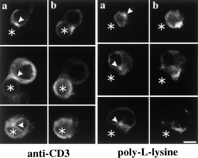
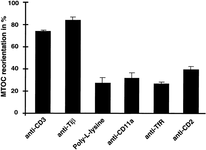
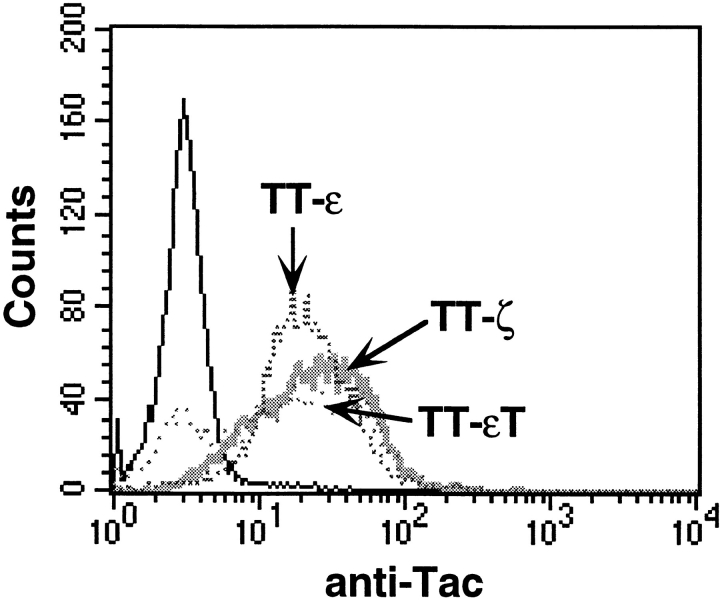
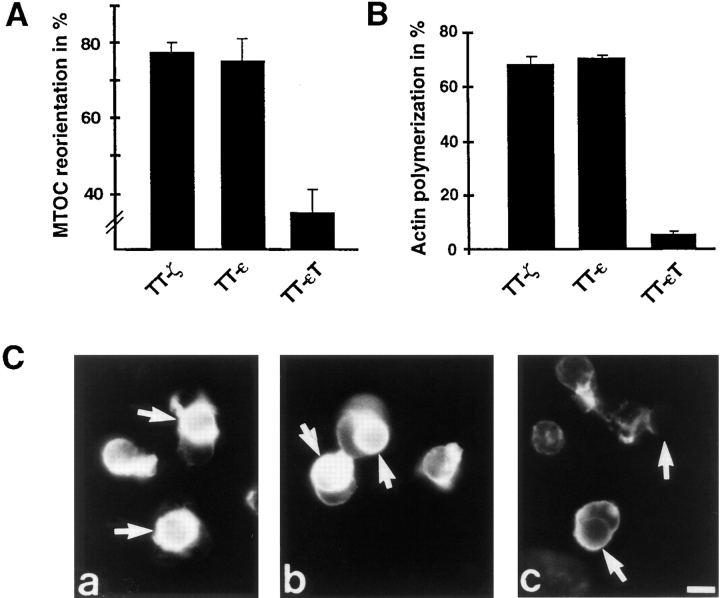
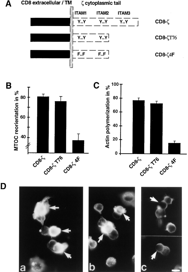
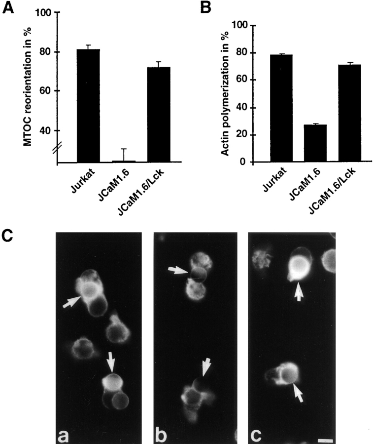
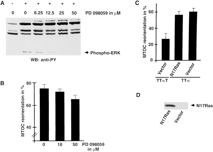
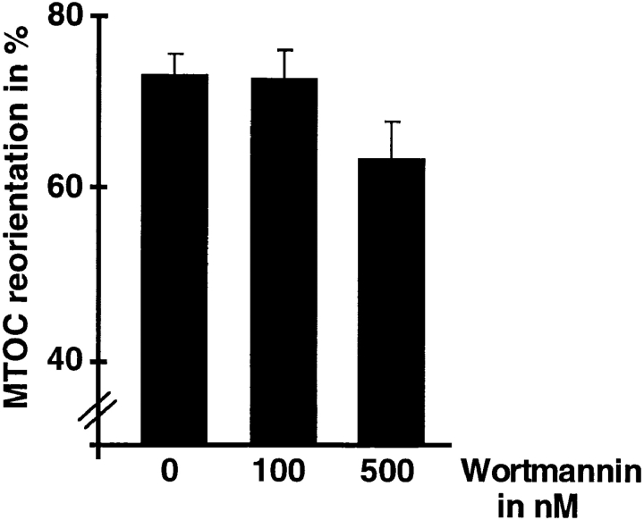
Binding of a T cell to an appropriate antigen-presenting cell (APC) induces the rapid reorientation of the T cell cytoskeleton and secretory apparatus towards the cell-cell contact site in a T cell antigen receptor (TCR) and peptide/major histocompatibility complex-dependent process. Such T cell polarization directs the delivery of cytokines and cytotoxic mediators towards the APC and contributes to the highly selective and specific action of effector T cells. To study the signaling pathways that regulate cytoskeletal rearrangements in T lymphocytes, we set up a conjugate formation assay using Jurkat T cells as effectors and cell-sized latex beads coated with various antibodies as artificial APCs. Here, we report that beads coated with antibodies specific for the TCR-CD3 complex were sufficient to induce T cell polarization towards the bead attachment site, as judged by reorientation of the microtubule-organizing center (MTOC) and localized actin polymerization. Thus, these cytoskeletal changes did not depend on activation of additional coreceptors. Moreover, single subunits of the TCR complex, namely TCR-zeta and CD3epsilon, were equally effective in inducing cytoskeletal polarization. However, mutagenesis of the immunoreceptor tyrosine-based activation motifs (ITAMs), present three times in TCR-zeta and once in CD3epsilon, revealed that the induction of cytoskeletal rearrangements required the presence of at least one intact ITAM. In agreement with this result, lack of functional Lck, the protein tyrosine kinase responsible for ITAM phosphorylation, abolished both MTOC reorientation and polarized actin polymerization. Both inhibitor and transient overexpression studies demonstrated that MTOC reorientation could occur in the absence of Ras activation. Our results suggest that APC-induced T cell polarization is a TCR-mediated event that is coupled to the TCR by the same signaling motif as TCR-induced gene activation, but diverges in its distal signaling requirements.


Figures

Figure 1
Figure 1
Anti–TCR-coated latex beads induce MTOC reorientation and polarized actin polymerization in Jurkat T cells. Jurkat T cells were mixed at a 1:2 ratio with anti–CD3ε-coated latex beads (left) or poly-l-lysine-coated latex beads (right). After 30 min at 37°C, conjugates were stained with the antitubulin antibody YOL1/34 (a) and rhodamine-phalloidin to visualize F-actin (b). The position of cell-bound latex beads is indicated by an asterisk, the position of the MTOC by an arrowhead. Bar, 5 μm.
Figure 2
Figure 2
MTOC reorientation in Jurkat cells is specifically associated with TCR/CD3 cross-linking. Jurkat T cells were mixed at a 1:2 ratio with latex beads coated either with poly-l-lysine or antibodies against CD11a/LFA-1, the transferrin receptor, and CD2. Conjugates were stained with antitubulin antibody. Each column represents the average of at least four individual experiments in which >100 conjugates were scored for MTOC reorientation. MTOCs positioned between the bead contact site and the T cell nucleus, in close proximity to the T cell plasma membrane, were scored as positive for reorientation.
Figure 3
Figure 3
Chimera surface expression in stable Jurkat-derived clones expressing chimeras between Tac and the cytoplasmic tail of TCR-ζ (TT-ζ), CD3ε (TT-ε), or a truncated version of CD3ε (TT-εT). Stable clones were analyzed for chimera surface expression by FACS® analysis using an FITC-labeled Tac-specific antibody. The solid line represents untransfected Jurkat T cells.
Figure 4
Figure 4
Clustering of chimeras containing the cytoplasmic domain of CD3ε or TCR-ζ can induce MTOC reorientation and actin polymerization. Stable Jurkat-derived clones expressing chimeras between Tac and the cytoplasmic tail of TCR-ζ (TT-ζ), CD3ε (TT-ε), or a truncated version of CD3ε (TT-εT) were analyzed. Clones were mixed at a 1:2 ratio with latex beads coated with anti–Tac. After incubation for 10 min at 37°C, conjugates were processed as in Fig. 1. (A) Conjugates were scored for MTOC reorientation. Averages of four different experiments are shown. To better visualize the differences in MTOC reorientation, the scale of the graph starts at 25%, an arbitrarily set background value obtained with poly-l-lysine-coated beads. (B) Only conjugates between single latex beads and single Jurkat cells were scored for actin polymerization. Three different experiments were averaged. (C) Conjugates between anti–Tax latex beads and Jurkat clones expressing either TT-ζ (a), TT-ε (b), or TT-εT (c) were stained for F-actin. Latex beads are indicated by an arrow. Bar, 5 μm.
Figure 5
Figure 5
MTOC reorientation and actin polymerization require ITAM phosphorylation. (A) Schematic representation of the expressed CD8 chimeras. (B) Conjugates between the CD8 chimera–expressing clones and anti–CD8-coated latex beads were analyzed for MTOC reorientation as in Fig. 2. Results of six different experiments are averaged. (C) Actin polymerization in single bead–T cell conjugates was scored as in Fig. 4. Averages of three different experiments are shown. (C) Actin polymerization in response to CD8-ζ (a), CD8-ζT76 (b), and CD8-ζ4F (c) cross-linking was analyzed as in Fig. 4. The position of latex beads is indicated by an arrow. Bar, 5 μm.
Figure 6
Figure 6
CD8 chimera surface expression in stable Jurkat-derived clones. Stable clones were analyzed for chimera surface expression by FACS® analysis using an FITC-labeled CD8-specific antibody. The solid line represents untransfected Jurkat T cells.
Figure 7
Figure 7
MTOC reorientation and actin polymerization are abolished in the Lck-deficient cell line JCaM1.6. Jurkat T cells and the signaling mutant JCaM1.6 were bound to anti–CD3ε beads and scored as described in Fig. 2 for (A) MTOC reorientation and (B and C) actin polymerization. C shows actin polymerization in Jurkat T cells (a), the Lck-deficient mutant JCaM1.6 (b), and JCaM1.6/Lck reconstituted with murine Lck (c). Arrows point to cell-bound beads. Averages of seven (A) and three (B) experiments are shown. Bar, 5 μm.
Figure 8
Figure 8
Expression of dominant-negative ZAP-70 mutant impairs ITAM-mediated MTOC reorientation. Jurkat T cells were cotransfected with TT-εT or TT-ε (30 μg), together with 40 μg of empty vector or plasmids encoding different ZAP-70 mutants. 36 h later, cells were mixed with anti–Tac latex beads and incubated for 10 min at 37°C. Conjugates were fixed in paraformaldehyde and stained with antitubulin antibody. Percent MTOC inhibition equals XN/MN, where X is the percent MTOC reorientation triggered by TT-ε in the presence of dominant-negative ZAP-70, M is the percent MTOC reorientation triggered by TT-ε alone, and N is the percent MTOC reorientation triggered by TT-εT. Results are the average of three independent experiments. The averaged percent MTOC reorientation for each condition was 20 ± 6% for TT-εT plus vector, 67 ± 1% for TT-ε plus vector, 40 ± 6% for TT-ε plus SH2 (N+C), and 64 ± 5% for TT-ε plus SH2 (N+*C).
Figure 9
Figure 9
MTOC reorientation is mediated by Ras-independent pathways. (A) Jurkat T cells were incubated with the indicated concentrations of PD 098059 in DMSO for 30 min, followed by a 10-min stimulation with C305-coated latex beads (+). Untreated control cells were mixed with poly-l-lysine-coated beads (−). Cells were lysed and whole cell lysates were analyzed by immunoblotting with an antiphosphotyrosine antibody. (B) Jurkat T cells were incubated with the indicated concentrations of PD 098059 for 30 min. Subsequently, C305-coated latex beads were added at a 1:2 ratio and conjugates were incubated for 10 min. Conjugates were fixed and stained with antitubulin antibody. The average of four independent experiments is shown. (C) TAg Jurkat cells were cotransfected with TT-εT or TT-ε (30 μg) together with 40 μg of empty vector or a plasmid encoding the dominant-negative Ras mutant N17Ras. 36 h later, cells were mixed with anti–Tac latex beads and incubated for 10 min at 37°C. Conjugates were fixed in paraformaldehyde and stained with antitubulin antibody. The averaged percent MTOC reorientations of two independent experiments are shown. (D) Lysates from 106 Tag Jurkat cells either cotransfected with TT-ε and empty vector or N17Ras were analyzed for the presence of mutant Ras with a Ha-Ras-specific mAb.
Figure 10
Figure 10
PI-3 kinase is not required for TCR-mediated MTOC reorientation in Jurkat T cells. Jurkat T cells were incubated with the indicated concentrations of Wortmannin for 30 min. Subsequently, Leu4-coated latex beads were added at a 1:2 ratio and conjugates were incubated for 30 min. Conjugates were fixed and stained with antitubulin antibody. The average of four independent experiments is shown.

References

    1. Alberola-Ila J, Forbush KA, Seger R, Krebs EG, Perlmutter RM. Selective requirement for MAP kinase activation in thymocyte differentiation. Nature. 1995;373:620–623. - PubMed
    1. Anel A, Richieri GV, Kleinfeld AM. A tyrosine phosphorylation requirement for cytotoxic T lymphocyte degranulation. J Biol Chem. 1994;269:9506–9513. - PubMed
    1. Arpaia E, Shahar M, Dadi H, Cohen A, Roifman CM. Defective T cell receptor signaling and CD8+thymic selection in humans lacking ZAP-70 kinase. Cell. 1994;76:947–958. - PubMed
    1. Cardine AM, Kirchgessner H, Eckerskorn C, Meuer SC, Schraven B. Human T lymphocyte activation induces tyrosine phosphorylation of α-tubulin and its association with the SH2 domain of the p59fyn protein tyrosine kinase. Eur J Immunol. 1995;25:3290–3297. - PubMed
    1. Chan AC, Kadlecek TA, Elder ME, Filipovich AH, Kuo W-L, Iwashima M, Parslow TG, Weiss A. ZAP-70 deficiency in an autosomal recessive form of severe combined immunodeficiency. Science. 1994;264:1599–1601. - PubMed

Publication types

MeSH terms