. 1998 Feb 23;140(4):911-23.
doi: 10.1083/jcb.140.4.911.

The p75 neurotrophin receptor mediates neuronal apoptosis and is essential for naturally occurring sympathetic neuron death

Affiliations

The p75 neurotrophin receptor mediates neuronal apoptosis and is essential for naturally occurring sympathetic neuron death

S X Bamji et al. J Cell Biol. .

Abstract

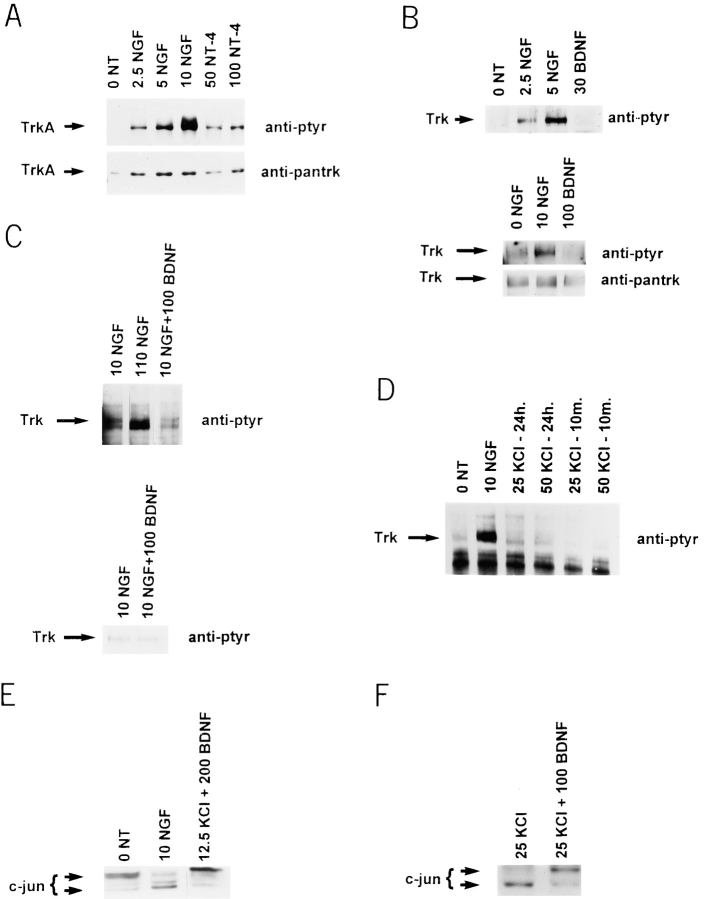
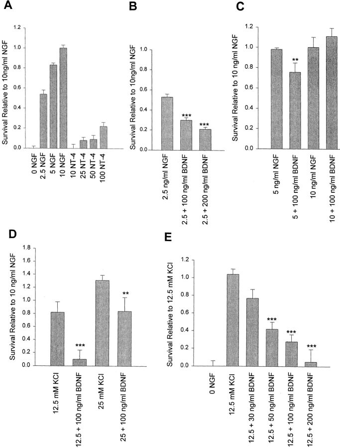
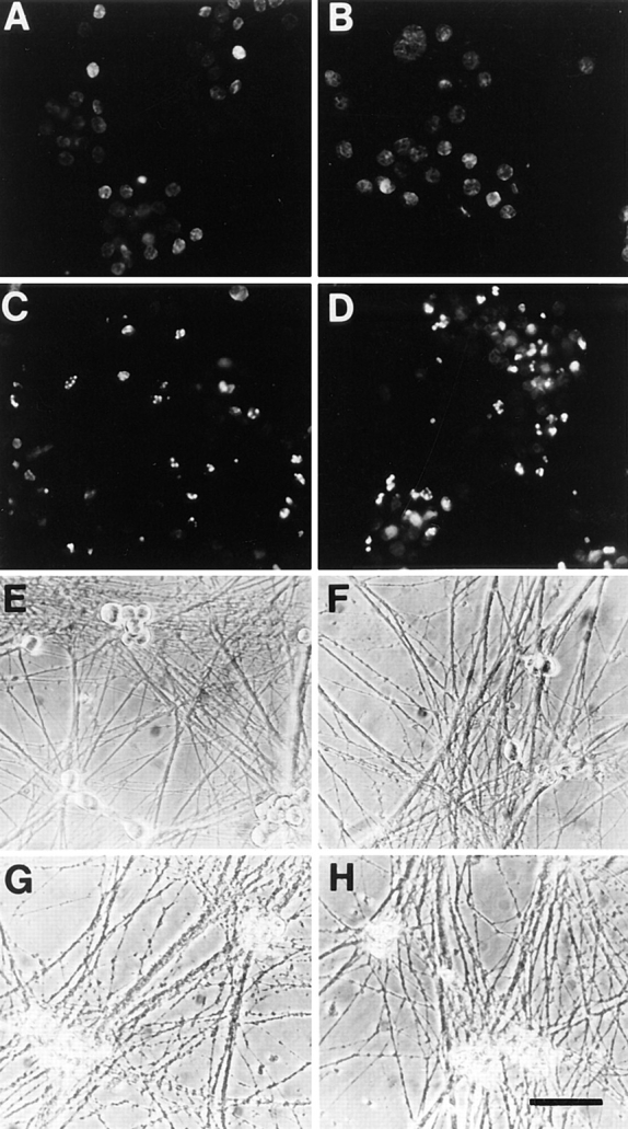
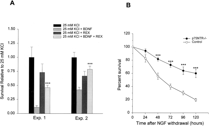
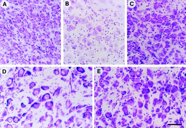
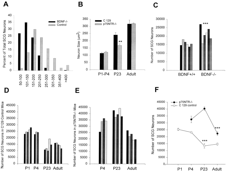
To determine whether the p75 neurotrophin receptor (p75NTR) plays a role in naturally occurring neuronal death, we examined neonatal sympathetic neurons that express both the TrkA tyrosine kinase receptor and p75NTR. When sympathetic neuron survival is maintained with low quantities of NGF or KCl, the neurotrophin brain-derived neurotrophic factor (BDNF), which does not activate Trk receptors on sympathetic neurons, causes neuronal apoptosis and increased phosphorylation of c-jun. Function-blocking antibody studies indicate that this apoptosis is due to BDNF-mediated activation of p75NTR. To determine the physiological relevance of these culture findings, we examined sympathetic neurons in BDNF-/- and p75NTR-/- mice. In BDNF-/- mice, sympathetic neuron number is increased relative to BDNF+/+ littermates, and in p75NTR-/- mice, the normal period of sympathetic neuron death does not occur, with neuronal attrition occurring later in life. This deficit in apoptosis is intrinsic to sympathetic neurons, since cultured p75NTR-/- neurons die more slowly than do their wild-type counterparts. Together, these data indicate that p75NTR can signal to mediate apoptosis, and that this mechanism is essential for naturally occurring sympathetic neuron death.


Figures

Figure 1
Figure 1
(A) NT-4 activates TrkA on sympathetic neurons. Sympathetic neurons were selected in 10 ng/ml NGF for 5 d, washed free of neurotrophin, and then exposed to various concentrations of NGF (from 2.5 to 10 ng/ml) or NT-4 (50 and 100 ng/ml) for 10 min. Cellular lysates were immunoprecipitated with an antibody specific to TrkA, and probed first with anti-phosphotyrosine (anti-ptyr) to visualize TrkA autophosphorylation, and then reprobed with anti-panTrk to monitor total TrkA levels (anti-pantrk). All samples were normalized for equal amounts of protein. (B and C) BDNF does not activate Trk on sympathetic neurons. (B) To examine short-term Trk autophosphorylation, sympathetic neurons were washed free of NGF and exposed to 2.5, 5, or 10 ng/ml NGF or to 30 or 100 ng/ml BDNF for 10 min. Total Trk protein was immunoprecipitated with anti-panTrk, and Trk autophosphorylation analyzed by blotting with anti-phosphotyrosine (anti-ptyr). In the bottom panel, the same blot was reprobed with panTrk antibody 203 (anti-pantrk) to ensure that similar amounts of total Trk protein were present in all lanes. (C) To examine long-term Trk autophosphorylation, sympathetic neurons were selected in 10 ng/ml NGF, and then switched to 10 ng/ml NGF (10 NGF), 110 ng/ml NGF (110 NGF), or 10 ng/ml NGF + 100 ng/ml BDNF (10 NGF + 100 BDNF). Analysis of Trk autophosphorylation was performed as in B. In all cases, samples were normalized for equal amounts of protein, and blots were reprobed for total Trk protein levels. (D) KCl treatment does not lead to Trk activation on sympathetic neurons. Sympathetic neurons were washed free of neurotrophin and switched to 10 ng/ml NGF for 10 min, or to 25 or 50 mM KCl for 10 min or 24 h. Trk autophosphorylation was analyzed as in B. (E and F) BDNF-mediated activation of p75 leads to increased phosphorylation of c-jun. Sympathetic neurons were selected in 50 ng/ml NGF, washed free of neurotrophin, and then switched to 10 ng/ml NGF, or 12.5 or 25 mM KCl plus or minus BDNF. 18 h later, cellular lysates were analyzed for c-jun on Western blots. (E) As previously reported (Ham et al., 1995), the size of c-jun increases in response to NGF withdrawal, a shift indicative of increased phosphorylation. 200 ng/ml BDNF caused a similar size shift in the presence of 12.5 mM KCl. (F) This size shift was specific to BDNF and not to KCl, as shown here with 25 mM KCl plus or minus 100 ng/ml BDNF.
Figure 2
Figure 2
(A) NT-4 is fivefold less efficient than NGF at supporting sympathetic neuron survival at equivalent levels of TrkA autophosphorylation. A comparison of sympathetic neuron survival in response to 2.5–10 ng/ml NGF versus 10–100 ng/ml NT-4, as monitored using MTT assays that measure mitochondrial function and cell survival. Neonatal sympathetic neurons were cultured in 10 ng/ml NGF for 5 d in 48-well dishes, washed free of neurotrophin-containing medium, and then incubated for 2 d in various concentrations of NGF or NT-4, as indicated on the x-axis. Each point represents the values pooled from three independent sets of survival assays, each of which was performed in quadruplicate. In these assays, absolute values are normalized so that the value obtained without neurotrophin is 0% survival, whereas that obtained with 10 ng/ml NGF is considered 100% survival. Error bars represent the standard error. (B–D) BDNF decreases sympathetic neuron survival in the presence of limiting quantities of NGF (B and C) or KCl (D). Neurons were selected in 50 ng/ml NGF for 5 d, and then switched to 2.5 ng/ml NGF, 5 ng/ml NGF, 10 ng/ml NGF, 12.5 mM KCl, or 25 mM KCL, with or without 100 or 200 ng/ml BDNF for 2 d. Survival was monitored by MTT assays. Results are expressed relative to those obtained with 10 ng/ml NGF, and represent the mean ± standard error. B and D include the combined data from three independent experiments performed in triplicate. C represents the data from one representative experiment performed in quadruplicate (**P < 0.05, ***P < 0.005 relative to NGF [B and C] or KCl [D] alone). (E) BDNF decreases sympathetic neuron survival in a concentration-dependent fashion. Neurons were selected in 50 ng/ml NGF for 5 d, and then switched to 12.5 mM KCl with or without concentrations of BDNF ranging from 30 to 200 ng/ml for 2 d. Survival was monitored by MTT assays. Results are normalized relative to those obtained with 12.5 mM KCl alone, and represent the mean ± standard error from the combined data of four independent experiments (***P < 0.005 relative to 12.5 mM KCl alone).
Figure 3
Figure 3
Sympathetic neurons undergo apoptosis in response to BDNF. (A–D) Fluorescence photomicrographs of sympathetic neurons analyzed by TUNEL labeling. Neurons were selected in 50 ng/ml NGF for 5 d, washed free of neurotrophin, and then switched to 12.5 mM KCl with (D) or without (B) 100 ng/ml BDNF for 18 h. As controls, sister cultures were maintained in 50 ng/ml NGF (A) or withdrawn from NGF (C). 18 h later, neurons were analyzed for apoptosis using TUNEL labeling for fragmented nuclear DNA. (E–H) Phase-contrast photomicrographs of cultured sympathetic neurons. Neurons were cultured as above, and then switched to 12.5 mM KCl with (H) or without (F) 100 ng/ml BDNF for 48 h. As controls, sister cultures were maintained in 50 ng/ml NGF (E) or withdrawn from NGF (G). Bar, 74 μm.
Figure 4
Figure 4
(A) The p75NTR antibody, REX, inhibits the BDNF-mediated apoptosis of sympathetic neurons. A comparison of sympathetic neuron survival, as monitored using MTT assays, in response to 25 mM KCl ± BDNF with and without the presence of the function-blocking anti-p75NTR, REX. Sympathetic neurons were cultured in 50 ng/ml NGF for 5 d, washed free of neurotrophin-containing medium, and then incubated for 2 d in 25 mM KCl or 25 mM KCl + 100 ng/ml BDNF with or without REX. Results of two representative experiments, each of which was performed in triplicate, are shown. In all cases, results represent the mean ± standard error, and are normalized so that the survival mediated by 25 mM KCl alone is 100%. REX by itself had no significant effect (P > 0.05) on sympathetic neuron survival as mediated by 25 mM KCl. (***P < 0.004 for the comparison between 25 mM KCl + REX and 25 mM KCL + 100 ng/ml BDNF + REX). (B) Death of cultured p75NTR−/− neurons is delayed after NGF withdrawal. Time course of survival of cultured p75NTR−/− and p75NTR+/+ (Control) neurons following NGF withdrawal. Representative fields of neurons were counted immediately upon NGF withdrawal, and again every 24 h. Results are normalized so that the number of neurons at the time of NGF withdrawal is 100%. Results represent the mean ± standard error of the combined data from five individual experiments each for the p75NTR−/− and control neurons; every individual experiment was performed in triplicate. (***P < 0.002 for the comparison between p75NTR−/− and control neurons at each time point).
Figure 5
Figure 5
Morphology of sympathetic neurons of the superior cervical ganglion in BDNF−/− and p75NTR−/− mice. Photomicrographs of cresyl violet-stained cross-sections of the SCG from P15 BDNF−/− mice (A), P15 BDNF+/+ littermates (B), P23 p75NTR−/− mice (C), adult C129 mice (D), and adult p75NTR−/− mice (E). Bar, 38.5 μm.
Figure 6
Figure 6
At P15 to P23, sympathetic neuron number is increased in the SCG of BDNF−/− and p75NTR−/− mice. (A) Cross-sectional area of P15 BDNF−/− neurons relative to control neurons from animals of the same background, plotted as a size distribution histogram, in bins of 50 μm2. Note that the entire population of BDNF−/− sympathetic neurons is shifted to a smaller size relative to BDNF+/+ (Control) neurons of the same age. (B) Mean cross-sectional areas of sympathetic neurons of the SCG in p75NTR−/− mice at various developmental time points relative to control C129 mice. Note that from P1 to P4, and in adulthood, the size of sympathetic neurons is similar in the presence or absence of p75NTR, but that at P23, neurons lacking p75NTR are significantly smaller. Neuron size is expressed as cross-sectional area in μm2, and error bars represent the standard error (**P < 0.05). (C) The number of sympathetic neurons of the SCG is increased in P15 BDNF−/− animals relative to their control littermates, as determined by counting. The number of neurons in each of six control BDNF+/+ and BDNF−/− animals are shown; each bar represents the number of neurons in the SCG of one animal of the indicated genotype. The means of these two groups are significantly different (***P < 0.01). The mean ± the standard error are summarized in Table I as “Total counted neuron number.” (D) Sympathetic neuron number in the SCG of control C129 mice at developmental time points encompassing the period of naturally occurring sympathetic neuron death. The number of neurons counted in the SCG at P1, P4, P23, and adulthood are shown; each bar represents the number of neurons in the SCG of one C129 animal of the indicated age. Five animals were analyzed at P1, three at P4, five at P23, and five at adulthood. The mean ± standard error of these groups are plotted in F. (E) Sympathetic neuron number in the SCG of p75NTR−/− mice at developmental time points encompassing the period of naturally occurring sympathetic neuron death. The number of neurons counted in the SCG at P4, P23, and adulthood are shown; each bar represents the number of neurons counted in the SCG of one p75NTR−/− animal of the indicated age. Four animals were analyzed at P4, five at P23, and five adults. The mean ± standard error of these groups are plotted in F. (F) Comparison of the time course of naturally occurring sympathetic neuron death in the SCG of p75NTR−/− versus control (C129 control) animals. The points represent the mean ± standard error of the counts shown in D and E. There is a highly significant decrease in neuronal number from P4 to P23 in the control C129 mice, and from P23 to adulthood in the p75NTR−/− mice (***P < 0.007). The means for all of these groups are summarized in Table I as “Total counted neuron number”.

References

    1. Anton ES, Weskamp G, Reichardt LF, Matthew WD. Nerve growth factor and its low-affinity receptor promote Schwann cell migration. Proc Natl Acad Sci USA. 1994;91:2795–2799. - PMC - PubMed
    1. Barbacid M. The trk family of neurotrophin receptors. J Neurobiol. 1994;25:1386–1403. - PubMed
    1. Barde Y-A. Trophic factors and neuronal survival. Neuron. 1989;2:1525–1534. - PubMed
    1. Barker PA, Shooter EM. Disruption of NGF binding to the low affinity neurotrophin receptor p75LNTR reduces NGF binding to TrkA on PC12 cells. Neuron. 1994;13:203–215. - PubMed
    1. Barrett GL, Bartlett PF. The p75 nerve growth factor receptor mediates survival or death depending on the stage of sensory neuron development. Proc Natl Acad Sci USA. 1994;91:6501–6505. - PMC - PubMed

Publication types

MeSH terms

Substances