. 1998 Feb 23;140(4):947-59.
doi: 10.1083/jcb.140.4.947.

Vascular endothelial growth factor induces endothelial fenestrations in vitro

Affiliations

Vascular endothelial growth factor induces endothelial fenestrations in vitro

S Esser et al. J Cell Biol. .

Abstract

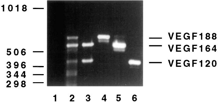
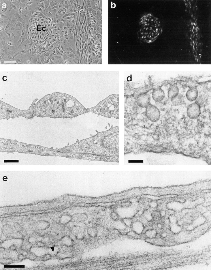
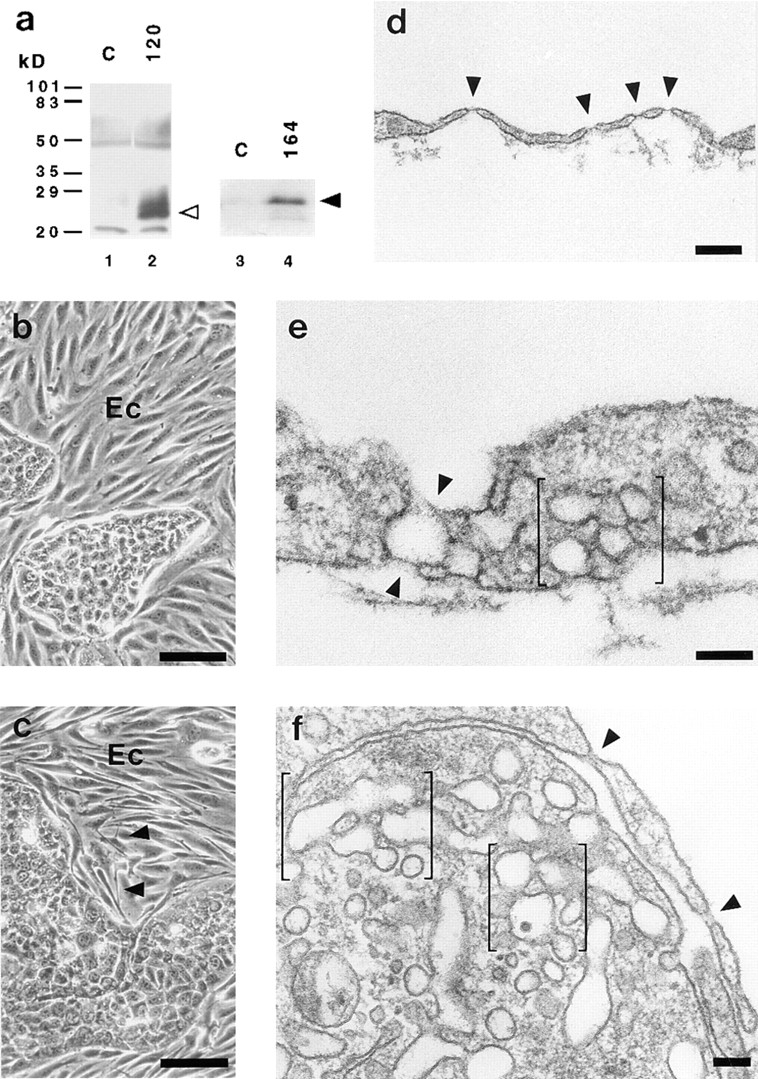
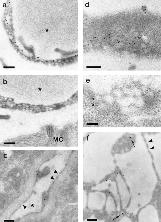
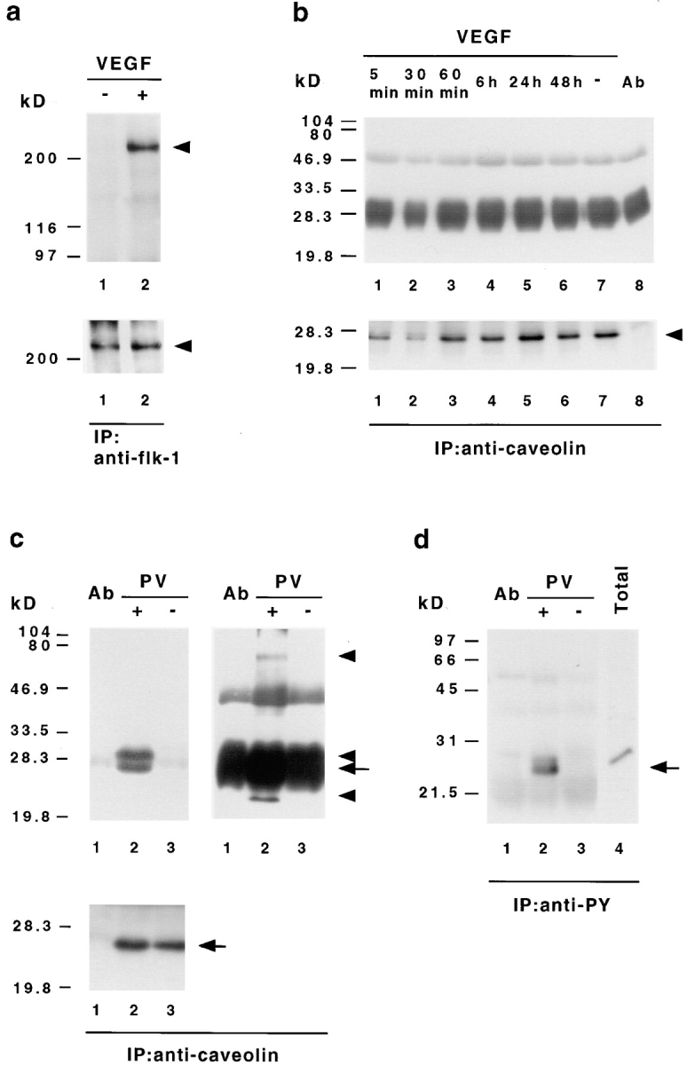
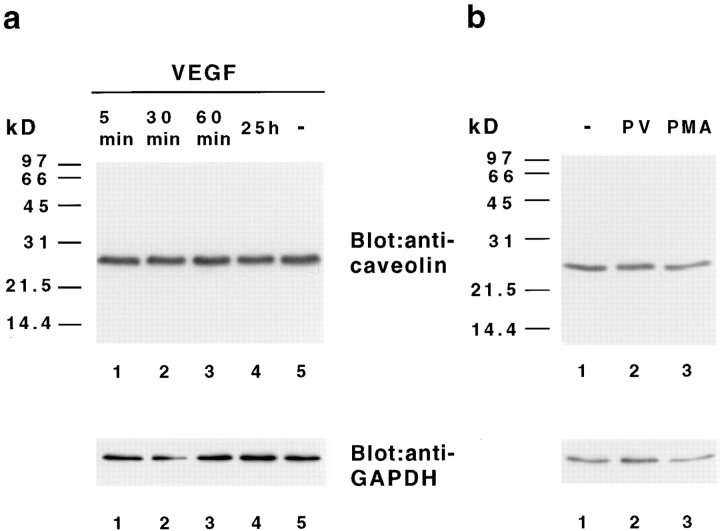
Vascular endothelial growth factor (VEGF) is an important regulator of vasculogenesis, angiogenesis, and vascular permeability. In contrast to its transient expression during the formation of new blood vessels, VEGF and its receptors are continuously and highly expressed in some adult tissues, such as the kidney glomerulus and choroid plexus. This suggests that VEGF produced by the epithelial cells of these tissues might be involved in the induction or maintenance of fenestrations in adjacent endothelial cells expressing the VEGF receptors. Here we describe a defined in vitro culture system where fenestrae formation was induced in adrenal cortex capillary endothelial cells by VEGF, but not by fibroblast growth factor. A strong induction of endothelial fenestrations was observed in cocultures of endothelial cells with choroid plexus epithelial cells, or mammary epithelial cells stably transfected with cDNAs for VEGF 120 or 164, but not with untransfected cells. These results demonstrate that, in these cocultures, VEGF is sufficient to induce fenestrations in vitro. Identical results were achieved when the epithelial cells were replaced by an epithelial-derived basal lamina-type extracellular matrix, but not with collagen alone. In this defined system, VEGF-mediated induction of fenestrae was always accompanied by an increase in the number of fused diaphragmed caveolae-like vesicles. Caveolae, but not fenestrae, were labeled with a caveolin-1-specific antibody both in vivo and in vitro. VEGF stimulation led to VEGF receptor tyrosine phosphorylation, but no change in the distribution, phosphorylation, or protein level of caveolin-1 was observed. We conclude that VEGF in the presence of a basal lamina-type extracellular matrix specifically induces fenestrations in endothelial cells. This defined in vitro system will allow further study of the signaling mechanisms involved in fenestrae formation, modification of caveolae, and vascular permeability.


Figures

Figure 1
Figure 1
Expression of VEGF isoforms in bovine choroid plexus. RT-PCR analysis with VEGF-specific primers of RNA extracted from bovine or mouse tissues demonstrates predominant expression of mRNAs for VEGF isoforms 120 and 164 in choroid plexus (lane 3), whereas the 188 isoform was additionally expressed in lung (lane 2). The VEGF amplification products of 432-, 564-, and 636-bp correspond to VEGF 120, VEGF 164, and VEGF 188, respectively. As a positive control, full-length cDNAs of the three VEGF isoforms were amplified (lanes 4–6). The negative control without DNA template is shown in lane 1. The identity of the PCR products was confirmed by Southern blot analysis with a VEGF-specific probe (data not shown).
Figure 2
Figure 2
Coculture of ACE cells with isolated choroid plexus epithelial cells. (a) Light micrograph illustrating the difference in morphology between epithelial and endothelial cells (Ec). (b) Immunofluorescence staining with an antibody against von Willebrand factor of the same microscopic area shown in a. Islands of labeled endothelial cells within the unlabeled epithelial cell monolayer can be seen. (c and d) Transmission electron micrographs of ACE cells showing cell contacts and apical–basal morphology (c) and caveolar invaginations at the plasma membrane (d). (e) ACE cell in coculture with choroid plexus epithelial cells showing fused clustered vesicles with a diaphragm (arrowhead). Bars: (a) 100 μm; (c) 1 μm; (d and e) 0.1 μm.
Figure 4
Figure 4
Electron micrograph of ACE cells cultured on basal-lamina type extracellular matrix after a 24-h treatment with VEGF. (a) ACE cells form long thin cellular processes that are interrupted by numerous fenestrations (arrowheads). (b) The endothelial cell is still connected to the underlying extracellular matrix (M). Fenestrations (arrowheads) are bridged by thin diaphragms, which sometimes contain a central knob (arrows). (c) ACE cell with a cluster of fused vesicles, which are separated only by thin diaphragms with a central knob (arrows). Bars: (a and b) 0.2 μm; (c) 0.1 μm.
Figure 3
Figure 3
Cocultures of ACE cells with epithelial cells transfected with VEGF. (a) Transfected epithelial cells secrete VEGF into the medium. Conditioned media of normal and transfected epithelial cells were subjected to immunoblot analysis using anti-VEGF antibodies recognizing all known mouse VEGF isoforms. EpH4 cells stably transfected with cDNAs for VEGF 120 and 164 (lanes 2 and 4) secrete high levels of VEGF 120 (open arrowhead) and 164 (closed arrowhead) into the medium when compared to untransfected control cells (C; lanes 1 and 3). The broad band around 60 kD corresponds to the mol wt of BSA. (b and c) Light micrographs of cocultures of ACE cells with EpH4 cells, which were either untransfected (b) or transfected with the cDNA for VEGF 120 (c). The endothelial cells (Ec) appear to be more elongated and have developed small branches and sprouts (arrowheads) in cocultures with transfected EpH4 cells. (d–f) Electron micrographs showing sections through ACE cells cocultivated with VEGF 120–transfected epithelial cells. (d) Fenestrae (arrowheads) are arranged similarly to the in vivo situation in extremely attenuated areas of the endothelial cell. (e) Vertical section through a cellular process. Note fused clustered vesicles (bracket) combined with a dilated transendothelial channel (arrowheads) surrounding a larger vacuole. (f) Clusters of diaphragmed vesicles (brackets) and fenestrae (arrowheads) in ACE cells are shown. Bars: (b and c) 100 μm; (d) 0.25 μm; (e and f) 0.1 μm.
Figure 5
Figure 5
Immunoelectron microscopy using anti–caveolin-1 antibody. (a) Many caveolae are labeled with gold particles in mouse skeletal muscle capillaries. (b) Controls show no labeling for caveolin-1. (c) In capillaries of the choroid plexus, caveolin-1 labeling is found in caveolae but not in fenestrations (arrowheads). (d–f) Electron micrographs of ACE cells cocultured with VEGF 120–transfected epithelial cells. (d) Tangential section of an endothelial cell showing labeling of caveolae. (e) Tangential section through a grape-like cluster of diaphragmed vesicles. Only one vesicle is labeled (arrow). (f) Endothelial cell processes with multiple unlabeled fenestrae (arrowheads) and several gold-labeled caveolae (arrows). Capillary lumina are indicated by an asterisk. MC indicates muscle cell. Bars: (a–d and f) 0.2 μm; (e) 0.1 μm.
Figure 6
Figure 6
VEGF-induced signal transduction does not stimulate tyrosine phosphorylation of caveolin-1. (a) Tyrosine phosphorylation of bovine VEGF receptor-2 (flk-1) in ACE cells after VEGF stimulation. Endothelial cells were stimulated with (+) or without (−)100 ng/ml VEGF 165 for 5 min, extracted with Triton buffer, and then immunoprecipitated with anti–flk-1 antibodies. The precipitates were analyzed by Western immunoblotting with an antibody against phosphotyrosine (top). The same membrane was afterwards reprobed with an antibody recognizing flk-1 (bottom). In contrast to unstimulated cells (lane 1) bovine flk-1 (arrowhead) is found to be specifically phosphorylated on tyrosine after VEGF treatment (lane 2). (b) VEGF does not induce caveolin-1 tyrosine phosphorylation. ACE cells grown on basal lamina-type extracellular matrix were stimulated with VEGF 165 for the indicated time, extracted with SDS lysis buffer, immunoprecipitated with VIP-21N antibody, and then subjected to immunoblot analysis with antiphosphotyrosine antibody (top). Subsequent reprobing of the same membrane with anti–caveolin-1 antibody (bottom) indicated that equal amounts of caveolin-1 (arrowhead) have been precipitated. Protein bands at 50 and 30–25 kD corresponding to the IgG heavy and light chains eluted from the Sepharose, together with the bound proteins, are also present in immunoprecipitations without cell lysates (top, lane 8). (c and d) Pervanadate-induced tyrosine phosphorylation of caveolin-1 and associated proteins. ACE cells were treated with (+) or without (−) pervanadate (PV), extracted with SDS lysis buffer, and then subjected to immunoprecipitation with antibodies against caveolin-1 (c) or phosphotyrosine (d), followed by Western immunoblotting with antiphosphotyrosine (c, top) or anticaveolin-1 (d) antibodies. Blots of caveolin-1 immunoprecipitates were reprobed with anti–caveolin-1 antibody (c, bottom). The bands at 50 and 30–25 kD correspond to the IgG heavy and light chains. To reveal all precipitated phosphoproteins, two different exposures for the antiphosphotyrosine blots are shown in c. Note that the anticaveolin-1 antibody detects only one band in antiphosphotyrosine- and anti–caveolin-1 precipitates (c and d, arrows), whereas the antiphosphotyrosine antibody recognizes several proteins in the anticaveolin-1 precipitates (c, top arrowheads). As controls, immunoprecipitations without cell lysates (lanes 1 in c and d) and total cellular extracts (d, lane 4) are shown.
Figure 7
Figure 7
VEGF does not alter the level of caveolin-1 protein. ACE cells were grown on basal-lamina matrix and treated with (a) VEGF 165 for the indicated time or (b) with pervanadate (PV, 20 min) or PMA (200 ng/ml, 1 h). Total cellular extracts were subjected to SDS-PAGE and immunoblot analysis with anti–caveolin-1 antibodies (top). The amount of caveolin-1 did not change after stimulation with VEGF, pervanadate, or PMA. Subsequent probing of the same membrane with anti-GAPDH antibody indicates equal amounts of protein in each lane (bottom).

References

    1. Anderson RG. Caveolae: where incoming and outgoing messengers meet. Proc Natl Acad Sci USA. 1993;90:10909–10913. - PMC - PubMed
    1. Bacic M, Edwards NA, Merrill MJ. Differential expression of vascular endothelial growth factor (vascular permeability factor) forms in rat tissues. Growth Factors. 1995;12:11–15. - PubMed
    1. Birkenhager R, Schneppe B, Rockl W, Wilting J, Weich HA, McCarthy JE. Synthesis and physiological activity of heterodimers comprising different splice forms of vascular endothelial growth factor and placenta growth factor. Biochem J. 1996;316:703–707. - PMC - PubMed
    1. Breier G, Albrecht U, Sterrer S, Risau W. Expression of vascular endothelial growth factor during embryonic angiogenesis and endothelial cell differentiation. Development (Camb) 1992;114:521–532. - PubMed
    1. Breier G, Clauss M, Risau W. Coordinate expression of vascular endothelial growth factor receptor-1 (flt-1) and its ligand suggests a paracrine regulation of murine vascular development. Dev Dyn. 1995;204:228–239. - PubMed

Publication types

MeSH terms