Comparative Study
. 1998 Feb;180(4):840-5.
doi: 10.1128/JB.180.4.840-845.1998.

tRNA(Arg) (fimU) and expression of SEF14 and SEF21 in Salmonella enteritidis

Affiliations
Comparative Study

tRNA(Arg) (fimU) and expression of SEF14 and SEF21 in Salmonella enteritidis

S C Clouthier et al. J Bacteriol. 1998 Feb.

Abstract

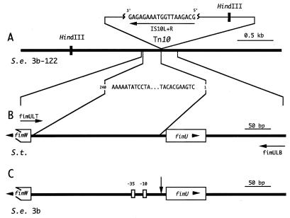
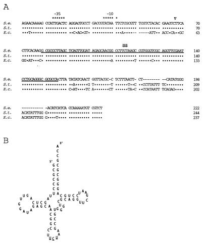
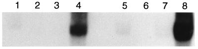
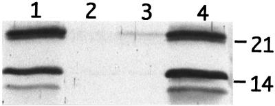
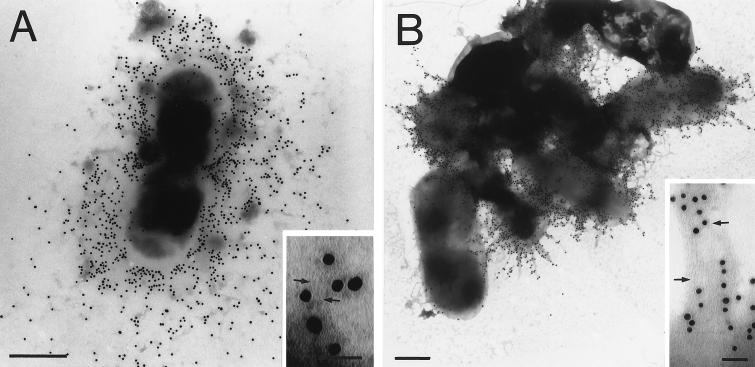
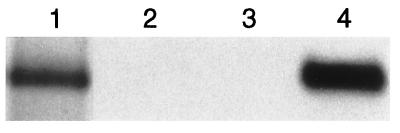
A Tn10 insertion affecting SEF14 fimbrial synthesis in Salmonella enteritidis was located 13 bp upstream of a gene designated fimU. The 77-bp DNA sequence of fimU from S. enteritidis was identical to that of fimU encoding tRNA(Arg) (UCU) from Salmonella typhimurium and 96% identical to that of the Escherichia coli argU homolog. Furthermore, the open reading frame adjacent to and overlapping the 3' end of fimU was similar to the prophage DLP12 integrase gene. The fimU-encoded transcript comigrated with total cellular tRNA and was predicted to form a tRNA-like cloverleaf structure containing the arginine anticodon UCU. Thus, fimU encoded a tRNA(Arg) specific for the rare codon AGA. fimU mapped to the SEF21 fim operon located 15 C's from the sef14 gene cluster. Although fimU was located within the SEF21 fim gene cluster, the fimU Tn10 insertion mutant of S. enteritidis was found to be defective in SEF14 as well as SEF21 (type 1) fimbria production. SEF17 and SEF18 fimbria production was not affected. Complementation of this mutant with plasmid-borne fimU restored normal production of the fimbrins SefA and FimA as well as their respective fimbriae SEF14 and SEF21. This is the first description of tRNA simultaneously controlling the production of two distinct fimbriae.


Figures

FIG. 1
FIG. 1
Location of Tn10 on the S. enteritidis 3b-122 chromosome and identification of the genes flanking the Tn10 insertion in S. enteritidis 3b. (A) Schematic diagram of S. enteritidis (S.e.) 3b-122 chromosomal DNA (black line) showing the Tn10 insert and the strategy used to obtain the chromosomal DNA sequence adjacent to one side of this insert. A 3-kb HindIII fragment comprising 3b-122 chromosomal DNA fused to one end of Tn10 was identified by hybridization with the Tn10 oligonucleotide IS10L+R. IS10L+R was also used as a sequencing primer to obtain 240 bp of DNA sequence from the subcloned HindIII fragment. (B) Schematic diagram of the S. typhimurium (S.t.) chromosome (black line) between fimW and fimU of the type I fimbrial gene cluster that was homologous to the 240 bp of S. enteritidis 3b-122 DNA sequence. Two 42-bp oligonucleotide primers, fimULT and fimULB (horizontal arrows), were made based on the S. typhimurium sequence previously deposited in GenBank (L19338) by Swenson and Clegg (39). (C) Segment of the S. enteritidis 3b chromosome (black line) amplified by PCR with the primers fimULT and fimULB. This amplified DNA segment was subcloned and sequenced (Fig. 2) to identify the DNA flanking the Tn10 insert. The Tn10 insertion point (vertical arrow) was determined to be between the −10 region and the start of the fimU gene. The presence of fimW, fimU, and the −35 region on the 3b chromosome is also noted.
FIG. 2
FIG. 2
Sequence comparison of fimU from S. enteritidis 3b (S.e.) with fimU of S. typhimurium (S.t.) and argU of E. coli (E.c.) as well as the predicted fimU RNA secondary structure. (A) Alignment of S. enteritidis fimU DNA sequence with both the S. typhimurium fimU (39) and E. coli (31) argU gene sequences. Symbols: •, DNA sequence identity; −, gaps introduced to maximize homology; ∗, bases constituting the −35 and −10 boxes; ⇓, bases constituting the anticodon; ▿, position of the Tn10 insertion on the S. enteritidis 3b-122 chromosome. The DNA sequence corresponding to the proposed mature tRNAArg (UCU) is underlined. (B) Diagram of the proposed secondary structure for tRNAArg (UCU) from S. enteritidis 3b. The anticodon bases are underlined.
FIG. 3
FIG. 3
Northern blot analysis of tRNAArg (UCU) production in S. enteritidis 3b strains. A fimU-specific probe was hybridized to PAGE-separated total RNA from S. enteritidis 3b (lanes 1 and 5), 3b-122 (lanes 2 and 6), 3b-122 pGEM-T1 (lanes 3 and 7), or 3b-122 pLU/TA 4-1 (lanes 4 and 8). Lanes 1 to 4 contain 25 μg of RNA, and lanes 5 to 8 contain 50 μg of RNA.
FIG. 4
FIG. 4
Complementation analysis of S. enteritidis 3b-122 Tn10 mutant with the fimU-containing recombinant plasmid pLU/TA 4-1. Whole-cell extracts were analyzed by Western blotting to determine the presence of SefA (21 kDa) and FimA (14 kDa) fimbrin proteins in S. enteritidis 3b (lane 1), 3b-122 (lane 2), 3b-122 pGEM-T1 (lane 3), and 3b-122 pLU/TA 4-1 (lane 4). Numbers at right indicate positions of SefA (21) and FimA (14).
FIG. 5
FIG. 5
Analysis of SEF14 and SEF21 fimbria assembly in S. enteritidis 3b-122 pLU/TA 4-1 by immunogold electron microscopy. S. enteritidis 3b-122 pLU/TA 4-1 was labeled with protein A-gold and negatively stained following incubation with immune serum generated to SEF14 (A) or SEF21 (B). Arrows indicate individual immunogold-labeled SEF14 and SEF21 fimbriae in panel A and B insets, respectively. The average diameter of the gold particles was 15 nm. Bar, 0.5 μm (electron micrograph) or 50 nm (inset).
FIG. 6
FIG. 6
Northern blot analysis of sefA transcription in S. enteritidis 3b strains. A sefA-specific probe was hybridized to 10 μg of total RNA from S. enteritidis 3b (lane 1), 3b-122 (lane 2), 3b-122 pGEM-T1 (lane 3), or 3b-122 LU/TA 4-1 (lane 4).

References

    1. Alm R A, Guerry P, Trust T J. Distribution and polymorphism of the flagellin genes among isolates of Campylobacter coli and Campylobacter jejuni. J Bacteriol. 1993;175:3051–3057. - PMC - PubMed
    1. Altschul S F, Gish W, Miller W, Myers E W, Lipman D J. Basic local alignment search tool. J Mol Biol. 1990;215:403–410. - PubMed
    1. Chen G-F, Inouye M. Role of the AGA/AGG codons, the rarest codons in global gene expression in Escherichia coli. Genes Dev. 1994;8:2641–2652. - PubMed
    1. Chou Q, Russell M, Birch D E, Raymond J, Bloch W. Prevention of pre-PCR mis-priming and primer dimerization improves low-copy-number amplification. Nucleic Acids Res. 1992;20:1717–1723. - PMC - PubMed
    1. Clouthier S C. Ph.D. thesis. Victoria, Canada: University of Victoria; 1995.

Publication types

MeSH terms

Associated data

LinkOut - more resources