. 1998 Feb;180(4):921-31.
doi: 10.1128/JB.180.4.921-931.1998.

Molecular characterization of a phage-inducible middle promoter and its transcriptional activator from the lactococcal bacteriophage phi31

Affiliations

Molecular characterization of a phage-inducible middle promoter and its transcriptional activator from the lactococcal bacteriophage phi31

S A Walker et al. J Bacteriol. 1998 Feb.

Abstract

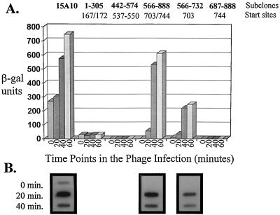
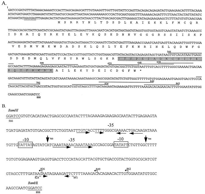
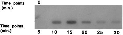
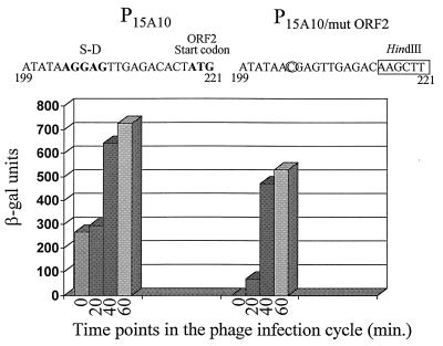
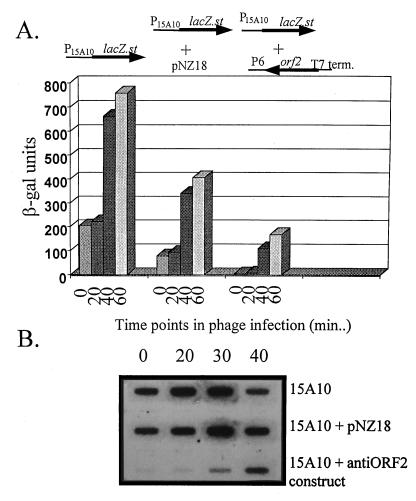
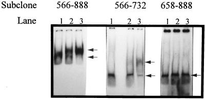
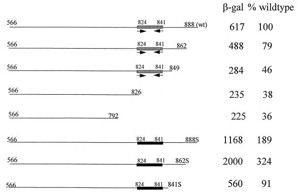
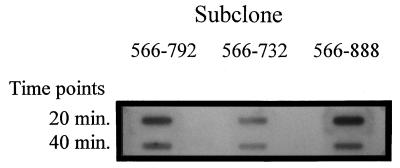
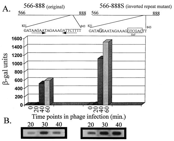
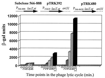
An inducible middle promoter from the lactococcal bacteriophage phi31 was isolated previously by shotgun cloning an 888-bp fragment (P15A10) upstream of the beta-galactosidase (beta-Gal) gene (lacZ.st) from Streptococcus thermophilus (D. J. O'Sullivan, S. A. Walker, S. G. West, and T. R. Klaenhammer, Bio/Technology 14:82-87, 1996). The promoter showed low levels of constitutive beta-Gal activity which could be induced two- to threefold over baseline levels after phage infection. During this study, the fragment was subcloned and characterized to identify a smaller, tightly regulated promoter fragment which allowed no beta-Gal activity until after phage infection. This fragment, defined within nucleotides 566 to 888 (P(566-888); also called fragment 566-888), contained tandem, phage-inducible transcription start sites at nucleotides 703 and 744 (703/744 start sites). Consensus -10 regions were present upstream of both start sites, but no consensus -35 regions were identified for either start site. A transcriptional activator, encoded by an open reading frame (ORF2) upstream of the 703/744 start sites, was identified for P(566-888). ORF2 activated P(566-888) when provided in trans in Escherichia coli. In addition, when combined with pTRK391 (P15A10::lacZ.st) in Lactococcus lactis NCK203, an antisense ORF2 construct was able to retard induction of the phage-inducible promoter as measured by beta-Gal activity levels. Finally, gel shift assays showed that ORF2 was able to bind to promoter fragment 566-888. Deletion analysis of the region upstream from the tandem promoters identified a possible binding site for transcriptional activation of the phage promoters. The DNA-binding ability of ORF2 was eliminated upon deletion of part of this region, which lies centered approximately 35 bp upstream of start site 703. Deletion analysis and mutagenesis studies also elucidated a critical region downstream of the 703/744 start sites, where mutagenesis resulted in a two- to threefold increase in beta-Gal activity. With these improvements, the level of expression achieved by an explosive-expression strategy was elevated from 3,000 to 11,000 beta-Gal units within 120 min after induction.


Figures

FIG. 1
FIG. 1
Representation of the phage-inducible promoter fragment P15A10 (27). The five putative transcription start sites determined by O’Sullivan et al. (27) are represented by vertical arrows (numbered 1 to 5). A complete open reading frame (ORF2; nucleotides 219 to 650) located upstream of start sites 4 and 5 is indicated. This fragment showed constitutive activity which was induced three- to fourfold upon phage infection of the host. P15A10 was subcloned by PCR into five different regions, as indicated. Fragment 1–305 was generated by using the universal −40 primer (on pTRK391) and a primer complementary to nucleotides 281 to 306 on P15A10. Fragment 442–574 was amplified by using one primer consisting of nucleotides 442 to 457 and one primer complementary to nucleotides 559 to 574. Subclone 566–888 was generated by using a primer consisting of nucleotides 566 to 582 and the lacZ primer described in Materials and Methods (on pTRK391). Subclone 687–888 was amplified by using a primer consisting of nucleotides 687 to 705 (T→A and A→C mutations at nucleotides 691 and 692, respectively) and the lacZ primer. Subclone 566–732 utilized the nucleotide 566 primer and a primer complementary to nucleotides 714 to 732. Addition of a 5′ BamHI site to at least one primer of each pair facilitated subsequent cloning procedures.
FIG. 2
FIG. 2
(A) β-Gal activity results of the five P15A10 regions subcloned into the promoter screening vector pTRK390. Time 0 is immediately before the addition of phage φ31 (cells at OD600 of ≅0.5). β-Gal assays were performed at least three different times. For each assay, time point determinations were performed in duplicate. (B) Northern analysis of RNA hybridized with a 32P-labeled lacZ.st probe. Northern analysis was not performed on subclones 1–305 and 442–574 because phage-inducible activity was associated exclusively with start sites 703 and 744.
FIG. 3
FIG. 3
(A) Sequence of the middle, phage-inducible promoter (P15A10) from the lytic, lactococcal bacteriophage φ31 (27). This fragment was shown to have a baseline level of constitutive activity before phage infection of the host. The amino acid sequence corresponding to the complete open reading frame (ORF2) present on the fragment is shown below the sequence. The Shine-Dalgarno sequence for ORF2 is underlined. The putative helix-turn-helix DNA binding motif is shaded. The PCR primers used in the 5′ and 3′ deletion analysis are marked over the sequence. Primers used in the 5′ deletion analysis are designated by the number of the first nucleotide, while primers used in the 3′ deletion analysis are designated by the number of the final nucleotide. These 3′ primers (3′-792, 3′-826, 3′-849, and 3′-862) are complementary to the sequence shown. (B) Sequence of the tightly regulated phage promoter P566–888. The phage-inducible transcription start sites (703 and 744) are marked by vertical arrows. The consensus −10 promoter sequences are boxed. No consensus −35 sequences were observed for either start site. Instead, inverted or direct repeats were observed in the −35 regions for both start sites. The critical region for activation by phage φ31 (between nucleotides 648 and 658) contained a pair of inverted repeats, marked by solid horizontal arrows. The inverted repeat downstream of the 703/744 start sites is marked by broken horizontal arrows. Small, leftward arrows above the sequence mark the positions of the subclones used to determine the importance of the downstream region (subclones 566–826, 566–849, and 566–862).
FIG. 4
FIG. 4
ORF2 mRNA levels during a phage φ31 lytic cycle of the sensitive host, L. lactis subsp. lactis NCK203. Time 0 is immediately before phage infection (cells at OD600 of ≅0.5).
FIG. 5
FIG. 5
Site-directed mutagenesis of the translational signals of ORF2 on P15A10. P15A10 was amplified by PCR into two separate fragments. Fragment 1–215 was amplified by using the universal −40 primer (on pTRK391) and a primer complementary to nucleotides 194 to 215 (G→C mutation at nucleotide 204). Fragment 222–888 was generated by using a primer consisting of nucleotides 222 to 243 and the lacZ primer (on pTRK391). Nucleotides 216 to 221, containing the ATG start codon for ORF2, were replaced with a HindIII site on both PCR fragments (1–215HindIII and HindIII222–888) to allow fusion. The changes made to ORF2 are indicated above the graph. The graph represents β-Gal results of P15A10 and P15A10/mutORF2. β-Gal assays were performed at least three separate times. For each assay, time point determinations were performed in duplicate.
FIG. 6
FIG. 6
Effect of an antisense construct of ORF2 on activation of P15A10. ORF2 was amplified from P15A10 by using one primer consisting of nucleotides 200 to 221 and one primer complementary to nucleotides 640 to 655 and was cloned behind the strong, constitutive P6 promoter (10) in pNZ18. The T7 terminator was cloned behind the P6::anti-ORF2 cassette. The T7 terminator was amplified from the E. coli expression vector pET28a (Novagen) by using a 5′ primer consisting of 5′-GAGAAGCCCGAAAGGAAGC-3′ and a 3′ primer consisting of 5′-ATCCGGATATAGTTCCTC-3′. (A) β-Gal activity when the antisense construct was combined with pTRK391 (P15A10::lacZ.st) (27) in L. lactis subsp. lactis NCK203 both before and after phage infection. β-Gal levels reported are the average of assays performed at least three separate times. For each assay, time point determinations were performed in duplicate. (B) Slot blot Northern analysis of RNA isolated at various points in the φ31 lytic cycle and probed with 32P-labeled lacZ.st.
FIG. 7
FIG. 7
Results of gel retardation assays performed with the ORF2 gene product. The labeled fragments used in the gel retardation assay are indicated at the top of the gels. For each fragment, lane 1 represents the control (no ORF2 added), lane 2 represents the effects of nonlabeled, competitive DNA on DNA binding (unlabeled subclone 588–888 added with ORF2), and lane 3 represents the ability of ORF2 to bind each fragment with no unlabeled 566–888 fragment present. The arrows to the right of each panel indicate the shift in mobility of each fragment upon addition of ORF2. For all three fragments, the ORF2 gene product was added from the same in vitro transcription-translation tube to ensure that the amount was identical between reactions.
FIG. 8
FIG. 8
Importance of sequences downstream of start sites 703/744 in promoter function. The β-Gal levels shown are for the time point 60 min after infection with phage φ31, just before cell lysis. The wild-type, downstream inverted repeat (hatched rectangles with inverted arrows) and the mutated, downstream region that disrupted the inverted repeat (solid rectangles) are indicated.
FIG. 9
FIG. 9
Slot blot analysis of RNA isolated at various time points in the phage φ31 lytic cycle of subclones 566–888, 566–792, and 566–732 and probed with 32P-labeled lacZ.st. lacZ.st mRNA was not detectable before phage infection (time 0; see Fig. 2B).
FIG. 10
FIG. 10
Effects of mutagenizing the downstream inverted repeat (nucleotides 824 to 841) on β-Gal activity and lacZ.st mRNA levels. A site-directed mutation to eliminate the inverted repeat was constructed by amplification of the 566–888 fragment into two separate fragments. Fragment 566–835 was amplified by using the universal −40 primer (on pTRK391) and a primer complementary to nucleotides 817 to 835 (A→G mutation at nucleotide 825). Fragment 842–888 was generated by using a primer consisting of nucleotides 842 to 858 and the lacZ primer (on pTRK391). Nucleotides 836 to 841 were replaced with a SalI site, which allowed fusion of the two fragments (566–835SalI and SalI842–888) to yield fragment 566–888S. Mutations made to the inverted repeat are indicated above the graph. β-Gal levels are the average of at least three separate assays. For each assay, time point determinations were performed in duplicate.
FIG. 11
FIG. 11
β-Gal activity of the new expression vector, pTRK480, compared to those of pTRK392 (27) and subclone 566–888 (pTRK477). Subclone 566–888 was constructed in the high-copy-number promoter screening vector, pTRK390. pTRK392 is based on the low-copy-number replicon pSA3 and contains the phage φ31 origin of replication (ori31) (27) and lacZ.st under the control of P15A10. The new expression vector was constructed by replacing P15A10 with P566–862S. The β-Gal results shown represent the averages of at least three different assays, with each time point determination being performed in duplicate.

Similar articles

Cited by

References

    1. Alatossava T, Klaenhammer T R. Molecular characterization of three small isometric-headed bacteriophages which vary in their sensitivity to the lactococcal phage resistance plasmid pTR2030. Appl Environ Microbiol. 1991;57:1346–1353. - PMC - PubMed
    1. Altschul S F, Gish W, Miller W, Myers E W, Lipman D J. Basic local alignment search tool. J Mol Biol. 1990;215:403–410. - PubMed
    1. Bölker M, Wulczyn F G, Kahmann R. Role of bacteriophage Mu C protein in activation of the mom gene promoter. J Bacteriol. 1989;171:2019–2027. - PMC - PubMed
    1. Casabadan M J, Cohen S N. Analysis of gene control signals by DNA fusion and cloning in Escherichia coli. J Mol Biol. 1980;138:179–207. - PubMed
    1. Chung D K, Chung S K, Batt C A. Antisense RNA directed against the major capsid protein of Lactococcus lactis subsp. cremoris bacteriophage F4-1 confers partial resistance to the host. Appl Microbiol Biotechnol. 1992;37:79–83. - PubMed

Publication types

LinkOut - more resources