. 1998 Mar 2;187(5):753-62.
doi: 10.1084/jem.187.5.753.

B lymphocyte chemotaxis regulated in association with microanatomic localization, differentiation state, and B cell receptor engagement

Affiliations

B lymphocyte chemotaxis regulated in association with microanatomic localization, differentiation state, and B cell receptor engagement

C C Bleul et al. J Exp Med. .

Abstract

Migration of mature B lymphocytes within secondary lymphoid organs and recirculation between these sites are thought to allow B cells to obtain T cell help, to undergo somatic hypermutation, to differentiate into effector cells, and to home to sites of antibody production. The mechanisms that direct migration of B lymphocytes are unknown, but there is evidence that G protein-coupled receptors, and possibly chemokine receptors, may be involved. Stromal cell- derived factor (SDF)-1alpha is a CXC chemokine previously characterized as an efficacious chemoattractant for T lymphocytes and monocytes in peripheral blood. Here we show with purified tonsillar B cells that SDF-1alpha also attracts naive and memory, but not germinal center (GC) B lymphocytes. Furthermore, GC B cells could be converted to respond to SDF-1alpha by in vitro differentiation into memory B lymphocytes. Conversely, the migratory response in naive and memory B cells was significantly reduced after B cell receptor engagement and CD40 signaling. The receptor for SDF-1, CXC chemokine receptor 4 (CXCR4), was found to be expressed on responsive as well as unresponsive B cell subsets, but was more rapidly downregulated on responsive cells by ligand. Finally, messenger RNA for SDF-1 was detected by in situ hybridization in a layer of cells surrounding the GC. These findings show that responsiveness to the chemoattractant SDF-1alpha is regulated during B lymphocyte activation, and correlates with positioning of B lymphocytes within a secondary lymphoid organ.


Figures
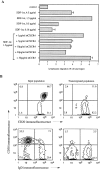
Figure 1
Figure 1
SDF-1α induces chemotaxis in CD38 naive and memory B lymphocytes, but not GC B cells from human tonsil. (A) Human tonsillar B lymphocytes were assayed in the bare filter chemotaxis assay with polycarbonate filters as part of a Transwell insert for migration to different concentrations of SDF-1α. Control shows migration to buffer alone. For antibody inhibition, human tonsillar B lymphocytes were preincubated for 15 min at the given concentrations of mAbs against the chemokine receptors CXCR4 (12G5 mAb) and CCR3 (7B11 mAb), respectively, before addition to the chemotaxis assay. Results are indicated as percent of total input, and error bars show the range of duplicates. The results are representative of four independent experiments. (B) Human tonsillar B lymphocytes were subjected to chemotaxis, and input and transmigrated populations were phenotyped by flow cytometry. Percentages of cells in the respective quadrants are indicated.
Figure 2
Figure 2
SDF-1α efficiently attracts purified CD38 naive and memory B lymphocytes by a pertussis toxin–sensitive mechanism. (A) B lymphocyte subsets were isolated by negative selection with an mAb specific for CD38 (to obtain naive and memory B cells) or CD44 (to obtain GC B cells) and tested in the chemotaxis assay to an optimal concentration of SDF-1α of 1.5 μg/ml. Results are shown as specific migration, i.e., migration above background migration to medium alone, and represent mean and standard deviation of three independent experiments. (B) Isolated CD38 naive and memory B lymphocytes were transmigrated to an optimal concentration of SDF-1α after pretreatment with or without pertussis toxin (PT) over 2 h at 37°C. Control shows migration of sham treated cells to buffer alone. Columns and error bars show the mean and standard deviation of three independent experiments.
Figure 2
Figure 2
SDF-1α efficiently attracts purified CD38 naive and memory B lymphocytes by a pertussis toxin–sensitive mechanism. (A) B lymphocyte subsets were isolated by negative selection with an mAb specific for CD38 (to obtain naive and memory B cells) or CD44 (to obtain GC B cells) and tested in the chemotaxis assay to an optimal concentration of SDF-1α of 1.5 μg/ml. Results are shown as specific migration, i.e., migration above background migration to medium alone, and represent mean and standard deviation of three independent experiments. (B) Isolated CD38 naive and memory B lymphocytes were transmigrated to an optimal concentration of SDF-1α after pretreatment with or without pertussis toxin (PT) over 2 h at 37°C. Control shows migration of sham treated cells to buffer alone. Columns and error bars show the mean and standard deviation of three independent experiments.
Figure 3
Figure 3
Naive and memory, but not GC B lymphocytes respond to SDF-1α by reorganization of the actin skeleton. Intracellular F-actin was measured using FITC-labeled phalloidin in purified CD38 naive and memory B cells (boxes) and CD38+ GC B cells (circles) after addition of SDF-1α at time 0 at a concentration of 1.5 μg/ml or in untreated CD38 naive and memory B cells (diamonds). Results are shown as percent of intracellular F-actin relative to the value before addition of chemokine, and are the mean and standard deviation of three independent experiments.
Figure 4
Figure 4
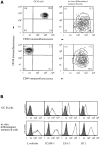
In vitro differentiation of GC B lymphocytes into memory B cells restores responsiveness to SDF-1α. (A) Two-color flow cytometry of isolated GC B lymphocytes (left) and in vitro generated memory B cells (right). GC B lymphocytes were cultured on CD40L expressing NIH3T3 cells in the presence of IL-2 and IL-10 for 7 d. Percentages of cells in the respective quadrants are indicated. (B) Flow cytometric analysis of GC and in vitro–generated memory B lymphocytes for homing receptors, adhesion, and costimulatory molecules. Histograms show the number of cells (y-axis) at each fluorescence intensity (log scale, x-axis). Specific mAb staining and nonbinding control IgG1 staining are shown as open and filled curves, respectively. (C) Migratory response of GC and in vitro–generated memory B lymphocytes. Results are shown as specific migration to 1.5 μg/ml of SDF-1α after subtraction of nonspecific migration to medium alone. The phenotype (Fig. 4, A and B) and migratory response (Fig. 4 C) are from a single experiment representative of three independent experiments.
Figure 4
Figure 4
In vitro differentiation of GC B lymphocytes into memory B cells restores responsiveness to SDF-1α. (A) Two-color flow cytometry of isolated GC B lymphocytes (left) and in vitro generated memory B cells (right). GC B lymphocytes were cultured on CD40L expressing NIH3T3 cells in the presence of IL-2 and IL-10 for 7 d. Percentages of cells in the respective quadrants are indicated. (B) Flow cytometric analysis of GC and in vitro–generated memory B lymphocytes for homing receptors, adhesion, and costimulatory molecules. Histograms show the number of cells (y-axis) at each fluorescence intensity (log scale, x-axis). Specific mAb staining and nonbinding control IgG1 staining are shown as open and filled curves, respectively. (C) Migratory response of GC and in vitro–generated memory B lymphocytes. Results are shown as specific migration to 1.5 μg/ml of SDF-1α after subtraction of nonspecific migration to medium alone. The phenotype (Fig. 4, A and B) and migratory response (Fig. 4 C) are from a single experiment representative of three independent experiments.
Figure 5
Figure 5
B cell receptor engagement reduces the migratory response in CD38 naive and memory B lymphocytes. (A) CD38 naive and memory B cells were added to the chemotaxis assay after a 2-h incubation on FcγRII (CD32) expressing NIH3T3 cells in the presence of the indicated mAbs and migrated to an optimal concentration of SDF-1α of 1.5 μg/ml. mAbs to lambda and kappa light chain and the control IgG1 were used at 2 μg/ml, and the mAb to CD40 at 10 μg/ml. Results are expressed relative to the migration after incubation with control IgG1 and represent mean and standard deviation of three independent experiments. (B) CD38 naive and memory B cells were incubated on CD32-NIH3T3 cells in the presence of 2 μg/ml mAb to lambda light chain and transmigrated to an optimal concentration of SDF-1α of 1.5 μg/ml. Input (left) and transmigrated cells (right) were stained for expression of kappa light chain.
Figure 5
Figure 5
B cell receptor engagement reduces the migratory response in CD38 naive and memory B lymphocytes. (A) CD38 naive and memory B cells were added to the chemotaxis assay after a 2-h incubation on FcγRII (CD32) expressing NIH3T3 cells in the presence of the indicated mAbs and migrated to an optimal concentration of SDF-1α of 1.5 μg/ml. mAbs to lambda and kappa light chain and the control IgG1 were used at 2 μg/ml, and the mAb to CD40 at 10 μg/ml. Results are expressed relative to the migration after incubation with control IgG1 and represent mean and standard deviation of three independent experiments. (B) CD38 naive and memory B cells were incubated on CD32-NIH3T3 cells in the presence of 2 μg/ml mAb to lambda light chain and transmigrated to an optimal concentration of SDF-1α of 1.5 μg/ml. Input (left) and transmigrated cells (right) were stained for expression of kappa light chain.
Figure 6
Figure 6
CXCR4 is more rapidly downregulated on responsive B lymphocytes than on GC B cells. Phenotyping of human tonsillar B lymphocytes after treatment without (A) or with (B) 2 μg/ml SDF-1α for 30 min at 37°C. The CD38+ population corresponds to GC B cells, and the CD38 population to naive and memory B cells. A reference dashed line at 102 fluorescence units is shown for comparison between the panels.
Figure 7
Figure 7
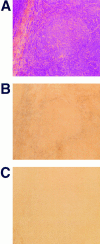
Detection of SDF-1 mRNA in human tonsil by in situ hybridization. Parallel frozen sections of human tonsil were either stained with hematoxylin and eosin (A) or hybridized with digoxigenin-labeled antisense (B) and sense (C) RNA probes specific for both SDF-1α and SDF-1β. mRNA expression was visualized using alkaline phosphatase–conjugated antidigoxigenin antibody and nitroblue tetrazolium salt/5-bromo-4-chloro-3-indolyl phosphate. Sections are shown using ×10 magnification.

Similar articles

Cited by

References

    1. Nieuwenhuis P, Ford WL. Comparative migration of B- and T-lymphocytes in the rat spleen and lymph-nodes. Cell Immunol. 1976;23:254–267. - PubMed
    1. Howard JC, Hunt SV, Gowans JL. Identification of marrow-derived small lymphocytes in the lymphoid tissue and thoracic duct lymph of normal rats. J Exp Med. 1972;135:200–219. - PMC - PubMed
    1. MacLennan ICM. Germinal centers. Annu Rev Immunol. 1994;12:117–139. - PubMed
    1. Rajewsky K. Clonal selection and learning in the antibody system. Nature. 1996;381:751–758. - PubMed
    1. Kelsoe G. Life and death in germinal centers (Redux) Immunity. 1996;4:107–111. - PubMed

Publication types