. 1998 Mar 2;187(5):813-8.
doi: 10.1084/jem.187.5.813.

Recognition of human histocompatibility leukocyte antigen (HLA)-E complexed with HLA class I signal sequence-derived peptides by CD94/NKG2 confers protection from natural killer cell-mediated lysis

Affiliations

Recognition of human histocompatibility leukocyte antigen (HLA)-E complexed with HLA class I signal sequence-derived peptides by CD94/NKG2 confers protection from natural killer cell-mediated lysis

F Borrego et al. J Exp Med. .

Abstract

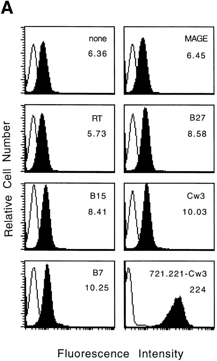
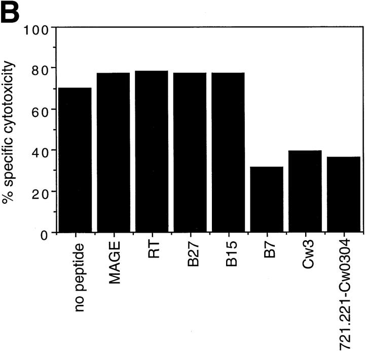
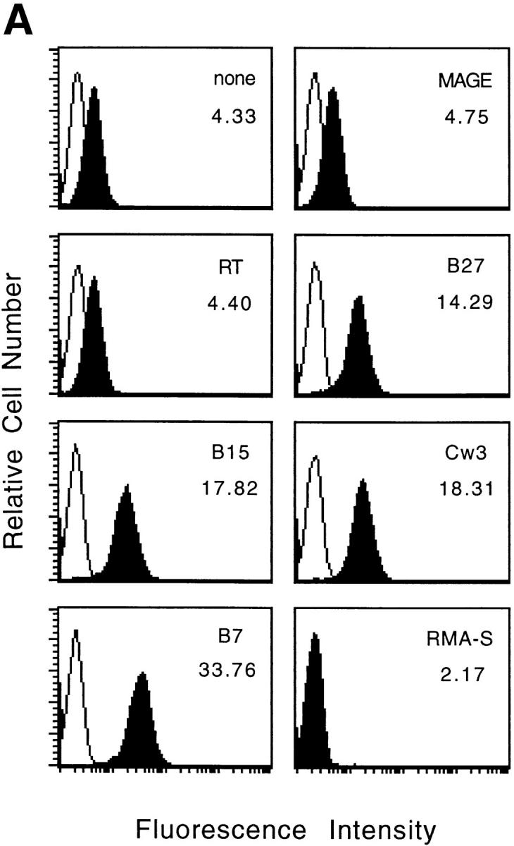
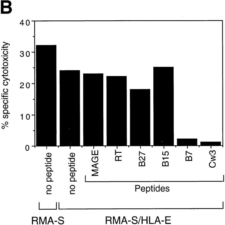
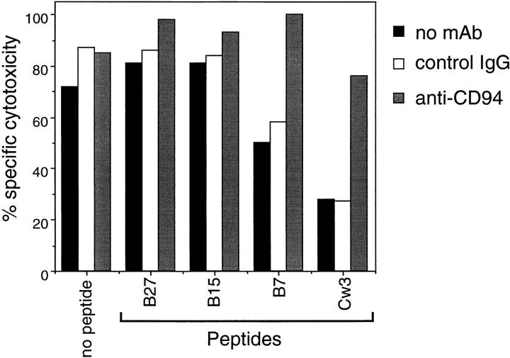
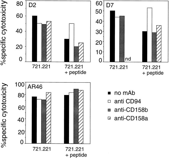
Human histocompatibility leukocyte antigen (HLA)-E is a nonclassical HLA class I molecule, the gene for which is transcribed in most tissues. It has recently been reported that this molecule binds peptides derived from the signal sequence of HLA class I proteins; however, no function for HLA-E has yet been described. We show that natural killer (NK) cells can recognize target cells expressing HLA-E molecules on the cell surface and this interaction results in inhibition of the lytic process. Furthermore, HLA-E recognition is mediated primarily through the CD94/NKG2-A heterodimer, as CD94-specific, but not killer cell inhibitory receptor (KIR)-specific mAbs block HLA-E-mediated protection of target cells. Cell surface HLA-E could be increased by incubation with synthetic peptides corresponding to residues 3-11 from the signal sequences of a number of HLA class I molecules; however, only peptides which contained a Met at position 2 were capable of conferring resistance to NK-mediated lysis, whereas those having Thr at position 2 had no effect. Interestingly, HLA class I molecules previously correlated with CD94/NKG2 recognition all have Met at residue 4 of the signal sequence (position 2 of the HLA-E binding peptide), whereas those which have been reported not to interact with CD94/NKG2 have Thr at this position. Thus, these data show a function for HLA-E and suggest an alternative explanation for the apparent broad reactivity of CD94/NKG2 with HLA class I molecules; that CD94/NKG2 interacts with HLA-E complexed with signal sequence peptides derived from "protective" HLA class I alleles rather than directly interacting with classical HLA class I proteins.


Figures

Figure 1
Figure 1
Cell surface expression of HLA-E molecules complexed with HLA class I signal-sequence derived peptides confers protection against NK cell– mediated lysis. (A) 721.221 target cells were incubated in the absence or presence of the indicated peptides at 26°C and analyzed for HLA class I expression. The empty histograms correspond to the secondary antibody (goat anti–mouse) alone and the filled histograms show the binding of the HLA class I–specific mAb B9.12.1. The histogram in the lower right hand corner shows the HLA class I expression on 721.221 cells transfected with HLA-Cw*0304. The mean fluorescence channel of B9.12.1 staining is shown inside the boxes. (B) 221707 NK cells were assayed for cytotoxicity against 721.221 target cells incubated with or without the indicated peptides at 26°C or 721.221-Cw3 cells. The E/T ratio was 15:1 and the percentage of specific lysis shown is the average of three experiments. The standard deviation was <10%.
Figure 1
Figure 1
Cell surface expression of HLA-E molecules complexed with HLA class I signal-sequence derived peptides confers protection against NK cell– mediated lysis. (A) 721.221 target cells were incubated in the absence or presence of the indicated peptides at 26°C and analyzed for HLA class I expression. The empty histograms correspond to the secondary antibody (goat anti–mouse) alone and the filled histograms show the binding of the HLA class I–specific mAb B9.12.1. The histogram in the lower right hand corner shows the HLA class I expression on 721.221 cells transfected with HLA-Cw*0304. The mean fluorescence channel of B9.12.1 staining is shown inside the boxes. (B) 221707 NK cells were assayed for cytotoxicity against 721.221 target cells incubated with or without the indicated peptides at 26°C or 721.221-Cw3 cells. The E/T ratio was 15:1 and the percentage of specific lysis shown is the average of three experiments. The standard deviation was <10%.
Figure 2
Figure 2
Expression of HLA-E by RMA-S cells with appropriate peptides confers resistance to NK-mediated lysis. (A) RMA-S/HLA-E cells were incubated in the absence or presence of the indicated peptide at 37°C and analyzed for cell surface HLA class I expression. The empty histograms represent secondary Ab controls (goat anti–mouse) and the filled histograms show the binding of the B9.12.1 mAb. The histogram in the lower right hand corner shows staining of RMA-S cells transfected with human β2-microglobulin alone. The mean fluorescence channel of B9.12.1 staining is shown inside the boxes. (B) 221707 NK cells were assayed for cytotoxicity against RMA-S and RMA-S/ HLA-E cells incubated in the presence or absence of the indicated peptides at 37°C. The E/T ratio was 40:1.
Figure 2
Figure 2
Expression of HLA-E by RMA-S cells with appropriate peptides confers resistance to NK-mediated lysis. (A) RMA-S/HLA-E cells were incubated in the absence or presence of the indicated peptide at 37°C and analyzed for cell surface HLA class I expression. The empty histograms represent secondary Ab controls (goat anti–mouse) and the filled histograms show the binding of the B9.12.1 mAb. The histogram in the lower right hand corner shows staining of RMA-S cells transfected with human β2-microglobulin alone. The mean fluorescence channel of B9.12.1 staining is shown inside the boxes. (B) 221707 NK cells were assayed for cytotoxicity against RMA-S and RMA-S/ HLA-E cells incubated in the presence or absence of the indicated peptides at 37°C. The E/T ratio was 40:1.
Figure 3
Figure 3
HLA-E mediated protection is blocked by anti-CD94 mAb. 721.221 target cells were incubated with or without the indicated peptides at 26°C and then used in a cytotoxicity assay with 221707 NK cells in the presence of anti-CD94 or control antibodies at a final concentration of 10 μg/ml. The E/T ratio was 15:1.
Figure 4
Figure 4
HLA-E is not recognized by CD158a and CD158b KIR receptors. 721.221 target cells were incubated at 26°C with or without the Cw3 peptide and then used in a cytotoxicity assay with the indicated NK clones (D2, D7, and AR46) in the presence of anti-CD94, anti-CD158a, or anti-CD158b mAb at a final concentration of 10 μg/ml. The E/T ratio was 5:1. nd, not determined.

Similar articles

Cited by

References

    1. Madden DR. The three-dimensional structure of peptide-MHC complex. Annu Rev Immunol. 1995;13:587–622. - PubMed
    1. Shawar SM, Vyas JV, Rodgers JR, Rich RR. Antigen presentation by major histocompatibility complex class I-bmolecules. Annu Rev Immunol. 1994;12:839–880. - PubMed
    1. Shawar SM, Cook RG, Rodgers JR, Rich RR. Specialized functions of MHC class I molecules. I. An N-formyl peptide receptor is required for construction of the class I antigen Mta. J Exp Med. 1990;171:897–912. - PMC - PubMed
    1. Lee N, Malacko AR, Ishitani A, Chen MC, Bajorath J, Marquardt H, Geraghty DE. The membrane-bound and soluble forms of HLA-G bind identical sets of peptides but differ with respect to TAP association. Immunity. 1995;3:591–600. - PubMed
    1. Koller BH, Geraghty DE, Shimizu Y, DeMars R, Orr HT. HLA-E: a novel HLA class I gene expressed in resting T-lymphocytes. J Immunol. 1988;141:897–904. - PubMed

MeSH terms