Review
. 1998 Mar 15;18(6):1953-62.
doi: 10.1523/JNEUROSCI.18-06-01953.1998.

Myelin-associated glycoprotein is a myelin signal that modulates the caliber of myelinated axons

Affiliations
Review

Myelin-associated glycoprotein is a myelin signal that modulates the caliber of myelinated axons

X Yin et al. J Neurosci. .

Abstract

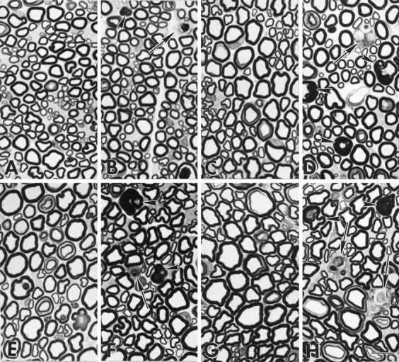
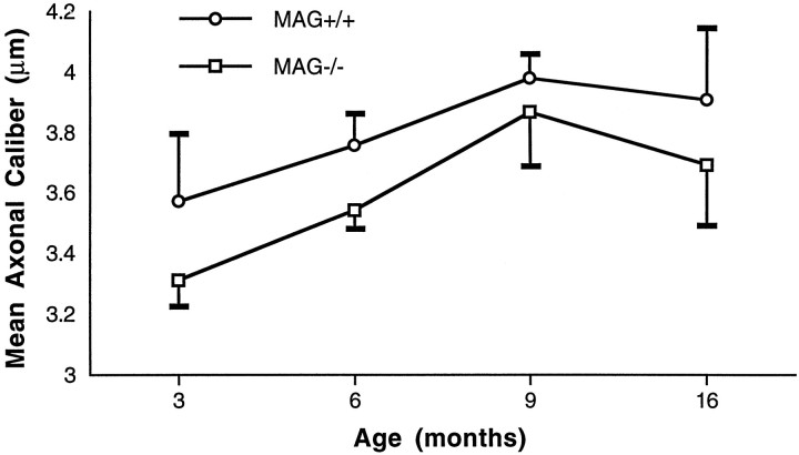
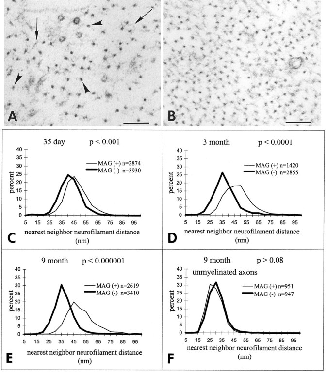
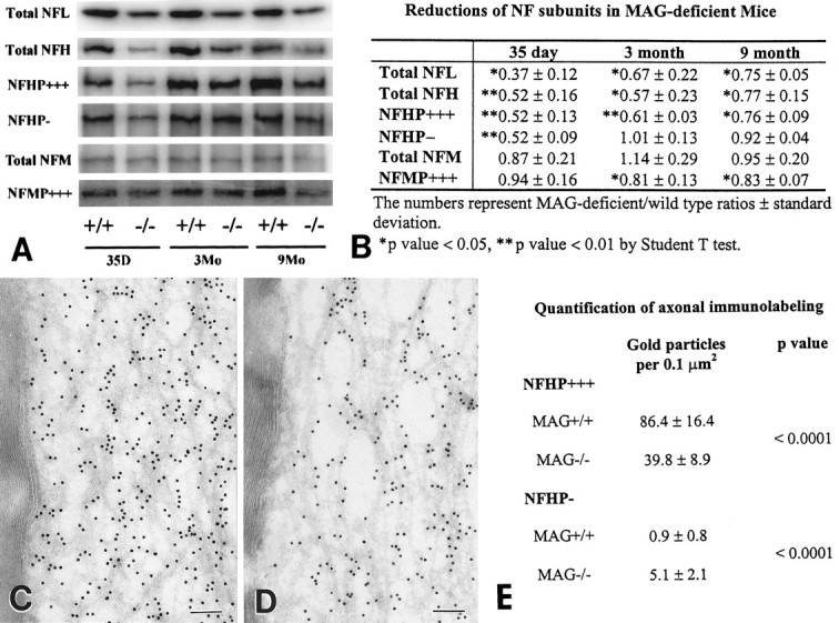
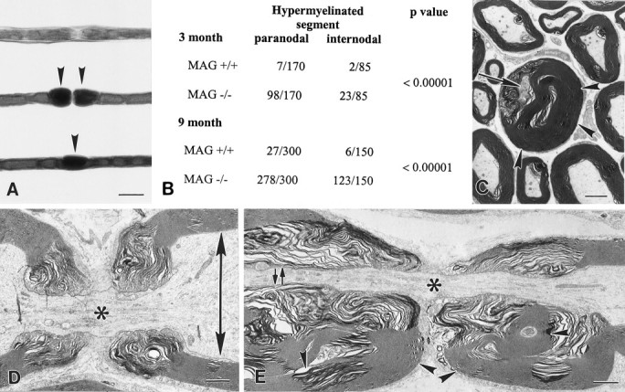
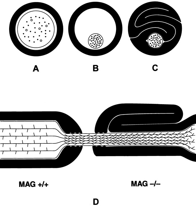
Myelination increases neuronal conduction velocity through its insulating properties and an unidentified extrinsic effect that increases axonal caliber. Although it is well established that demyelination can cause axonal atrophy, the myelin molecule that regulates axonal caliber is not known. Loss of the structural proteins of compact peripheral nervous system (PNS) myelin, P0 protein, and myelin basic protein does not lead to axonal atrophy. This study demonstrates that mice with a null mutation of the myelin-associated glycoprotein (MAG) gene have a chronic atrophy of myelinated PNS axons that results in paranodal myelin tomaculi and axonal degeneration. Absence of MAG was correlated with reduced axonal calibers, decreased neurofilament spacing, and reduced neurofilament phosphorylation. Because axonal atrophy and degeneration in MAG-deficient mice occur in the absence of inflammation, hypomyelination, significant demyelination-remyelination, or gain of function mutations, these data support a functional role for MAG in modulating the maturation and viability of myelinated axons.


Figures

Fig. 1.
Fig. 1.
MAG-deficient sciatic nerves contain degenerating myelin internodes and redundant myelin.AD, Epon sections of sciatic nerves from P35 (A = MAG+/+, B = MAG−/−) and 3-month-old mice (C = MAG+/+,D = MAG−/−). Occasional MAG-deficient myelinated fibers are degenerating (B, D,arrows), and some axons are surrounded by redundant or excessive myelin (B, D,arrowheads). EH, At 9 months (E = MAG+/+, F = MAG−/−) and 16 months (G = MAG+/+,H = MAG−/−) of age, degenerating myelin internodes (arrows) and redundant myelin (arrowheads) persist in MAG-deficient sciatic nerves. Compared with control nerves, myelinated fibers in MAG-deficient nerves appear to have reduced axonal calibers. Scale bar, 10 μm.
Fig. 2.
Fig. 2.
Average axonal calibers of myelinated fibers in control and MAG-deficient sciatic nerves. MAG-deficient myelinated axons have reduced calibers. Axons surrounded by normal-appearing myelin in MAG-deficient nerves are significantly smaller than those in control nerves (p < 0.005, for all ages combined). Statistical analysis was performed by the two-tailed Student’s t test. Values are mean ± SD.
Fig. 3.
Fig. 3.
MAG-deficient nerves have reduced neurofilament spacing. A, B, In electron micrographs, neurofilaments (arrowheads) appear as 10-nm-thick structures from which sidearms (arrows) extend. Compared with myelinated axons in 9-month-old control nerves (A), neurofilaments are spaced closer together in 9-month-old MAG-deficient myelinated axons (B). Scale bar, 0.1 μm. CE, Compared with myelinated axons in control nerves, the neurofilament nearest neighbor distance in myelinated axons in MAG-deficient nerves was significantly reduced at P35 (C), 3 months (D), and 9 months (E). Statistical analysis by Student’s t test analysis;n = number of neurofilaments analyzed.F, Neurofilament spacing in unmyelinated axons was similar in control and MAG-deficient nerves from 9-month-old mice;n = number of neurofilaments analyzed.
Fig. 4.
Fig. 4.
Neurofilament proteins and their phosphorylation states are reduced in sciatic nerves of MAG-deficient mice. A shows comparison between quantitative Western blots of neurofilament epitopes in control (+/+) and MAG-deficient (−/−) sciatic nerve homogenates from P35 and 3- and 9-month old mice. NFHP−, Poorly or nonphosphorylated NFH; NFHP+++ and NFMP+++, highly phosphorylated NFH and NFM. Three MAG-deficient and three control nerves were analyzed in triplicate at each time point, and representative bands were quantitated and expressed as MAG-deficient/wild-type ratios ± SD (B). Total NFL, total NFH, and NFHP+++ were significantly reduced at all ages. NFHP− was reduced in 35-d-old MAG-deficient homogenates but not at later time points. Total NFM remained unchanged at all ages, whereas NFMP+++ was significantly reduced in 3- and 9-month-old MAG-deficient homogenates. CE, Ultrathin cryosections of 9-month-old control (C) and MAG-deficient (D) sciatic nerves were immunostained with NFHP+++ antibodies by immunogold procedures. Compared with control nerves (C), MAG-deficient nerves (D) contained a 50% decrease in NFHP+++ labeling (E). In contrast, NFHP− labeling was significantly increased in MAG-deficient nerves (E). Scale bars, 0.1 μm.
Fig. 5.
Fig. 5.
Tomaculi result from axonal shrinkage and myelin collapse. A, Teased fibers from 9-month-old control (top) and MAG-deficient (center andbottom) sciatic nerves. Thick paranodal myelin was rare in control fibers but abundant in MAG-deficient fibers (center, arrowheads). Internodal segments also had thick myelin in MAG-deficient fibers (bottom,arrowhead). Scale bar, 20 μm. B, Thick-appearing myelin segments were quantified, grouped by location (paranodal and internodal), and found at significantly greater numbers in MAG-deficient fibers when compared with control fibers.CE, Electron micrographs of paranodal regions of MAG-deficient (C, E) and control (D) myelin internodes. In transverse section, thick redundant myelin (C,arrowheads) partially surrounds an axon (C, arrows) with remarkably small caliber. In longitudinal orientation, the difference between nodal (*) and internodal (arrows) axonal caliber is much greater in control (D) than in MAG-deficient (E) fibers. Redundant paranodal myelin surrounds paranodal axons with reduced caliber in MAG-deficient fibers (E, arrowheads). Scale bars:C, 2.0 μm; D, E, 1.0 μm.
Fig. 6.
Fig. 6.
Schematic representation of tomaculi formation in MAG-deficient paranodal regions. AC, Relationship between axonal caliber and myelin sheath in transverse section of normal myelinated fiber (A). In MAG-deficient nerves, paranodal axonal calibers shrink (B) and result in myelin sheath collapse and tomaculi formation (C). D, In MAG+/+ paranodal regions, increased neurofilament spacings correlate with large axonal caliber. In MAG−/− paranodal regions surrounded by tomaculi, neurofilament spacings and axonal calibers are similar to those in nodal regions.

References

    1. Adlkofer K, Frei R, Neuberg DH-H, Zielasek J, Toyka KV, Suter U. Heterozygous peripheral myelin protein 22-deficient mice are affected by a progressive demyelinating tomaculous neuropathy. J Neurosci. 1997;17:4662–4671. - PMC - PubMed
    1. Aguayo AJ, Epps J, Charron L, Bray GM. Multipotentiality of Schwann cells in cross anastomosed and grafted myelinated and unmyelinated nerves: quantitative microscopy and radioautography. Brain Res. 1976;104:1–20. - PubMed
    1. Arbuthnott ER, Boyd IA, Kalu KU. Ultrastructural dimensions of myelinated peripheral nerve fibres in the cat and their relation to conduction velocity. J Physiol (Lond) 1980;308:125–157. - PMC - PubMed
    1. Arquint M, Roder J, Chia L-S, Down J, Wilkinson O, Bayley H, Braun P, Dunn R. Molecular cloning and primary structure of myelin-associated glycoproteins. Proc Natl Acad Sci USA. 1987;84:600–604. - PMC - PubMed
    1. Carenini S, Montag D, Cremer H, Schachner M, Martini R. Absence of the myelin-associated glycoprotein (MAG) and the neural cell adhesion molecule (N-CAM) interferes with the maintenance, but not with the formation of peripheral myelin. Cell Tissue Res. 1997;287:3–9. - PubMed

Publication types

MeSH terms

Substances