Review
. 1998 Mar 15;18(6):1987-95.
doi: 10.1523/JNEUROSCI.18-06-01987.1998.

Inhibition by ATP of hippocampal synaptic transmission requires localized extracellular catabolism by ecto-nucleotidases into adenosine and channeling to adenosine A1 receptors

Affiliations
Review

Inhibition by ATP of hippocampal synaptic transmission requires localized extracellular catabolism by ecto-nucleotidases into adenosine and channeling to adenosine A1 receptors

R A Cunha et al. J Neurosci. .

Abstract

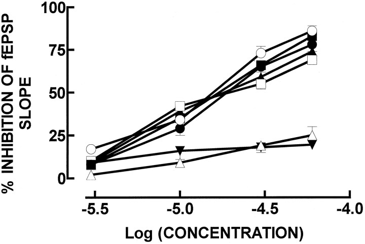
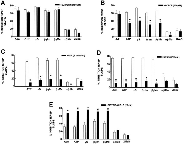
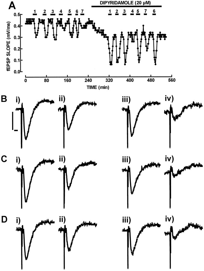
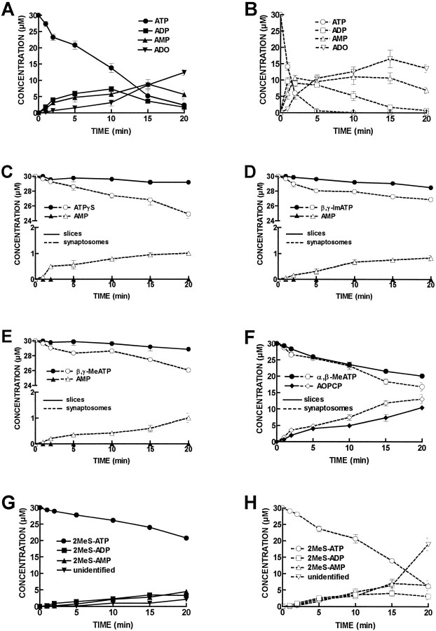
ATP analogs substituted in the gamma-phosphorus (ATPgammaS, beta, gamma-imido-ATP, and beta,gamma-methylene-ATP) were used to probe the involvement of P2 receptors in the modulation of synaptic transmission in the hippocampus, because their extracellular catabolism was virtually not detected in CA1 slices. ATP and gamma-substituted analogs were equipotent to inhibit synaptic transmission in CA1 pyramid synapses (IC50 of 17-22 microM). The inhibitory effect of ATP and gamma-phosphorus-substituted ATP analogs (30 microM) was not modified by the P2 receptor antagonist suramin (100 microM), was inhibited by 42-49% by the ecto-5'-nucleotidase inhibitor and alpha,beta-methylene ADP (100 microM), was inhibited by 74-85% by 2 U/ml adenosine deaminase (which converts adenosine into its inactive metabolite-inosine), and was nearly prevented by the adenosine A1 receptor antagonist 1,3-dipropyl-8-cyclopentylxanthine (10 nM). Stronger support for the involvement of extracellular adenosine formation as a main requirement for the inhibitory effect of ATP and gamma-substituted ATP analogs was the observation that an inhibitor of adenosine uptake, dipyridamole (20 microM), potentiated by 92-124% the inhibitory effect of ATP and gamma-substituted ATP analogs (10 microM), a potentiation similar to that obtained for 10 microM adenosine (113%). Thus, the present results indicate that inhibition by extracellular ATP of hippocampal synaptic transmission requires localized extracellular catabolism by ecto-nucleotidases and channeling of the generated adenosine to adenosine A1 receptors.


Figures

Fig. 1.
Fig. 1.
Inhibition of the fEPSP slope, recorded extracellularly in hippocampal CA1 pyramids, by exogenously added adenosine (○), ATP (•), ATPγS (▪), β,γ-imido-ATP (□), β,γ-methylene-ATP (▴), 2-methylthio-ATP (▵), and α,β-methylene-ATP (▾). The ordinates represent the percentage of inhibition of fEPSP slope produced by adenosine, ATP, or ATP analogs in relation to the fEPSP slope in control conditions (i.e., in the absence of any added drug to the perfusion solution). 0% corresponds to the fEPSP slope in control conditions (i.e., without any added drug), and 100% corresponds to blockade of fEPSP. The results are mean ± SEM of two to five experiments. The SEMs are shown when they exceed the symbols in size.
Fig. 2.
Fig. 2.
Modification of the inhibition by adenosine, ATP, and ATP analogs of fEPSPs by 100 μm suramin, a P2 receptor antagonist (A), by 100 μm α,β-methylene ADP (AOPCP), an inhibitor of ecto-5′-nucleotidase (B), by 2 U/ml adenosine deaminase (ADA), the enzyme that converts adenosine into its inactive metabolite, inosine (C), by 10 nm1,3-dipropyl-8-cyclopentylxanthine (DPCPX), an adenosine A1receptor antagonist (D), and by 20 μm dipyridamole, an inhibitor of adenosine uptake (E). The ordinates represent the percentage inhibition of fEPSP slope by adenosine or ATP, ATPγS (γS), β,γ-imido-ATP (β,γ-Im), β,γ-methylene-ATP (β,γ-Me), α, β-methylene-ATP (αβMe), and 2-methylthio-ATP (2MeS) in the absence (open bars) and presence (filled bars) of the drugs indicated in each panel. 0% corresponds to the fEPSP slope in control conditions (i.e., without any added drug or after addition of suramin, AOPCP, adenosine deaminase, DPCPX, or dipyridamole), and 100% corresponds to blockade of fEPSPs. In each experiment, suramin, AOPCP, adenosine deaminase, DPCPX, or dipyridamole were applied to the preparations 30–45 min before the effect of adenosine, ATP, or ATP analogs was tested in their presence. The effect of adenosine, ATP, or ATP analogs in the absence and presence of suramin, AOPCP, adenosine deaminase, DPCPX, or dipyridamole was always compared in the same experiment. * p < 0.05 (paired Student’s t test) when comparing with the effect of adenosine, ATP, or ATP analogs alone. The results are mean ± SEM of three to four experiments. The SEMs are shown when they exceed the symbols in size.
Fig. 3.
Fig. 3.
Effect of the inhibitor of adenosine uptake, dipyridamole to potentiate the inhibitory effect of ATPγS (1), β,γ-imido-ATP (2), β,γ-methylene-ATP (3), 2-methylthio-ATP (4), ATP (5), adenosine (6), and α,β-methylene-ATP (7) on fEPSP slope. In A is shown the time course of the slope of averages of eight consecutive fEPSPs recorded from the CA1 area of the hippocampus. The hippocampal slice was perfused with adenosine, ATP, or ATP analogs (10 μm) either in the absence or in the presence of dipyridamole (20 μm), as shown in the top bars. In B, C, andD are shown recordings of fEPSPs, corresponding to the absence of any added drug in the perfusion medium (i inB, C, and D), the presence of ATP (B, ii), ATPγS (C, ii), and adenosine (D,ii), the presence of dipyridamole (iii inB, C, and D), the simultaneous presence of dipyridamole and ATP (B,iv), the simultaneous presence of dipyridamole and ATPγS (C, iv), and the simultaneous presence of dipyridamole and adenosine (D,iv). Each recording is composed of a stimulus artifact followed by the presynaptic volley and the fEPSP and corresponds to the average of eight consecutive responses. Calibration bars (shown inB for B–D): 500 μV, 5 msec. Note that dipyridamole (20 μm) potentiated to a similar extent the inhibitory effect of γ-phosphorus-substituted ATP analogs (1, 2, and 3), ATP (5), and adenosine (6), whereas the inhibitory effect of 2-methylthio-ATP (4) and α,β-methylene-ATP (7) was not modified appreciably.
Fig. 4.
Fig. 4.
Progress curves of ATP and ATP analog catabolism in CA1 hippocampal slices and synaptosomes. ATP or ATP analogs (30 μm) were incubated at zero time with CA1 slices or synaptosomes. Samples (50 μl) were collected from the bath (600 μl) at the times indicated in the abscissa and analyzed by HPLC. InA is shown the catabolism of ATP (•) into ADP (▪), AMP (▴), and adenosine (▾) in CA1 slices. In B is shown the catabolism of ATP (○) into ADP (□), AMP (▵), and adenosine (▿) in CA1 synaptosomes. In C is shown the catabolism of ATPγS (•, ○) into AMP (▴,▵) in CA1 slices (filled symbols, filled lines) and CA1 synaptosomes (open symbols, broken lines). InD is shown the catabolism of β,γ-imido-ATP (•, ○) into AMP (▴,▵) in CA1 slices (filled symbols, filled lines) and CA1 synaptosomes (open symbols, broken lines). In E is shown the catabolism of β,γ-methylene-ATP (•, ○) into AMP (▴,▵) in CA1 slices (filled symbols, filled lines) and CA1 synaptosomes (open symbols, broken lines). InF is shown the catabolism of α,β-methylene ATP (•, ○) into α,β-methylene ADP (♦, ⋄) in CA1 slices (filled symbols, filled lines) and CA1 synaptosomes (open symbols, broken lines). InG is shown the catabolism of 2-methylthio-ATP (•) into 2-methylthio-ADP (▪), 2-methylthio-AMP (▴) and into an unidentified compound (▾) in CA1 slices. In H is shown the catabolism of 2-methylthio-ATP (○) into 2-methylthio-ADP (□), 2-methylthio-AMP (▵) and into an unidentified compound (▿) in CA1 synaptosomes. Each point is the average of four to five experiments. The vertical bars represent the SEM and are shown when they exceed the symbols in size. The concentrations of inosine and adenosine detected under control conditions, without addition of initial substrate, were subtracted. The concentration of any purine metabolite present at time 0 was also subtracted.

References

    1. Bailey SJ, Hickman D, Hourani SMO. Characterization of the P1-purinoceptors mediating contraction of the rat colon muscularis mucosae. Br J Pharmacol. 1992;105:400–404. - PMC - PubMed
    1. Bruns RF. Adenosine receptors. Roles and pharmacology. Ann NY Acad Sci. 1990;603:211–226. - PubMed
    1. Buell G, Lewis C, Collo G, North RA, Surprenant A. An antagonist-insensitive P2X receptor expressed in epithelia and brain. EMBO J. 1996;15:55–62. - PMC - PubMed
    1. Cascalheira JF, Sebastião AM. Adenine nucleotide analogues, including γ-phosphate-substituted analogues, are metabolised extracellularly in innervated frog sartorius muscle. Eur J Pharmacol. 1992;222:49–59. - PubMed
    1. Collo G, North RA, Kawashima E, Merlo-Pich E, Neidhart S, Surprenant A, Buell G. Cloning of P2X5 and P2X6 receptors and the distribution and properties of an extended family of ATP-gated ion channels. J Neurosci. 1996;16:2495–2507. - PMC - PubMed

Publication types

MeSH terms