Review
. 1998 Mar 15;18(6):2118-28.
doi: 10.1523/JNEUROSCI.18-06-02118.1998.

Insulin-like growth factor-I is a differentiation factor for postmitotic CNS stem cell-derived neuronal precursors: distinct actions from those of brain-derived neurotrophic factor

Affiliations
Review

Insulin-like growth factor-I is a differentiation factor for postmitotic CNS stem cell-derived neuronal precursors: distinct actions from those of brain-derived neurotrophic factor

Y Arsenijevic et al. J Neurosci. .

Abstract

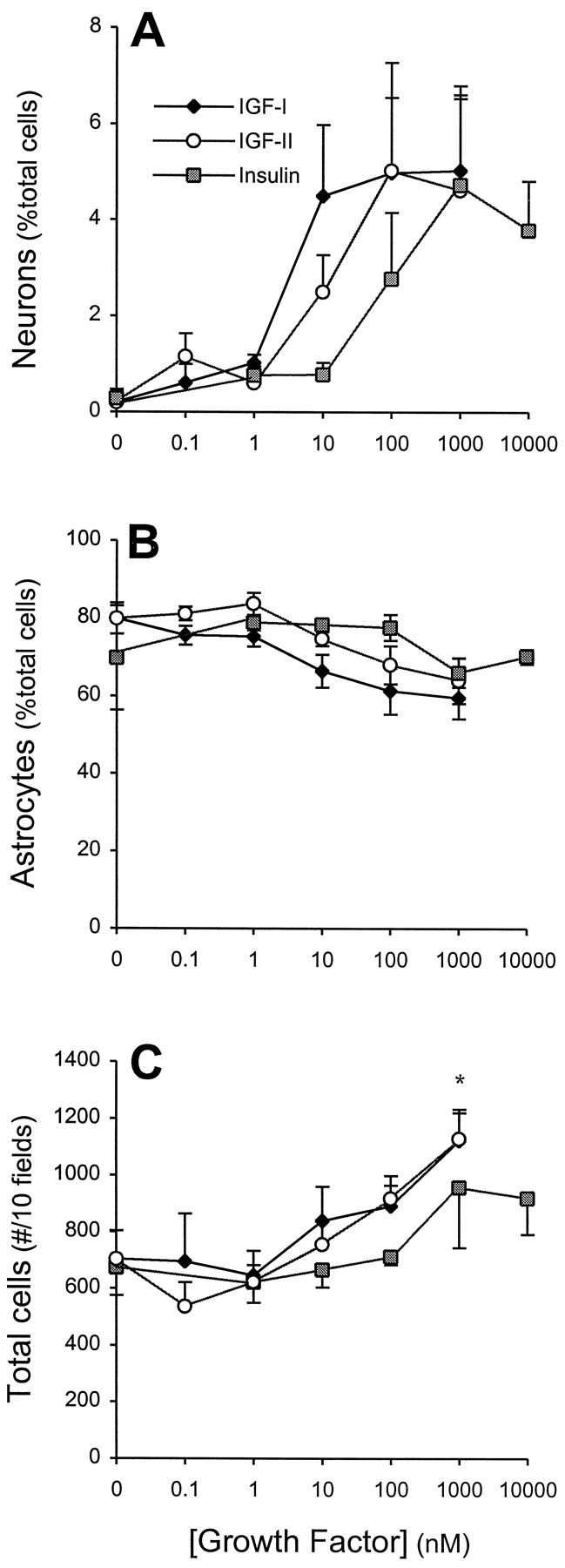
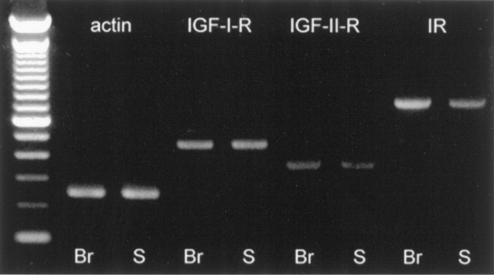
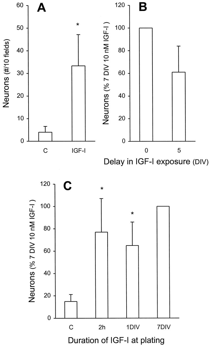
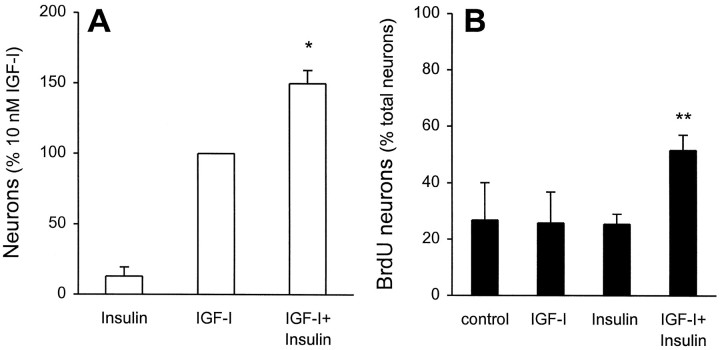
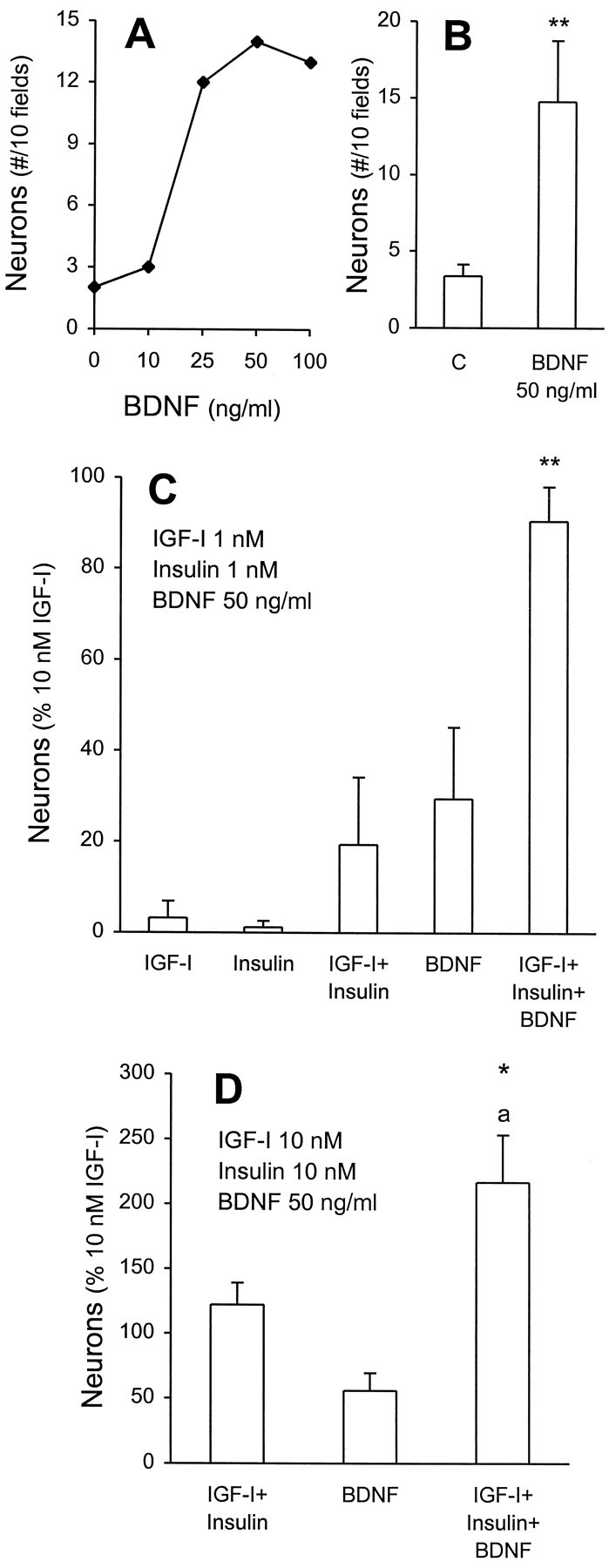
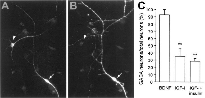
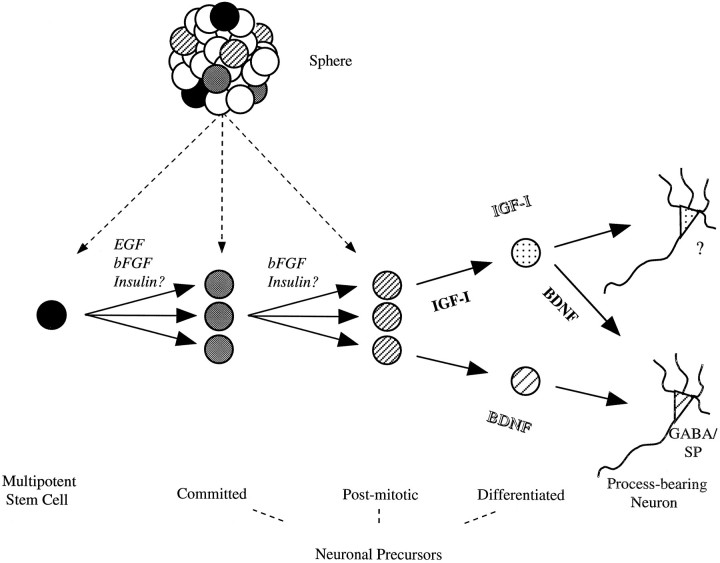
Insulin-like growth factor-I (IGF-I) has been reported previously to promote the proliferation, survival, and maturation of sympathetic neuroblasts, the genesis of retinal neurons, and the survival of CNS projection and motor neurons. Here we asked whether IGF-I could promote the in vitro differentiation of postmitotic mammalian CNS neuronal precursors derived from multipotent epidermal growth factor (EGF)-responsive stem cells. In the absence of IGF-I, virtually no neurons were present in cultured stem cell progeny, whereas IGF-I increased neuron number by eight- to 40-fold. Brief exposures (2 hr) to IGF-I were sufficient to allow for neuronal differentiation without affecting proliferation or survival. IGF-I actions could be mimicked by insulin and IGF-II at concentrations that correspond to the pharmacology of the IGF-I receptor, the latter for which the mRNA was detected in undifferentiated stem cell progeny. Although ineffectual alone at low concentrations (10 nM) that would activate its own receptor, insulin was able to potentiate the actions of IGF-I by acting on mitotically active neural precursors. When neuronal precursor differentiation by IGF-I was examined in relation to brain-derived neurotrophic factor (BDNF), two important observations were made: (1) BDNF could potentiate the differentiating actions of IGF-I plus insulin, and (2) BDNF could act on a separate population of precursors that did not require IGF-I plus insulin for differentiation. Taken together, these results suggest that IGF-I and BDNF may act together or sequentially to promote neuronal precursor differentiation.


Figures

Fig. 1.
Fig. 1.
Effects of IGFs on neuron numbers in cultures of EGF-generated precursors. A, Increasing concentrations of IGF-I, IGF-II, and insulin induced a dose-dependent increase in neuron (β-tubulin-immunoreactive) production, expressed as a percentage of total cells derived from EGF-generated spheres. The rank order of potency, IGF-I > IGF-II > insulin, is characteristic of IGF-I receptor pharmacology (LeRoith et al., 1993) (see Results for further discussion). Growth factors were added at plating and were present for the entire culture period. Under the identical experimental conditions the percentages of astrocytes (GFAP-immunoreactive (B) and total cell number (C) were not affected significantly, other than total cell numbers at supersaturating (1 μm) concentrations of IGF-I or IGF-II. Data represent the mean ± SEM of four independent cultures; *p < 0.05.
Fig. 2.
Fig. 2.
Detection of IGF receptor subtypes in embryonic brain and EGF-generated spheres. Reverse transcription and PCR of total RNA extracted revealed that IGF-I, insulin, and IGF-II receptor mRNA are present in the E14 brain (Br) and in the EGF-generated spheres of undifferentiated precursors (S). First-strand cDNA was amplified by using specific primers, as described in Materials and Methods. The ladder is the 100 bp ladder (from BRL). Amplified products were run out on a 1.5% agarose/Tris-acetate-EDTA gel and visualized with ethidium bromide. No genomic amplification was observed from any extracted RNA samples.
Fig. 3.
Fig. 3.
IGF-I increases neuron number without an apparent survival action. A, EGF-generated precursors were cultured in the absence or presence of 10 nm IGF-I for 7 DIV. The total number of neurons in 10 fields (total of 600–900 cells) was determined. In subsequent experiments, to ensure standardization with multiple comparisons, the number of neurons present after 7 DIV in the presence of 10 nm IGF-I (15–50 β-tubulin-immunoreactive neurons within 600–900 cells) serves as 100%. *p < 0.05 compared with control.B, A 5 d delay (from plating) in exposure of precursors to 10 nm IGF-I yielded neuron numbers that were not significantly different from a 7 DIV exposure starting at plating.C, Brief exposure to IGF-I was sufficient to induce neuronal differentiation. IGF-I (10 nm) was added for 2 hr (2 h) at plating or for 1 DIV, followed by extensive washing and a total 7 DIV incubation, and then compared with control (*significantly different, p < 0.05) or to a 7 DIV exposure (not significantly different). All data represent the mean ± SEM of three independent culture experiments.
Fig. 4.
Fig. 4.
IGF-I and insulin cooperate to produce enhanced numbers of neurons. EGF-generated precursors were cultured with 1 μm BrdU in the absence or presence of 10 nminsulin, 10 nm IGF-I, or the two combined, for 7 DIV.A, With the numbers of β-tubulin-immunoreactive neurons produced by 7 DIV of 10 nm IGF-I serving as 100% (see legend to Fig. 3 and Materials and Methods for details), the combined actions of 10 nm IGF-I plus 10 nminsulin are found to be more than additive (*p < 0.05; n = 4 independent cultures).B, In each condition, 100 β-tubulin-immunoreactive neurons were examined for incorporation of BrdU, and the percentage of double-labeled cells was calculated. The combined actions of 10 nm IGF-I plus 10 nm insulin result in a twofold increase in newly generated neurons (**p < 0.01 relative to control or insulin; p < 0.05 relative to IGF-I; n = 4 independent cultures). Taken together, these data suggest that increased neuronal numbers caused by combined insulin plus IGF-I is attributable to actions on mitotically active neuronal precursors.
Fig. 5.
Fig. 5.
Dual labeling for β-tubulin and BrdU provides evidence for newly generated neurons. EGF-generated spheres were dissociated and plated for 7 DIV with 1 μm BrdU in the presence of 10 nm IGF-I (A, C) or 10 nm IGF-I plus 10 nm insulin (B, D). Cells were dual-labeled for β-tubulin (A, B) and BrdU (C, D) immunoreactivity.Arrows illustrate β-tubulin-immunoreactive neurons that had incorporated BrdU. Despite the use of two monoclonal antibodies, our incubation procedure did not generate nonspecific overlap, as demonstrated by both coincident and noncoincident immunolabeling (with steady-state immunofluorescent intensity) in the same field (see Materials and Methods for details). Scale bar, 20 μm.
None
Figure 6. Independent and cooperative actions of BDNF and IGF-I plus insulin in neuronal differentiation. A, EGF-generated precursors were cultured without IGF-I or insulin and with increasing concentrations of BDNF for 7 DIV. A representative experiment illustrates a dose-dependent increase in neuron numbers (total cell number was unaffected; data not shown). B, Cumulative data for saturating concentrations (50 ng/ml) of BDNF illustrate a significant three- to fourfold increase in neurons relative to control (**p < 0.01;n = 8 independent cultures). C, BDNF potentiates the actions of low concentrations of insulin and IGF-I. As described in the legend to Figure 3 and Materials and Methods, the number of neurons generated by 7 DIV incubation with 10 nmIGF-I (15–50 β-tubulin-immunoreactive neurons within 600–900 cells) serves as 100%. The number of neurons generated by the combined actions of 1 nm insulin plus 1 nm IGF-I plus 50 ng/ml BDNF was significantly greater than either insulin plus IGF-I or BDNF alone (**p = 0.02). When the action of BDNF was subtracted, the resultant insulin plus IGF-I response was 3.85-fold greater than that generated in its absence (p < 0.05). D, BDNF acts on a distinct population of neuronal precursors. Effects of 10 nm IGF-I and/or insulin were tested with 50 ng/ml of BDNF. An additive action of IGF-I and insulin with BDNF was observed. *p = 0.03 in comparison to 10 nm IGF-I;ap < 0.01 in comparison to BDNF or to IGF-I coincubated with insulin (n = 5 independent cultures).
Fig. 7.
Fig. 7.
BDNF induces differentiation of GABAergic neurons, whereas IGF-I-generated neurons are principally non-GABAergic.A, B, IGF-I generated a population of non-GABAergic neurons. The arrow illustrates a cell that is immunoreactive for β-tubulin (A), but not for GABA (B, arrowhead). C, Quantitative analysis of the GABAergic neurons present after differentiation by BDNF, IGF-I, or IGF-I plus insulin. The results reveal that BDNF generated only GABAergic neurons, whereas IGF-I-generated and IGF-I plus insulin-generated neurons were principally non-GABAergic. In all, 100–120 neurons were examined per condition and within each experiment (n = 3); **p < 0.01.
Fig. 8.
Fig. 8.
A model for neurogenesis by multipotent stem cellsin vitro. Our working hypothesis is that multipotent stem cells produce neurons in four steps during which neuron number and differentiation are regulated. Both EGF and bFGF induce multipotent stem cells to proliferate. Basic FGF can stimulate committed neuronal precursors (e.g., neuron/astrocyte or neuron only), which are derived from multipotent stem cells, to produce neurons. In either case, insulin may act to recruit multipotential or bipotential precursors to a postmitotic neuronal fate. Subsequently, two possible models (lineage model in italic type and birthdate model in bold type) may serve to explain how IGF-I and BDNF act to induce the differentiation of postmitotic neuronal precursors. See Discussion for details of the two models.

References

    1. Adem A, Ekblom J, Gillberg P-G. Growth factor receptors in amyotrophic lateral sclerosis. Mol Neurobiol. 1994;9:225–231. - PubMed
    1. Ahmed S, Reynolds BA, Weiss S. BDNF enhances the differentiation but not the survival of CNS stem cell-derived neuronal precursors. J Neurosci. 1995;15:5765–5778. - PMC - PubMed
    1. Aizenman Y, de Vellis J. Brain neurons develop in a serum and glial free environment: effects of transferrin, insulin, insulin-like growth factor-I, and thyroid hormone on neuronal survival, growth, and differentiation. Brain Res. 1987;406:32–42. - PubMed
    1. Barbe MF. Tempting fate and commitment in the developing forebrain. Neuron. 1996;16:1–4. - PubMed
    1. Bartlett WP, Li X-S, Williams M, Benkovic S. Localization of insulin-like growth factor-I mRNA in murine central nervous system during postnatal development. Dev Biol. 1991;147:239–250. - PubMed

Publication types

MeSH terms