Review
. 1998 Mar 15;18(6):2188-99.
doi: 10.1523/JNEUROSCI.18-06-02188.1998.

Temporal cortex activation in humans viewing eye and mouth movements

Affiliations
Review

Temporal cortex activation in humans viewing eye and mouth movements

A Puce et al. J Neurosci. .

Abstract

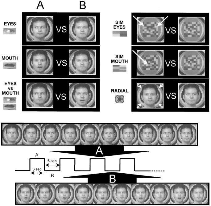
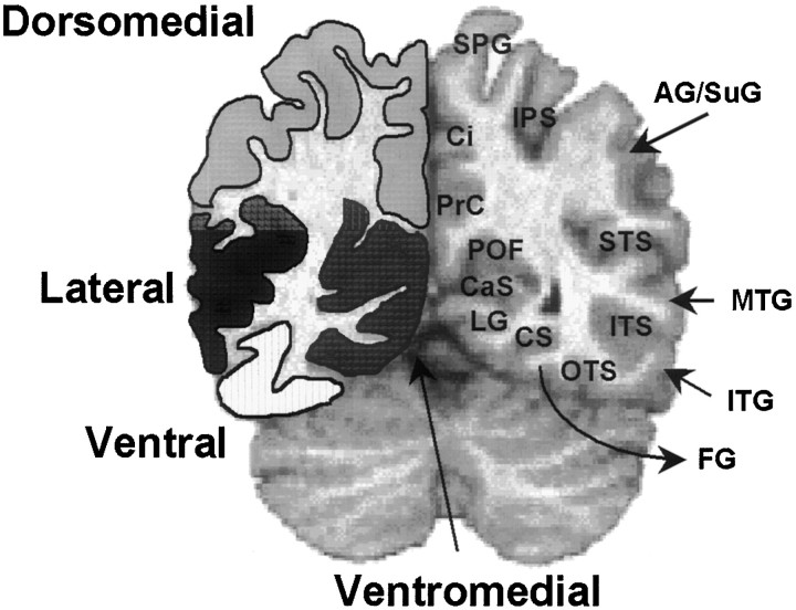
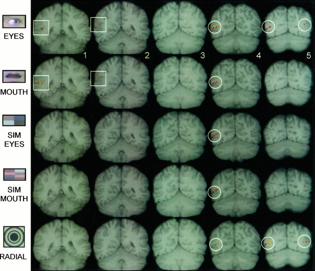
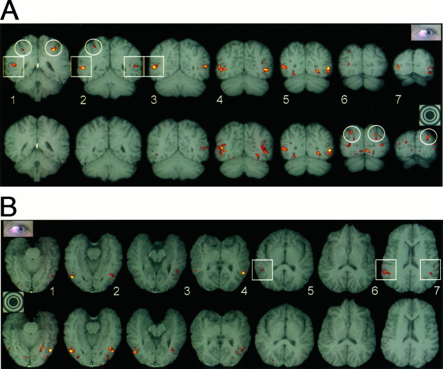
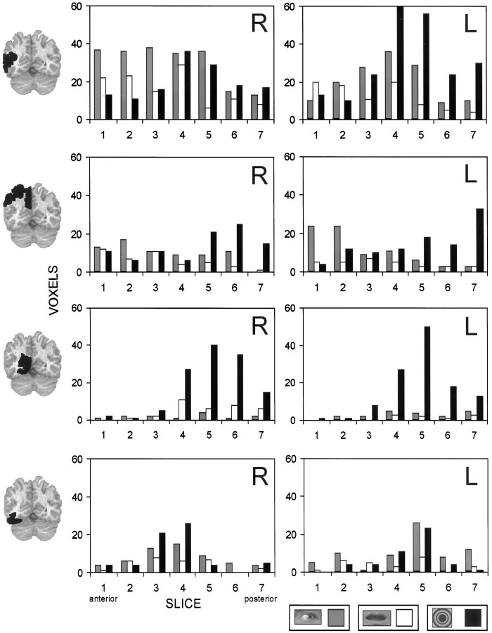
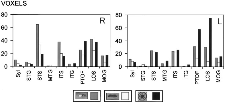
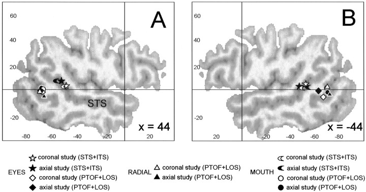
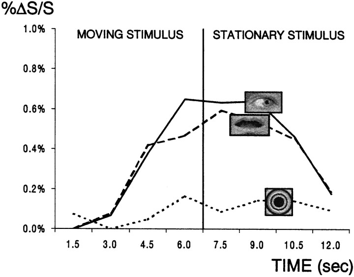
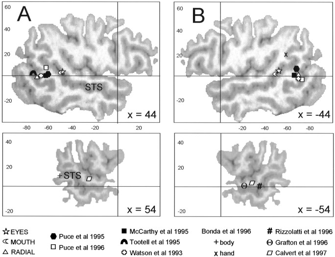
We sought to determine whether regions of extrastriate visual cortex could be activated in subjects viewing eye and mouth movements that occurred within a stationary face. Eleven subjects participated in three to five functional magnetic resonance imaging sessions in which they viewed moving eyes, moving mouths, or movements of check patterns that occurred in the same spatial location as the eyes or mouth. In each task, the stimuli were superimposed on a radial background pattern that continually moved inward to control for the effect of movement per se. Activation evoked by the radial background was assessed in a separate control task. Moving eyes and mouths activated a bilateral region centered in the posterior superior temporal sulcus (STS). The moving check patterns did not appreciably activate the STS or surrounding regions. The activation by moving eyes and mouths was distinct from that elicited by the moving radial background, which primarily activated the posterior-temporal-occipital fossa and the lateral occipital sulcus-a region corresponding to area MT/V5. Area MT/V5 was also strongly activated by moving eyes and to a lesser extent by other moving stimuli. These results suggest that a superior temporal region centered in the STS is preferentially involved in the perception of gaze direction and mouth movements. This region of the STS may be functionally related to nearby superior temporal regions thought to be involved in lip-reading and in the perception of hand and body movement.


Figures

Fig. 1.
Fig. 1.
The top panel illustrates the six experimental tasks. In EYES, lateral eye movements were contrasted to a static face with the eyes looking straight ahead. In MOUTH, an open mouth was contrasted to a closed mouth in a static face. Eye movements were contrasted with mouth movements in the EYES versus MOUTH task. In SIMULATED (SIM) EYES and SIMULATED MOUTH, colored checkerboard patterns with checks reversing position in spatially equivalent positions (white arrows) to the real eyes and mouth were contrasted to a static checkerboard. In all of these tasks the radial background moved continuously in an inward direction (small white arrows) during the entire duration of the imaging run. In RADIAL, the face remained static, and the radial background either moved in the direction indicated by the white arrows or remained static. The effect of an inwardly moving radial background was generated by changing the color of the concentric rings on each frame (see bottom panel). Thebottom panel depicts a schematic of a single cycle in the ABAB alternating design for the EYES versus MOUTH task. The duration of each subtask (A or B) was 6 sec. During each subtask, a series of 10 images (600 msec duration) was shown. In subtask A, the eyes shifted their position from the center to either left or right and back to center in a random manner. In subtask B, the mouth closed on alternate frames.
Fig. 2.
Fig. 2.
Four anatomical regions for classification of activated voxels (lateral, dorsomedial, ventromedial, and ventral) and their borders are outlined on the left side of a coronal anatomical image. Some of the structures falling within each region are shown on the right. STS, Superior temporal sulcus; MTG, middle temporal gyrus;ITS, inferior temporal sulcus; ITG, inferior temporal gyrus; OTS, occipitotemporal sulcus;FG, fusiform gyrus; CS, collateral sulcus; LG, lingual gyrus; CaS, calcarine sulcus; POF, parieto-occipital fissure;PrC, precuneus; Ci, cingulate gyrus and sulcus; SPG, superior parietal gyrus;IPS, intraparietal sulcus; AG/SuG, angular or supramarginal gyri.
Fig. 3.
Fig. 3.
Individual subject activation data overlaid on T1-weighted coronal anatomical images. Slice 1 is the most anterior. In EYES and MOUTH, focal activation was observed in the right lateral cortex of the two most anterior slices (framed by white squares). No activation was seen in the same regions for the other tasks. Activation in all tasks (framed by white circles) was seen in another region of right lateral cortex posterior and inferior to that seen to EYES and MOUTH. In this and Figure 4, the right hemisphere appears on the left side of the image, and the red to yellow color scale indicates lower to higher t values of activation. In this and Figure 4, activation data have been scaled, translated, and interpolated to fit their anatomical counterparts.
Fig. 4.
Fig. 4.
Individual subject activation data for EYES (top) and RADIAL (bottom) overlaid on T1-weighted anatomical images. A, Coronal slices 1–7. Slice 1 is the most anterior. A region of activation in the right lateral cortex is seen in slices 1–3 to EYES but not to RADIAL (white squares). Extensive activation of lateral cortex bilaterally occurs in slices 4–7 for both EYES and RADIAL. Activation in the IPS (white circles) was also seen to EYES anteriorly in slices1 and 2 and posteriorly to RADIAL in slices 6 and 7. B, Oblique axial slices (1–7) for the same subject and tasks. Slice 1 is the most ventral. Activation to EYES (white squares) but not to RADIAL is seen in slices5 and 7, as in A.
Fig. 5.
Fig. 5.
Voxel counts as a function of hemisphere (R, right; L, left) and slice for each region in 11 subjects. Lateral (top), dorsomedial (second from top), ventromedial (second frombottom), and ventral (bottom) for EYES (gray histograms), MOUTH (white histograms) and RADIAL (black histograms). Slice1 is the most anterior, and slice 7 is the most posterior. EYES elicited more activation in slices1–3 than the other two tasks, whereas RADIAL elicited the most prominent activation in slices 4–7 in the left hemisphere. In the dorsomedial region, the most prominent activation was elicited to EYES in slices 1 and 2 of the left hemisphere and to RADIAL in slices 5–7 of both hemispheres. In the ventromedial region, RADIAL elicited the most prominent activation in slices 4–7 of both hemispheres. The least activation was seen in the ventral region and was not different across tasks.
Fig. 6.
Fig. 6.
Voxel counts as a function of hemisphere and anatomical structure for the lateral region for EYES (gray histograms), MOUTH (white histograms), and RADIAL (black histograms) in 11 subjects. In the right hemisphere, EYES produced the most activation in the STS, whereas in the left hemisphere the most activation occurred in the PTOF and LOS to radial. Syl, Sylvian fissure;STG, superior temporal gyrus; STS, superior temporal sulcus; MTG, middle temporal gyrus;ITS, inferior temporal sulcus; ITG, inferior temporal gyrus; PTOF, parieto-temporo-occipital fossa; LOS, lateral occipital sulcus;MOG, middle occipital gyrus.
Fig. 7.
Fig. 7.
Activation centroids to EYES, MOUTH, and RADIAL. Two centroids are shown: anteriorly for the STS/ITS and posteriorly for the PTOF/LOS for coronal and axial fMRI studies. A, Right hemisphere. B, Left hemisphere. Centroids are superimposed on a sagittal view of a representative brain, 44 mm from the midline. In this and Figure 9, coordinates in they-axis (horizontal) andz-axis (vertical) are in the system of Talairach and Tournoux (1988), and the anterior commissure–posterior commissure line (horizontal line) and the anterior commissure at y = 0 (vertical line) are shown. The SEs around the centers of activation (x, y, z) for the coronal studies were EYES anterior (left, 1, 1, 2; right, 1, 1, 2), MOUTH anterior (left, 1, 3, 2; right, 3, 2, 2), EYES posterior (left, 1, 1, 2;right, 2, 2, 2), MOUTH posterior (left, 9, 2, 2; right, 3, 4, 2), and RADIAL posterior (left 1, 2, 1; right 2, 2, 2).
Fig. 8.
Fig. 8.
Time course of activation of the right STS for a single 12 sec cycle averaged over all cycles in each task for 10 subjects. Percent signal change (%ΔS/S) is shown on the y-axis for EYES, MOUTH, and RADIAL. For the first 6 sec of the cycle the relevant stimulus is in motion, whereas for the second half of the cycle it is stationary. The right STS is activated by EYES (solid line) and MOUTH (broken line) but not by RADIAL (dotted line).
Fig. 9.
Fig. 9.
Centroids of activation (STS/ITS for EYES and MOUTH and PTOF/LOS for RADIAL) in this study compared with centroids of activation for the perception of hand action or body movement (Bonda et al., 1996), hand grasping (Rizzolatti et al., 1996; Grafton et al., 1996), silent lip-reading of numbers (Calvert et al., 1997), nonanimate movement (Watson et al., 1993; McCarthy et al., 1995; Tootell et al., 1995), and the perception of static faces (Puce et al., 1995, 1996).A, Right hemisphere. B, Left hemisphere. Centroids of activation are superimposed on two sagittal views of a representative brain.

References

    1. Allison T, Ginter H, McCarthy G, Nobre A, Puce A, Luby M, Spencer DD. Face recognition in the human extrastriate cortex. J Neurophysiol. 1994a;71:821–825. - PubMed
    1. Allison T, McCarthy G, Belger A, Puce A, Luby M, Spencer DD, Bentin S. What is a face?: electrophysiological responsiveness of human extrastriate visual cortex to human faces, face components, and animal faces. Soc Neurosci Abstr. 1994b;20:316.
    1. Allison T, McCarthy G, Nobre A, Puce A, Belger A. Human extrastriate visual cortex and the perception of faces, words, numbers, and colors. Cereb Cortex. 1994c;5:544–554. - PubMed
    1. Baizer JS, Ungerleider LG, Desimone R. Organization of visual inputs to the inferior temporal and parietal cortex in macaques. J Neurosci. 1991;11:168–190. - PMC - PubMed
    1. Bentin S, Allison T, Puce A, Perez E, McCarthy G. Electrophysiological studies of face perception in humans. J Cognit Neurosci. 1996;8:551–565. - PMC - PubMed

Publication types

LinkOut - more resources