Review
. 1998 Mar 15;18(6):2231-8.
doi: 10.1523/JNEUROSCI.18-06-02231.1998.

Alterations in dopamine release but not dopamine autoreceptor function in dopamine D3 receptor mutant mice

Affiliations
Review

Alterations in dopamine release but not dopamine autoreceptor function in dopamine D3 receptor mutant mice

T E Koeltzow et al. J Neurosci. .

Abstract

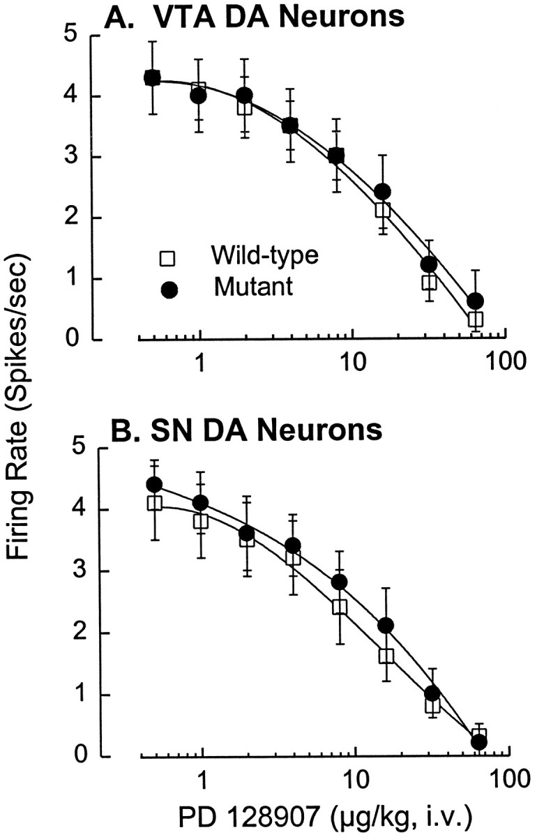
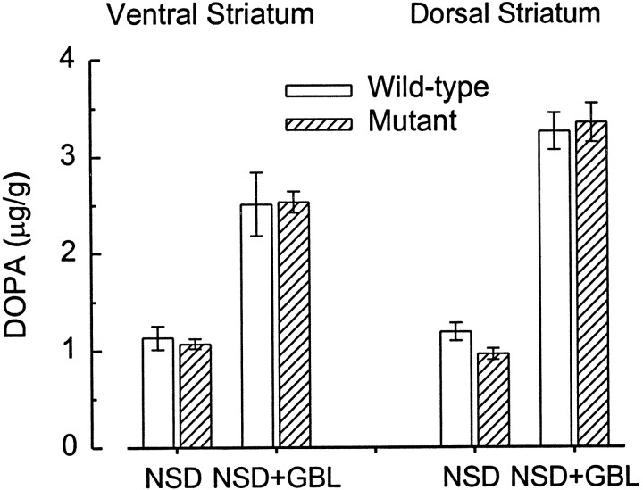
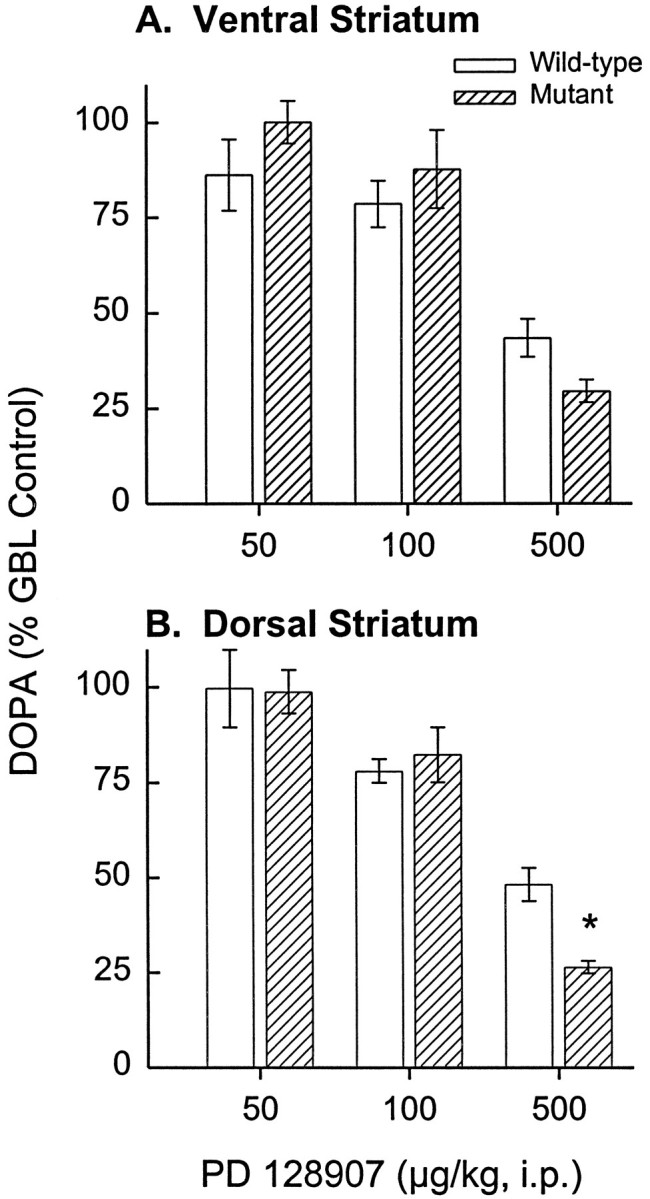
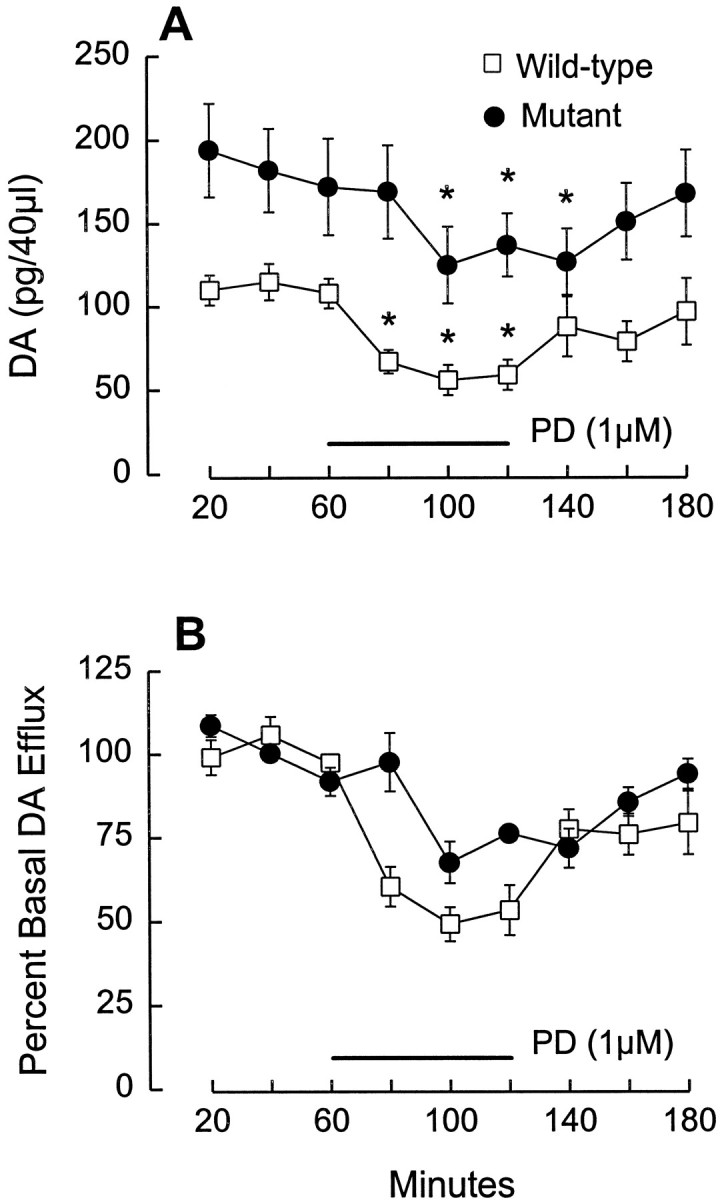
Dopamine (DA) autoreceptors expressed along the somatodendritic extent of midbrain DA neurons modulate impulse activity, whereas those expressed at DA nerve terminals regulate both DA synthesis and release. Considerable evidence has indicated that these DA autoreceptors are of the D2 subtype of DA receptors. However, many pharmacological studies have suggested an autoreceptor role for the DA D3 receptor. This possibility was tested with mice lacking the D3 receptor as a result of gene targeting. The basal firing rates of DA neurons within both the substantia nigra and ventral tegmental area were not different in D3 receptor mutant and wild-type mice. The putative D3 receptor-selective agonist R(+)-trans-3,4,4a, 10b-tetrahydro-4-propyl-2H,5H-(1)benzopyrano(4,3-b)-1,4-oxazin+ ++-9-ol (PD 128907) was equipotent at inhibiting the activity of both populations of midbrain DA neurons in the two groups of mice. In the gamma-butyrolactone (GBL) model of DA autoreceptor function, mutant and wild-type mice were identical with respect to striatal DA synthesis and its suppression by PD 128907. In vivo microdialysis studies of DA release in ventral striatum revealed higher basal levels of extracellular DA in mutant mice but similar inhibitory effects of PD 128907 in mutant and wild-type mice. These results suggest that the effects of PD 128907 on dopamine cell function reflect stimulation of D2 as opposed to D3 receptors. Although D3 receptors do not seem to be significantly involved in DA autoreceptor function, they may participate in postsynaptically activated short-loop feedback modulation of DA release.


Figures

Fig. 1.
Fig. 1.
Similar inhibition of midbrain DA neurons by PD 128907 in D3 receptor mutant and wild-type mice.A, Cumulative dose–response curves showing the significant dose-dependent suppression of VTA DA neuronal activity by PD 128907 (F6,120 = 62.5;p < 0.001) and the lack of difference between the wild-type and mutant mice with respect to this effect. There was also no significant difference between the groups with respect to basal firing rates, 4.3 ± 0.6 spikes/sec (mean ± SEM) in wild-type mice (n = 12) and 4.3 ± 0.5 spikes/sec in the mutant mice (n = 10). Eticlopride reversed the inhibition to 85–110% of the basal firing rate in every cell tested. B, Similar dose–response curves indicating dose-dependent inhibition of SN DA neurons by PD 128907 (F6,138 = 51.5; p < 0.001) and the lack of difference between wild-type and mutant mice with respect to this effect. There was also no significant difference in the basal firing rates for SN DA neurons in wild-type (4.1 ± 0.6 spikes/sec; n = 12) and mutant (4.4 ± 0.4 spikes/sec; n = 13) mice. Eticlopride reversed the agonist-induced inhibition to 82–112% of the basal firing rate in every cell tested. Each point represents the mean ± SEM.
Fig. 2.
Fig. 2.
Recording sites for DA neurons within the mouse midbrain. Approximate recording sites (filled circles) for DA neurons within the ventral tegmental area (VTA) and substantia nigra (SN) of wild-type (left) and D3 receptor mutant (right) mice. Coordinates are expressed as anterior to lambda. RN, Red nucleus.
Fig. 3.
Fig. 3.
Similar GBL-induced increase in striatal DOPA formation in D3 receptor mutant and wild-type mice. Basal levels of DOPA, measured after inhibition of l-aromatic amino acid decarboxylase with NSD 1015, did not significantly differ in wild-type and D3 receptor mutant mice in either the ventral or dorsal striatum. GBL significantly increased DOPA formation in both ventral (F1,20 = 58.58;p < 0.001) and dorsal (F1,19 = 261.79; p < 0.001) striatum, as a result of the cessation of impulse-dependent DA release and the relief of tonic autoreceptor-mediated inhibition of tyrosine hydroxylase activity. Again, there were no significant differences in the GBL-induced increase in the two groups of mice. Error bars indicate SEM. Sample size is six for all groups except for the NSD alone group (dorsal striatum in mutants) wheren = 5.
Fig. 4.
Fig. 4.
Similar inhibition of GBL-induced striatal DOPA formation by PD 128907 in D3 receptor mutant and wild-type mice. A, Reversal of GBL-induced DOPA synthesis by PD 128907 (F3,57 = 29.97; p< 0.001) in the ventral striatum was not significantly different in D3 receptor mutant and wild-type mice. Data are presented as the percent of DOPA levels measured in mice administered GBL plus NSD without PD 128907 (wild type, 2.51 μg/gm/30 min; mutant, 2.53 μg/gm/30 min). Sample sizes for the 0, 0.05, 0.1, and 0.5 mg/kg doses of PD 128907 are 16, 4, 6, and 6 for the mutant and 19, 3, 5, and 6 for the wild-type mice, respectively. B, Reversal of GBL-induced DOPA synthesis by PD 128907 (F3,57 = 25.23; p < 0.001) in the dorsal striatum was also not significantly different in the two groups of mice; basal levels were 3.26 ng/gm/30 min for the wild-type and 3.40 ng/gm/30 min for the mutant mice. Error bars indicate SEM. Sample sizes for the 0, 0.05, 0.1, and 0.5 mg/kg doses of PD 128907 are 21, 4, 6, and 6 for the mutant and 14, 3, 5, and 5 for the wild-type mice, respectively. Note that the effect of the highest dose of PD 128907 on DA synthesis within the dorsal striatum was actually significantly greater in the mutant than in the wild-type mice (*p < 0.05; Dunnett’s test).
Fig. 5.
Fig. 5.
Localization of microdialysis probe in the ventral striatum. In this coronal section of the mouse forebrain, thetract left by the microdialysis probe can be seen traversing the ventral striatum. Only the bottom 2 mm of the probe (located between the arrowheads) consisted of open dialysis membrane. NAc, Nucleus accumbens;CPu, caudate-putamen; LV, lateral ventricle; ICj, island of Calleja; ac, anterior commisure; cc, corpus callosum.
Fig. 6.
Fig. 6.
Regulation of basal DA efflux, but not PD 128907-induced suppression of DA release, is altered in D3receptor mutant mice. A, After 2–3 hr of perfusion, three baseline fractions (20 min each) were collected (0–60 min). Basal DA efflux was significantly higher in mutant mice (F1,9 = 26.9; p = 0.0008). Values for basal levels in wild-type mice were ∼13 nm, which is comparable with values obtained in our previously published studies of rat NAc (e.g., see Wolf et al., 1994). PD 128907 was applied for 60 min (horizontal bar), beginning after collection of the third baseline fraction. PD 128907 significantly reduced DA efflux in both wild-type and mutant mice (F1,10 = 7.798; p = 0.019).Asterisks indicate points that differ significantly (p < 0.01; Dunnett’s test) when compared with the weighted mean of the three predrug baseline values. B, When the results shown in A are expressed as percent of basal DA efflux, the inhibitory effect of PD 128907 appears blunted in the mutant mice. However, ANCOVA indicated that this apparent effect was solely attributable to the initial differences in basal DA efflux, i.e., there was no difference between the two groups with respect to the release-suppressing effects of PD 128097 when basal DA levels were controlled as a covariate. Results are expressed as mean ± SEM (n = 6/group).

Similar articles

Cited by

References

    1. Accili D, Fishburn CS, Drago J, Steiner H, Lachowicz JE, Park BH, Gauda EB, Lee EJ, Cool MH, Sibley DR, Gerfen CR, Westphal H, Fuchs S. A targeted mutation of the D3 dopamine receptor gene is associated with hyperactivity in mice. Proc Natl Acad Sci USA. 1996;93:1945–1949. - PMC - PubMed
    1. Aghajanian GK, Bunney BS. Dopamine “autoreceptors”: pharmacological characterization by microiontophoretic single unit recording studies. Naunyn Schmiedebergs Arch Pharmacol. 1977;297:1–7. - PubMed
    1. Ahlenius S, Salmi P. Behavioral and biochemical effects of the dopamine D3 receptor-selective ligand, 7-OH-DPAT, in the normal and the reserpine-treated rat. Eur J Pharmacol. 1994;260:177–181. - PubMed
    1. Aretha CW, Sinha A, Galloway MP. Dopamine D3-preferring ligands act at synthesis modulating autoreceptors. J Pharmacol Exp Ther. 1995;274:609–613. - PubMed
    1. Bouthenet M-L, Souil E, Martres M-P, Sokoloff P, Giros B, Schwartz J-C. Localization of dopamine D3 receptor mRNA in the rat brain using in situ hybridization histochemistry: comparison with dopamine D2 receptor mRNA. Brain Res. 1991;564:203–219. - PubMed

Publication types

MeSH terms