. 1998 Mar 3;95(5):2067-72.
doi: 10.1073/pnas.95.5.2067.

A highly conserved lysine residue on the head domain of type II keratins is essential for the attachment of keratin intermediate filaments to the cornified cell envelope through isopeptide crosslinking by transglutaminases

Affiliations

A highly conserved lysine residue on the head domain of type II keratins is essential for the attachment of keratin intermediate filaments to the cornified cell envelope through isopeptide crosslinking by transglutaminases

E Candi et al. Proc Natl Acad Sci U S A. .

Abstract

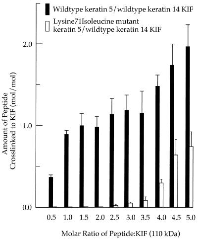
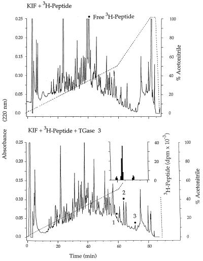
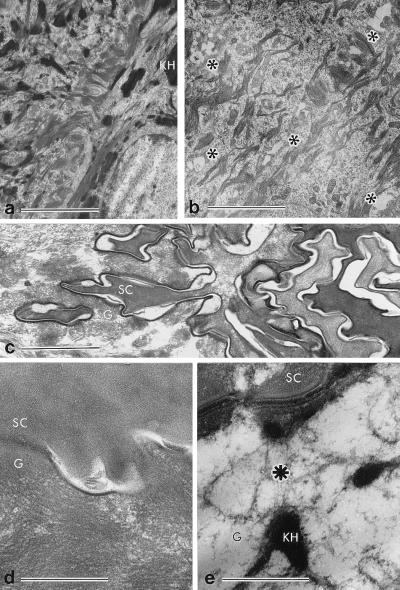
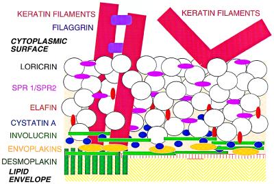
We have addressed the question of how keratin intermediate filaments are associated with the cell envelope at the periphery of cornified epidermal cells. Many peptides from human epidermal cell envelopes containing isopeptide crosslinks inserted by transglutaminases in vivo have been characterized. A major subset involves the type II keratin chains keratin 1, 2e, 5, or 6 crosslinked to several protein partners through a lysine residue located in a conserved region of the V1 subdomain of their head domains. This sequence specificity was confirmed in in vitro crosslinking experiments. Previously the causative mutation in a family with diffuse nonepidermolytic palmar-plantar keratoderma was shown to be the loss in one allele of the same lysine residue of the keratin 1 chain. Ultrastructural studies of affected palm epidermis have revealed abnormalities in the organization of keratin filaments subjacent to the cell envelope and in the shape of the cornified cells. Together, these data suggest a mechanism for the coordination of cornified cell structure by permanent covalent attachment of the keratin intermediate filament cytoskeleton to the cell envelope by transglutaminase crosslinking. Furthermore, these studies identify the essential role of a conserved lysine residue on the head domains of type II keratins in the supramolecular organization of keratin filaments in cells.


Figures

Figure 1
Figure 1
The conserved Lys residue 71 of the keratin 5 chain is not essential for KIF assembly in vitro. KIF were assembled from equimolar mixtures of: wild-type keratins 5 and 14 (a); the Lys-71 → Ile-substituted form of keratin 5 and wild-type keratin 14 (b); and the Leu-174 → Pro-substituted form of keratin 5 and wild-type keratin 14 (c). (Bar = 100 nm.)
Figure 2
Figure 2
Stoichiometry of crosslinking of a synthetic loricrin peptide to KIF in vitro. Wild-type keratin 5/14 KIF (solid bars) or with KIF assembled from the keratin 5 chain with Lys-71 → Ile substitution and wild-type keratin 14 (open bars) were mixed with the synthetic peptide in various molar ratios as shown, crosslinked with the TGase 3 enzyme, and assayed.
Figure 3
Figure 3
Recovery by HPLC of tryptic peptides containing crosslinks introduced by the TGase 3 enzyme in vitro between keratin 1/10 KIF and a synthetic loricrin peptide. (Lower, Inset) The labeled peptides shown were isolated for quantitation and sequencing (Table 3).
Figure 4
Figure 4
Ultrastructural analysis of palmar epidermis from a normal individual (a and d) and an affected patient with diffuse NEPPK disease (b, c, and e). In the patient, the basal and lower spinous layers appeared normal (data not shown). However, in cell layers extending from the upper spinous to granular layers, where CE assembly is initiated, separations of KIF tonofibrils from desmosomes and the cell periphery occur in the patient, leading to microclefts, which are marked by asterisks in b. In addition, at the interface between the uppermost granular and first cornified cell, a highly convoluted interface is evident in the patient (c), which is absent in normal epidermis (d). At higher resolution, the KIF appear withdrawn from the desmosomal and interdesomomal regions (asterisk) in many places in the patient (e), unlike in normal (d). [Bars = 2.5 μm (a and b), 1 μm (c), 0.5 μm (d and e)]. SC, stratum corneum; G, granular layer; KH, keratohyalin granule.
Figure 5
Figure 5
Model showing two different modes of association of KIF with the foreskin epidermal CE. At left is the possible organization of KIF near the vicinity of a desmosomal remnant including the cytoplasmic domain of desmoplakin with envoplakin and involucrin. At right is shown the possible association of KIF with the intracellular surface of the CE, which is enriched in loricrin and SPRs. This model is not drawn to scale, because the molecular dimensions of no CE structural proteins are known. Note that although few direct data are available, it is to be expected that foreskin and palmar-plantar epidermal CEs will be generally similar, especially because they both contain high amounts of loricrin and have similar masses per unit area (30); however, they are different from other internal stratified squamous epithelia, which contain little or no loricrin.

Similar articles

Cited by

References

    1. Hohl D. Dermatologica. 1990;180:201–211. - PubMed
    1. Greenberg C S, Birckbichler P J, Rice R H. FASEB J. 1991;5:3071–3077. - PubMed
    1. Reichert U, Michel S, Schmidt R. In: Molecular Biology of the Skin. Darmon M, Blumenberg M, editors. New York: Academic; 1993. pp. 107–150.
    1. Holbrook K A, Wolff K. In: Dermatology in General Medicine. Fitzpatrick T B, Eisen A Z, Wolff K, Freedberg I M, Austen K F, editors. New York: McGraw–Hill; 1993. pp. 97–145.
    1. Elias P M. J Dermatol. 1996;23:756–758. - PubMed

MeSH terms

LinkOut - more resources