. 1998 Mar 3;95(5):2083-8.
doi: 10.1073/pnas.95.5.2083.

Retrotransposon R1Bm endonuclease cleaves the target sequence

Affiliations

Retrotransposon R1Bm endonuclease cleaves the target sequence

Q Feng et al. Proc Natl Acad Sci U S A. .

Abstract

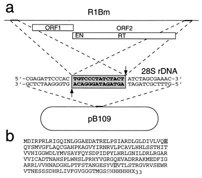
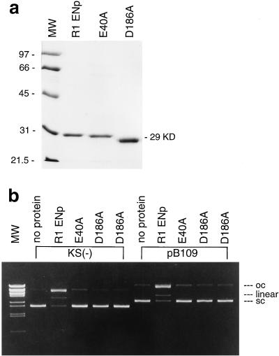
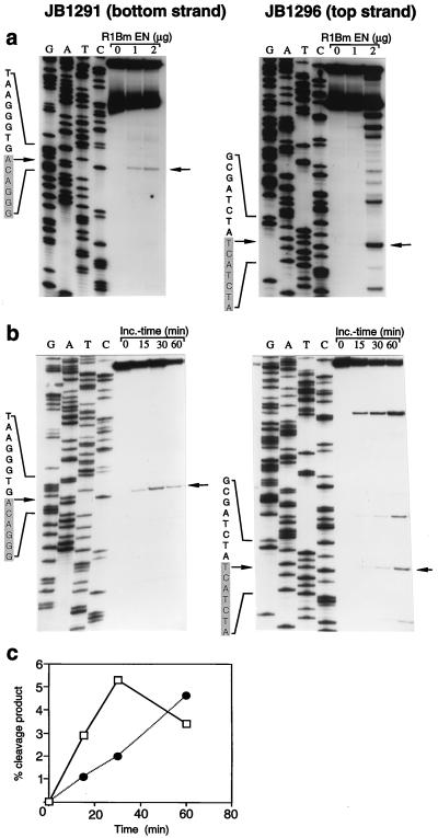
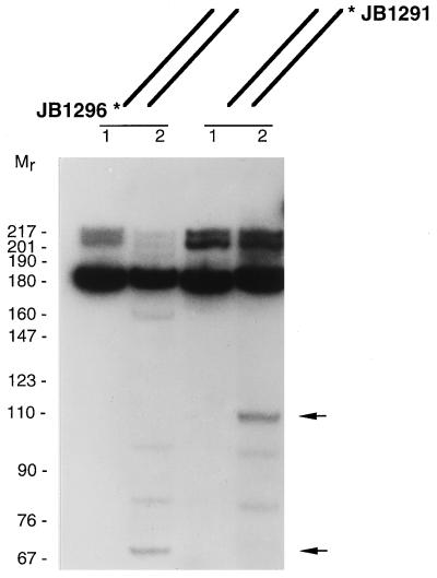
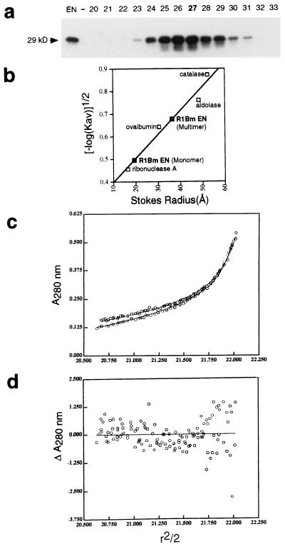
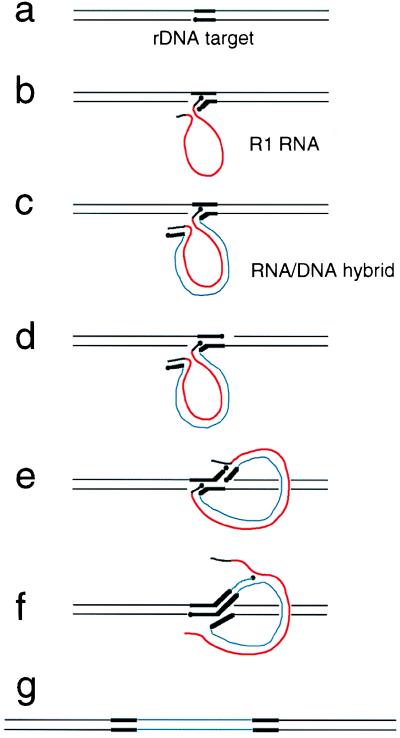
The R1Bm element, found in the silkworm Bombyx mori, is a member of a group of widely distributed retrotransposons that lack long terminal repeats. Some of these elements are highly sequence-specific and others, like the human L1 sequence, are less so. The majority of R1Bm elements are associated with ribosomal DNA (rDNA). R1Bm inserts into 28S rDNA at a specific sequence; after insertion it is flanked by a specific 14-bp target site duplication of the 28S rDNA. The basis for this sequence specificity is unknown. We show that R1Bm encodes an enzyme related to the endonuclease found in the human L1 retrotransposon and also to the apurinic/apyrimidinic endonucleases. We expressed and purified the enzyme from bacteria and showed that it cleaves in vitro precisely at the positions in rDNA corresponding to the boundaries of the 14-bp target site duplication. We conclude that the function of the retrotransposon endonucleases is to define and cleave target site DNA.


Figures

Figure 1
Figure 1
Structure of R1Bm element and its target site. (a) The genome organization of R1Bm is indicated; the ORFs are shown as boxes, and the positions of the EN and RT domains in ORF2 are indicated. tsd is shown as bold lines (top) or nucleotide sequence (shaded box, middle) in target plasmid pB109. (b) The sequence of the R1Bm EN domain expressed is indicated, with the putative active site residues mutagenized to alanine codons shaded. The expressed protein contained 43 amino acids derived from the pET15b vector at the C terminus, including 6 His residues (the remainder are designated X33).
Figure 2
Figure 2
Expression, purification, and activity of R1Bm EN protein. (a) R1Bm EN protein and its mutant versions E40A and D186A were expressed from the phage T7 promoter. The proteins were purified by nickel chelate chromatography and run on SDS/PAGE; the predicted Mr is 29,000. The unusual electrophoretic mobility of the D186A mutant was observed for two independent isolates. (b) The activity of the R1Bm EN protein was optimized and tested on two plasmid substrates, pBluescript (KS) vector and pB109. The positions of open circle (oc), linear, and supercoiled (sc) plasmid controls are indicated on the right. Note that the mutant proteins are inactive. MW, molecular weight standards.
Figure 3
Figure 3
Sequence-specific cleavage of the R1Bm target DNA. (a) A 175-bp fragment centered around the R1Bm insertion site was synthesized by PCR with individually 5′ end-labeled primers JB1291 (bottom strand) or 1296 (top strand) and pB109 template. The radioactive full-length double-stranded target DNAs were incubated without or with 1 or 2 μg of R1Bm EN protein. Arrows indicate the major sites of cleavage that correspond to the tsd boundaries indicated in Fig. 1a; tsd sequences are shaded. The molecular weight standards were dideoxy sequencing reactions primed with the same radioactive oligonucleotides on pB109 template. The exact positions of the major cleavages were confirmed by mixing experiments in which the products were mixed with the molecular weight standards (not shown). The faster moving band in the substrate in part a was shown to correspond to incompletely denatured double-stranded DNA and could be eliminated from subsequent experiments by denaturing by boiling for 10 min in 95% formamide. (b) The cleavage of the bottom strand at lower enzyme concentrations than the top strand (part a above) suggested that bottom strand cleavage might precede top strand cleavage kinetically. This was confirmed in a time course experiment. The experiment was performed as above except with 2 μg of R1Bm EN, and samples were removed at 0, 15, 30, and 60 min. Note that cleavage at the target sequence boundary (arrow) on the bottom strand (JB1291) peaks at 30 min whereas top strand cleavage (JB1296) peaks later. (c) The radioactivity in the bands representing the target sequence boundary cleavages was measured by PhosphorImager analysis, expressed as percent of initial substrate, and plotted as a function of time. Late in the reaction, the signal begins to decrease, suggesting the presence of a small amount of a contaminating random nuclease activity. Solid line, bottom strand; dotted line, top strand.
Figure 4
Figure 4
Double-strand cleavage of target DNA by R1Bm EN. The 175-bp substrates described in Fig. 3a were incubated without (lanes 1) or with 2 μg of R1Bm EN enzyme (lanes 2). The arrows indicate the size of the double-stranded DNA products resulting from cleavage on both strands at the R1Bm insertion site. The expected cleavage products would be 67 and 94 bp long with 3′ overhangs of 14 nt, the effect of which on electrophoretic mobility is uncertain. Molecular weight standards (Mr) are radiolabeled pBR322 DNA MspI fragments.
Figure 5
Figure 5
R1Bm EN behaves as a multimer. (a) Purified R1Bm EN protein (24 μg) was applied to a sizing column with molecular weight standards, and the fractions were immunoblotted with the anti-tag antibody to detect EN protein. Nearly all of the EN protein (as well as bulk absorbance) peaked at fractions 23–31, but a second small peak was observed on a long exposure at fraction 42 (not shown) and is assumed to represent monomeric enzyme. (b) Determination of Stokes radius of R1Bm EN monomer and multimer by gel filtration chromatography at 4°C with the indicated standards. The EN monomer peak runs with a Stokes radius of 19 Å corresponding to a globular protein of 19 kDa even though the actual mass is 29 kDa, suggesting that the EN has weak affinity for the column matrix. The Stokes radius of the multimer (36 Å) corresponds to a globular protein of 81 kDa and thus is consistent with a spherical tetramer with weak affinity for the column matrix. (c and d) Sedimentation equilibrium data for R1Bm EN at 12,000 and 15,000 rpm at 4°C. c represents the actual data (open dots) and the results of a global fit of these two data sets to a monomer–tetramer–dodecamer model (lines; see Materials and Methods). d presents a composite residual plot for the global fit in c; r, radius. A random distribution of actual data points (dots) about the predicted value (line) indicates very good agreement with the model. The data rule out a pure monomeric state for the R1Bm EN. A monomer–tetramer–dodecamer model with a tetramer as the predominant species (92%) fit these data best. Thus both gel filtration and sedimentation equilibrium methods are consistent with a predominantly tetrameric enzyme.
Figure 6
Figure 6
Proposed model for R1Bm retrotransposition. The model described here is based on the TPRT model developed for the R2Bm element (3), an element that like R1 is sequence-specific for rDNA but unlike R1Bm has no clearly definable EN domain. Also, unlike R2Bm, which deletes a few base pairs of target DNA as part of the retrotransposition process, R1Bm creates a 14-bp tsd. The bifunctional EN/RT protein encoded by ORF2 (Fig. 1a) is presumably required for retrotransposition of R1, as is known to be the case for the human L1 element (1). For clarity, the protein has been omitted from the diagram; in principle, the ORF2 protein could carry out all of the diagrammed steps. As the protein is predominantly multimeric, we propose that the two target nicks could be made by separate subunits. (a) The rDNA target for R1 is symbolized by thin black lines with the 14-bp target site recognized by R1 (Fig. 1a) shown as bold black lines. The R1Bm EN domain creates a nick in the rDNA bottom strand; 3′-hydroxyls are indicated by black dots. (b) R1 RNA sequences (thick red line) are expressed as rDNA cotranscripts (RNA segments derived from rDNA are shown in black). Complementary base pairing between the cotranscribed rRNA sequence and the rDNA target allows formation of a primer–template complex, which is then (c) extended by the R1 RT activity to form an RNA/DNA hybrid R1 intermediate; the R1 DNA strand is shown as a thin blue line. (d) Nicking of the target top strand is carried out by the R1 EN, generating a primer for R1 second strand synthesis. (e) The newly synthesized rDNA sequences to the left of the R1 sequences can serve as a template for such priming; the R1 RNA could be displaced during continued polymerization, presumably by the R1 RT activity (f), or could be degraded by host RNase H; in either case this would result in completion of the R1 element insertion (g).

Similar articles

Cited by

References

    1. Feng Q, Moran J, Kazazian H, Boeke J D. Cell. 1996;87:905–916. - PubMed
    1. Barzilay G, Hickson I D. BioEssays. 1995;17:713–719. - PubMed
    1. Luan D D, Korman M H, Jakubczak J L, Eickbush T H. Cell. 1993;72:595–605. - PubMed
    1. Xiong Y, Eickbush T H. Mol Cell Biol. 1988;8:114–123. - PMC - PubMed
    1. Jakubczak J L, Burke W D, Eickbush T H. Proc Natl Acad Sci USA. 1991;88:3295–3299. - PMC - PubMed

Publication types

MeSH terms

LinkOut - more resources