. 1998 Mar 3;95(5):2152-7.
doi: 10.1073/pnas.95.5.2152.

Sequence-specific ligation of DNA using RecA protein

Affiliations

Sequence-specific ligation of DNA using RecA protein

L J Ferrin et al. Proc Natl Acad Sci U S A. .

Abstract

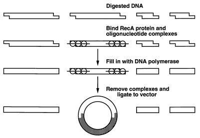
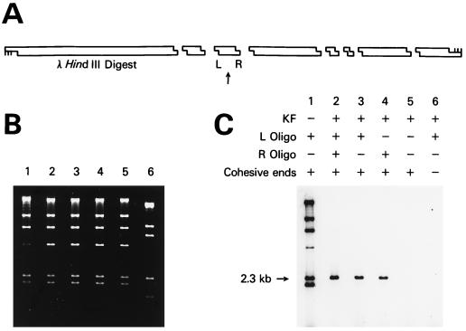
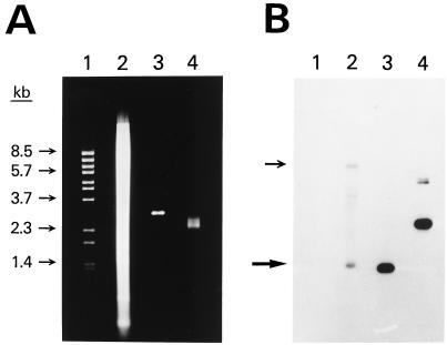
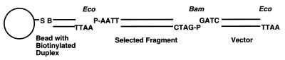
A method is described that allows the sequence-specific ligation of DNA. The method is based on the ability of RecA protein from Escherichia coli to selectively pair oligonucleotides to their homologous sequences at the ends of fragments of duplex DNA. These three-stranded complexes were protected from the action of DNA polymerase. When treated with DNA polymerase, unprotected duplex fragments were converted to fragments with blunt ends, whereas protected fragments retained their cohesive ends. By using conditions that greatly favored ligation of cohesive ends, a second DNA fragment could be selectively ligated to a previously protected fragment of DNA. When this second DNA was a vector, selected fragments were preferentially cloned. The method had sufficient power to be used for the isolation of single-copy genes directly from yeast or human genomic DNA, and potentially could allow the isolation of much longer fragments with greater fidelity than obtainable by using PCR.


Figures

Figure 1
Figure 1
Schematic of the strategy used for sequence-specific cloning of DNA. An explanation is given in the text.
Figure 2
Figure 2
Sequence-specific labeling of a fragment of λ DNA. (A) Schematic showing the position of the 2.3-kb λ DNA HindIII fragment labeled. The arrow points to the 2.3-kb fragment, and L and R show the positions of the two oligonucleotides used to direct sequence-specific ligation to a short radioactive duplex with a HindIII cohesive end. (B) Agarose gel stained with ethidium bromide showing the HindIII fragments of λ DNA in lanes 1–5 and a ScaI digest in lane 6. The two shortest HindIII fragments have run off the bottom of the gel. (C) Autoradiogram of the dried gel. Lane 1 shows λ DNA where KF was omitted and every fragment was labeled. The band at 4.4 kb is less intense because of ligation to the 23.1-kb band via the terminal λ cos sites. Lane 2 shows labeling of both ends of the 2.3-kb fragment. Lanes 3, 4, and 5 show the effect of omitting either one or both of the oligonucleotides. Lane 6 shows the results by using λ DNA fragments with blunt ends.
Figure 3
Figure 3
Sequence-specific cloning of a portion of the human int-2 gene. (A) Agarose gel stained with ethidium bromide. Lane 1 shows λ BstEII size standards. Lane 2 shows 20 μg of human genomic DNA that had been digested with EcoRI and BamHI. Lanes 3 and 4 show 0.25 μg of pooled plasmid DNA prepared by using the RAC procedure. The DNA in lane 3 has been digested with EcoRI and BamHI. (B) Southern blot of the gel in A. A 0.6-kb portion of the int-2 gene was used as the probe. Arrows show the positions of both of the alleles of the int-2 fragment. The amount of radioactivity in each band was quantitated by using a BAS 2000 Fujix Bio-Imaging Analyzer. Specifically, using the software of the instrument, each band was centered in a rectangle and the total amount of signal within the rectangle was determined. A rectangle was also placed just above each band to measure the background signal. All rectangles were the same size and shape. In lane 2, the lower allele band gave a reading of 1,420 with a background of 721. In lane 4, the two bands from the plasmid had a combined reading of 15,711 with a background of 1,076. After subtracting the background signal, the ratio of the plasmid signal in lane 4 to the lower allele signal in lane 2 was 20. Because 80 times more DNA was loaded in lane 2 than in lane 4, the total enrichment was 1,600.
Figure 4
Figure 4
Diagram of the construct made when selected fragment-vector molecules were separated from unligated vector. An explanation is given in the text. EcoRI and BamHI cohesive ends are shown. S, streptavidin; B, biotin; P, 5′ phosphate.

Similar articles

Cited by

References

    1. Ferrin L J, Camerini-Otero R D. Science. 1991;254:1494–1497. - PubMed
    1. Ferrin L J, Camerini-Otero R D. Nat Genet. 1994;6:379–383. - PubMed
    1. Ferrin L J. In: Genetic Engineering: Principles and Methods. Setlow J, editor. Vol. 17. New York: Plenum; 1995. pp. 21–30. and references therein. - PubMed
    1. Barton M C, Emerson B M. Genes Dev. 1994;8:2453–2465. - PubMed
    1. Boren J, Lee I, Callow M J, Rubin E M, Innerarity T L. Genome Res. 1996;6:1123–1130. - PubMed

MeSH terms

LinkOut - more resources