. 1998 Mar 3;95(5):2284-9.
doi: 10.1073/pnas.95.5.2284.

Stabilization and activation of p53 are regulated independently by different phosphorylation events

Affiliations

Stabilization and activation of p53 are regulated independently by different phosphorylation events

M V Chernov et al. Proc Natl Acad Sci U S A. .

Abstract

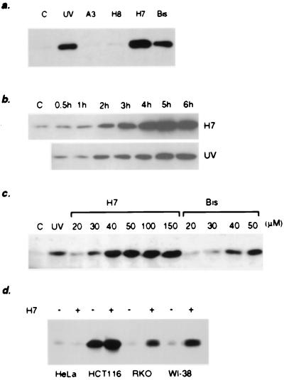
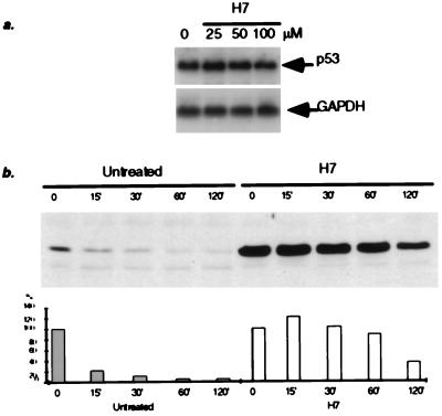
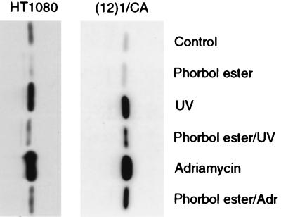
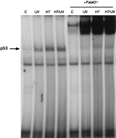
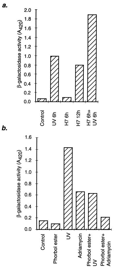
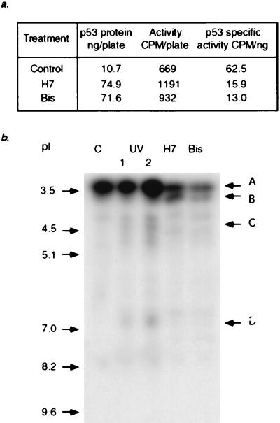
Treatment of mouse or human cells with the protein kinase C (PKC) inhibitors H7 or bisindolylmaleimide I induced an increase in the lifetime of p53, leading to its accumulation. In inhibitor-treated cells, p53 translocated to the nuclei and bound to DNA but was not competent to induce transcription. However, transactivation could be induced by subsequent DNA damage. Phorbol ester, a potent activator of PKC, significantly inhibited the accumulation of p53 after DNA damage. Therefore, constitutive PKC-dependent phosphorylation of p53 itself, or of a protein that interacts with p53, is required for the rapid degradation of p53 in untreated cells. Furthermore, an increase in the lifetime of p53 is not accompanied necessarily by its activation. Treatment with the PKC inhibitors decreased the overall level of p53 phosphorylation but led to the appearance of a phosphopeptide not seen in tryptic digests of p53 from untreated cells. Therefore, the lifetime and activities of p53 are likely to be regulated by distinct alterations of the phosphorylation pattern of p53, probably caused by the actions of different kinases.


Figures

Figure 1
Figure 1
The effect of kinase inhibitors on the level and activity of p53 in mouse and human cells. (a) The level of p53 in (12)1/CA cells treated for 6 hr with different kinase inhibitors. (b) Time course of p53 accumulation in mouse (12)1/CA cells treated with 50 μM H7 or irradiated with 25 J/m2 UV light. (c) The induction of p53 in mouse cells treated for 6 hr with different concentrations of H7 or Bis. (d) The induction of p53 by H7 in human cells.
Figure 2
Figure 2
Effect of H7 on the level of p53 mRNA and the stability of the p53 protein. (a) Analysis of p53 mRNA from human cells treated with different concentrations of H7. Total RNA was extracted 5 hr after treatment. (b) The effect of H7 on the lifetime of p53 in mouse cells. (12)1/CA cells were treated with H7 (50 μM) for 6 hr, followed by incubation in the presence of cycloheximide (CHX). The levels of p53 protein were analyzed at the times indicated. The amount of protein at zero time was scored as 100%.
Figure 3
Figure 3
The effect of phorbol ester on the basal and induced levels of p53 in mouse and human cells. (12)1/CA or HT1080 cells were treated with phorbol ester (80 ng/ml), UV light (25 J/m2), or Adriamycin (200 ng/ml) or were pretreated with phorbol ester for 6 hr followed by UV light or Adriamycin. Protein extracts were prepared 6 hr after the DNA-damaging treatment.
Figure 4
Figure 4
Nuclear accumulation of p53 in mouse cells treated with H7. Cells were irradiated with 25 J/m2 UV light or treated with H7 (50 μM). After 6 hr, the cells were fixed and probed with the p53-specific antibody PAb421 and with fluorescein-conjugated second antibody. Staining with 4′,6′-diamidino-2-phenylindole (DAPI) was used to reveal the nuclei.
Figure 5
Figure 5
DNA binding activity of p53 in cells treated with H7 or irradiated with UV light. p53-specific DNA binding activity in nuclear extracts was analyzed 5 hr after treatment. The last four lanes show the effect of the p53-specific antibody PAb421.
Figure 6
Figure 6
Effect of H7 or phorbol ester on p53-dependent expression of β-galactosidase in mouse cells, with or without DNA damage. (a) (12)1/CA cells were treated with 50 μM H7 or irradiated with UV light (25 J/m2), and β-galactosidase activity was measured at the times shown. Alternatively, the cells were treated with H7 for 6 hr and then irradiated with UV light before analysis 6 hr later. (b) (12)1/CA cells were treated with phorbol ester (80 ng/ml), UV light (25 J/m2), or Adriamycin (200 ng/ml) or pretreated with phorbol ester for 6 hr, followed by treatment with UV light or Adriamycin. β-galactosidase activity was analyzed 6 hr after the DNA damaging treatment.
Figure 7
Figure 7
Phosphorylation of p53 in mouse cells treated with H7 or Bis or irradiated with UV light. The treated cells were labeled for 5 hr with [32P]-orthophosphate, and p53 was immunoprecipitated with PAb421. (a) Specific activities of p53 from untreated or treated cells. (b) Tryptic peptide analysis of the labeled p53. The phosphopeptides were separated by isoelectric focusing. Equal amounts of radioactivity were loaded in each lane.

Similar articles

Cited by

References

    1. Yin Y, Tainsky M A, Bischoff F Z, Strong L C, Wahl G M. Cell. 1992;70:937–948. - PubMed
    1. Hollstein M, Sidransky D, Vogelstein B, Harris C C. Science. 1991;253:49–53. - PubMed
    1. Kastan M B, Onyekwere O, Sidransky D, Vogelstein B, Craig R W. Cancer Res. 1991;51:6304–6311. - PubMed
    1. Fritsche M, Haessler C, Brandner G. Oncogene. 1993;8:307–318. - PubMed
    1. Graeber T G, Peterson J F, Tsai M, Monica K, Fornace A, Jr, Giaccia A J. Mol Cell Biol. 1994;14:6264–6277. - PMC - PubMed

Publication types

MeSH terms