. 1998 Mar 3;95(5):2319-24.
doi: 10.1073/pnas.95.5.2319.

Hsp70 accumulation in chondrocytic cells exposed to high continuous hydrostatic pressure coincides with mRNA stabilization rather than transcriptional activation

Affiliations

Hsp70 accumulation in chondrocytic cells exposed to high continuous hydrostatic pressure coincides with mRNA stabilization rather than transcriptional activation

K Kaarniranta et al. Proc Natl Acad Sci U S A. .

Abstract

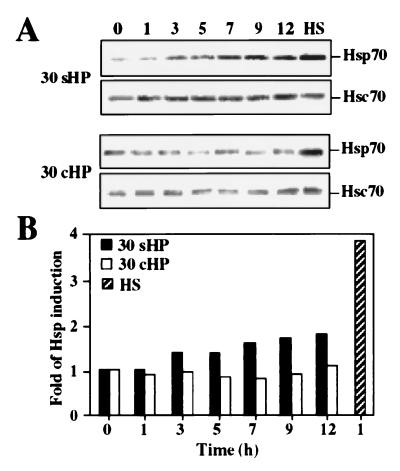
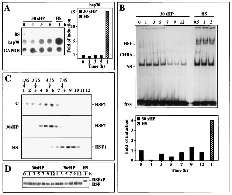
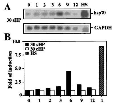
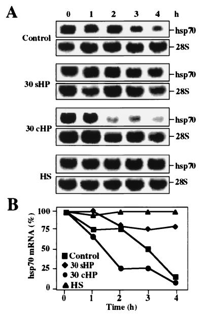
In response to various stress stimuli, heat shock genes are induced to express heat shock proteins (Hsps). Previous studies have revealed that expression of heat shock genes is regulated both at transcriptional and posttranscriptional level, and the rapid transcriptional induction of heat shock genes involves activation of the specific transcription factor, heat shock factor 1 (HSF1). Furthermore, the transcriptional induction can vary in intensity and kinetics in a signal- and cell-type-dependent manner. In this study, we demonstrate that mechanical loading in the form of hydrostatic pressure increases heat shock gene expression in human chondrocyte-like cells. The response to continuous high hydrostatic pressure was characterized by elevated mRNA and protein levels of Hsp70, without activation of HSF1 and transcriptional induction of hsp70 gene. The increased expression of Hsp70 was mediated through stabilization of hsp70 mRNA molecules. Interestingly, in contrast to static pressurization, cyclic hydrostatic loading did not result in the induction of heat shock genes. Our findings show that hsp70 gene expression is regulated posttranscriptionally without transcriptional induction in chondrocyte-like cells upon exposure to high continuous hydrostatic pressure. We suggest that the posttranscriptional regulation in the form of hsp70 mRNA stabilization provides an additional mode of heat shock gene regulation that is likely to be of significant importance in certain forms of stress.


Figures

Figure 1
Figure 1
(A) Western blot analysis with antibodies against Hsp70 and Hsc70. Samples were from whole cell extracts of nonstressed control cells (0 h), cells exposed to 30-MPa static (30 sHP) or cyclic (30 cHP) HP for 1–12 h, and cells exposed to heat shock (lane HS) at 43°C for 5 h. Experiments were repeated three times. (B) Analysis of Hsp70 protein accumulation was performed by chemiluminescence reaction, and protein levels were quantitated densitometrically.
Figure 2
Figure 2
Analysis of hsp70 transcription rate and HSF1 activation. (A) Transcription rates of hsp70 and GAPDH genes were analyzed by nuclear run-on assay. Equal number of nuclei were isolated from nonstressed control cells (0 h) and from cells exposed to 30-MPa HP (30 sHP) for 1–5 h or to heat shock (HS) at 42°C for 1 h. Band BS indicates the Bluescript plasmid that was used as a vector control, and GAPDH was used as a loading control. Quantitative analysis of the hsp70 transcription rate, relative to GAPDH, was performed by using a PhosphorImager (Molecular Dynamics). (B) Gel mobility shift assay of HSF1-binding activity to a 35-bp 32P-labeled oligonucleotide containing the HSE was performed with whole cell extracts isolated from nonstressed control cells (0 h) or cells exposed to 30-MPa static HP (30 sHP) for 1–12 h or to heat shock (lanes HS) at 43°C for 0.5, 1, and 2 h. Quantitative analysis of HSF1 DNA-binding activity, relative to levels of nonspecific HSE-binding activity (NS) was performed by using a PhosphorImager. CHBA, constitutive binding activity to HSE; Free, unbound labeled HSE oligonucleotide. (C) HSF1 in the whole cell extracts (500 μg of protein) was analyzed by 15–50% glycerol gradient sedimentation method, and the positions of HSF1 proteins were visualized by Western blot analysis. Cells were either untreated (blot C) or exposed to 30-MPa static HP (blot 30 sHP) for 1 h or to heat shock (blot HS) at 43°C for 1 h. The sedimentation positions of protein standards are indicated (cytochrome c, 1.9 S; BSA, 3.2 S; carbonic anhydrase, 4.3 S; alcohol dehydrogenase, 7.4 S). (D) HSF1 hyperphosphorylation analysis with antibody against HSF1 by Western blotting from control nonstressed cells (0 h) or cells exposed to 30-MPa static (30 sHP) or 30-MPa cyclic HP (30 cHP) for 1–12 h or to heat shock (lane HS) at 43°C for 1 h. HSF1+P indicates the position of the HSF1 after hyperphosphorylation.
Figure 3
Figure 3
Analysis of the steady-state level of hsp70 mRNA by Northern blot hybridization. (A) Samples were from control nonstressed cells (0 h) or cells exposed to 30-MPa static HP (30 sHP) up to 12 h or to heat shock (lane HS) at 43°C for 1 h. The RNA samples were hybridized with the 32P-labeled cDNA probes for hsp70 and GAPDH. The experiments were repeated three times. (B) Quantitative analysis of hsp70 mRNA levels, relative to GAPDH, was performed by using a PhosphorImager.
Figure 4
Figure 4
Analysis of hsp70 mRNA stability by Northern blot hybridization. (A) Control nontreated cells (blot C), cells exposed to 30-MPa static (30 sHP) or cyclic HP (30 cHP) for 3 h, and to heat shock (HS) at 43°C for 1 h were incubated in the presence of 5 μM actinomycin D for 0–4 h. The RNA samples were hybridized with hsp70 and 28S probe. The 28S rRNA served as a normalization control. (B) Quantitative analysis of hsp70 mRNA levels, relative to 28S rRNA, was performed by using a PhosphorImager.

Similar articles

Cited by

References

    1. Lindquist S, Craig E A. Annu Rev Genet. 1988;22:631–677. - PubMed
    1. Morimoto R I. Science. 1993;259:1409–1410. - PubMed
    1. Wu C. Annu Rev Cell Dev Biol. 1995;11:441–469. - PubMed
    1. Rabindran S K, Giorgi G, Clos J, Wu C. Proc Natl Acad Sci USA. 1991;88:6906–6910. - PMC - PubMed
    1. Sarge K D, Zimarino V, Holm K, Wu C, Morimoto R I. Genes Dev. 1991;5:1902–1911. - PubMed

Publication types

LinkOut - more resources