. 1998 Mar 3;95(5):2331-6.
doi: 10.1073/pnas.95.5.2331.

RNA-binding protein TIAR is essential for primordial germ cell development

Affiliations

RNA-binding protein TIAR is essential for primordial germ cell development

A R Beck et al. Proc Natl Acad Sci U S A. .

Abstract

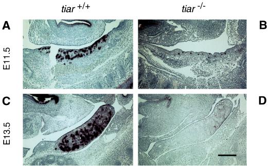
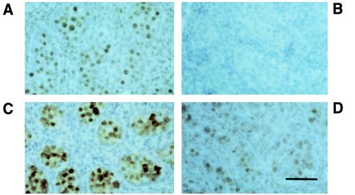
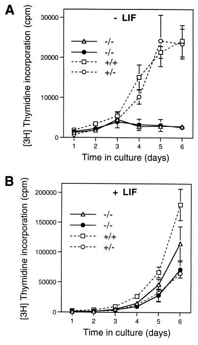
Primordial germ cells (PGCs) give rise to both eggs and sperm via complex maturational processes that require both cell migration and proliferation. However, little is known about the genes controlling gamete formation during the early stages of PGC development. Although several mutations are known to severely reduce the number of PGCs reaching and populating the genital ridges, the molecular identity of only two of these genes is known: the c-kit receptor protein tyrosine kinase and the c-kit ligand (the steel factor). Herein, we report that mutant mice lacking TIAR, an RNA recognition motif/ribonucleoprotein-type RNA-binding protein highly expressed in PGCs, fail to develop spermatogonia or oogonia. This developmental defect is a consequence of reduced survival of PGCs that migrate to the genital ridge around embryonic day 11.5 (E11.5). The numbers of PGCs populating the genital ridge in TIAR-deficient embryos are severely reduced compared to wild-type embryos by E11.5 and in the mutants PGCs are completely absent at E13.5. Furthermore, TIAR-deficient embryonic stem cells do not proliferate in the absence of exogenous leukemia inhibitory factor in an in vitro methylcellulose culture assay, supporting a role for TIAR in regulating cell proliferation. Because the development of PGCs relies on the action of several growth factors, these results are consistent with a role for TIAR in the expression of a survival factor or survival factor receptor that is essential for PGC development. TIAR-deficient mice thus provide a model system to study molecular mechanisms of PGC development and possibly the basis for some forms of idiopathic infertility.


Figures

Figure 1
Figure 1
Gene targeting at the tiar locus. (A) Schematic representation of the tiar gene exon–intron structure with restriction enzyme sites (11), the structure of the targeting vector, and the structure of the tiar locus after integration of the targeting vector. Correct gene targeting results in replacement of portions of intron 5 and exon 6 by the marker gene for positive selection (PGKneo). The hsv-TK (thymidine kinase) expression cassette was used for negative selection. The 3′ probe used for Southern blot analysis is shown as a stippled box. B, BamHI; H, HindIII; S, SalI; X, XbaI. (B) Protein immunoblot of total lysates of tiar−/− and tiar+/+ ES cells by using the anti-TIAR mAb 6E3 (11) confirming absence of TIAR protein in tiar−/− cells relative to wild-type cells. Use of an anti-TIAR mAb reactive with a different TIAR epitope (anti-3E6) (11) gave the same result (data not shown). (C) Southern blot analysis of DNA from offspring derived from heterozygous matings. Genomic DNA was digested with BamHI and analyzed by using the 3′ probe, yielding the >12-kb and 4.0-kb fragments expected for the wild-type allele and mutant allele, respectively. Southern blot analysis of ES cell DNA using a 5′ DNA probe and a neo DNA probe showed proper targeting and single insertion of the transfected vector (data not shown).
Figure 2
Figure 2
Reduced weight of tiar−/− mice during development. (A) Weights of tiar+/+ and tiar+/− embryos (open diamonds) or tiar−/− (solid diamonds) embryos are shown at different days of embryonic development. No significant weight difference was found between tiar+/+ and tiar+/− embryos. (B) Weights of tiar+/+ mice (solid line) and tiar−/− mice (dotted line) between 5 and 45 days postpartum. Each line represents the weight of an individual mouse.
Figure 3
Figure 3
Lack of germ cells in TIAR-deficient testes and ovaries. Hematoxylin and eosin-stained histological sections of tiar+/+ (A) and tiar−/− (B) adult testes at low magnification (60-fold), tiar+/+ (C) and tiar−/− (D) adult testes at high magnification (240-fold), tiar+/+ (E) and tiar−/− (F) adult ovaries at low magnification (60-fold), and tiar+/+ (G) and tiar−/− (H) adult ovaries at high magnification (240-fold). No significant difference was observed between tiar+/+ and tiar+/− adult gonads (data not shown). (Bar = 200 μm for low magnification and 50 μm for high magnification.)
Figure 4
Figure 4
TIAR expression is required for development of primordial germ cells. Staining for alkaline phosphatase activity (brown) of E11.5 tiar+/+ (A) and tiar−/− (B) gonads and E13.5 tiar+/+ (C) and tiar−/− (D) gonads. (Bar = 200 μm for paramedial sagittal sections.) The mucosal lining of the embryonic stomach lumen in the upper left corner of each panel also shows alkaline phosphatase activity. The number of alkaline phosphatase staining cells seen is representative of two tiar+/+ and nine tiar−/− E11.5 embryos. Sectioning of four entire tiar−/− E11.5 embryos and staining for alkaline phosphatase activity did not reveal any ectopic alkaline phosphatase staining cells (data not shown). The number of alkaline phosphatase staining cells in 3 tiar+/− E11.5 embryos was reduced slightly compared with tiar+/+ embryos.
Figure 5
Figure 5
Primordial germ cells express high levels of TIAR protein. Immunostaining of sections of E14.5 tiar+/+ gonads (A) and tiar−/− gonads (B) using the anti-TIAR 3E6 mAb (11). A neighboring section of the tiar+/+ gonad stained for GCNA1 (C), a PGC-specific antigen (24). (D) A section of a E12.5 tiar+/+ gonad stained with mAb 3E6. (Bar = 50 μm.) Staining with an Ig-type matched control mAb further confirmed the specificity of mAb 3E6 staining (data not shown).
Figure 6
Figure 6
tiar−/− ES cell proliferation defect. tiar−/− ES (solid lines) and control tiar+/+ or tiar+/− ES (dashed lines) cell lines were grown in media containing methylcellulose in the absence (A) or presence (B) of exogenous LIF. Cell growth was determined by using [3H]thymidine incorporation at the indicated times. Values represent mean values of triplicate samples with vertical bars indicating the SDs. Data are shown for two independent tiar−/− ES cell lines and control tiar+/+ and tiar+/− cell lines. Similar results were obtained in four additional experiments.

Similar articles

Cited by

References

    1. Hogan B, Beddington R, Costantini F, Lacy E. Manipulating the Mouse Embryo: A Laboratory Manual. Plainview, NY: Cold Spring Harbor Lab. Press; 1994.
    1. McLaren A. In: Organization of the Early Vertebrate Embryo. Zagris N, Duprat A M, Durston A J, editors. New York: Plenum; 1995. pp. 1–9.
    1. Lawson K A, Hage W J. CIBA Found Symp. 1994;182:68–84. - PubMed
    1. Ginsburg M, Snow M H L, McLaren A. Development (Cambridge, UK) 1990;110:521–528. - PubMed
    1. Eddy E M, Clark J M, Gong D, Fenderson B A. Gamete Res. 1981;4:333–362.

Publication types

MeSH terms