. 1998 Mar 3;95(5):2452-7.
doi: 10.1073/pnas.95.5.2452.

Human T cell lymphotropic virus type I Tax protein trans-activates interleukin 15 gene transcription through an NF-kappaB site

Affiliations

Human T cell lymphotropic virus type I Tax protein trans-activates interleukin 15 gene transcription through an NF-kappaB site

N Azimi et al. Proc Natl Acad Sci U S A. .

Abstract

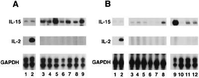
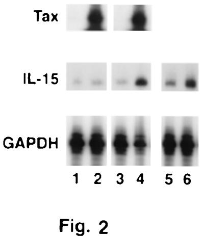
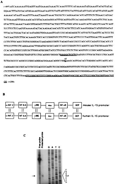
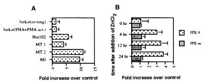
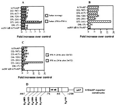
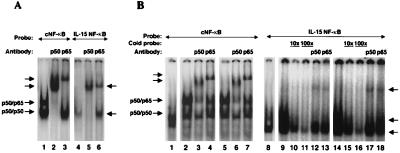
Interleukin 15 (IL-15) mRNA is expressed in a wide variety of tissue types. However, with the exception of some T cell lines, IL-15 transcript expression has not been described in T cells. Herein we demonstrate that IL-15 mRNA can be detected in freshly isolated normal T cells and T cell lines. Furthermore, its expression is 3- to 4-fold higher in human T cell lymphotropic virus type I (HTLV-I)-infected T cells. By using reporter constructs bearing the 5' regulatory region of the IL-15 gene, we observed a positive correlation between HTLV-I Tax protein expression and IL-15 promoter activity in HTLV-I-infected T cells. Additionally, by using a Jurkat T cell transfectant that expresses Tax under an inducible promoter, we demonstrated that the expression of IL-15 mRNA increased 3-fold as Tax was expressed, suggesting that the Tax protein activates IL-15 transcription. An NF-kappaB consensus sequence is located at the -75 and -65 region of the IL-15 5' regulatory region. Mutations in the NF-kappaB motif or deletion of this sequence abrogated the promoter activity in both HTLV-I-positive and Jurkat Tax-transfectant cells. These data represent evidence for trans-activation of the IL-15 gene by the HTLV-I Tax protein through an NF-kappaB motif and suggest a potential role for IL-15 in HTLV-I-associated diseases such as adult T cell leukemia and HTLV-I-associated myopathy/tropical spastic paraparesis.


Figures

Figure 1
Figure 1
IL-15 mRNA expression in various T cell populations, as examined by RPA. (A) IL-15 RNA from T cells from PB of a normal individual before (lane 1) and after (lane 2) activation (PHA at 1 μg/ml and PMA at 20 ng/ml for 4 h). Lanes 3–9 contain RNA from the T cells of seven HTLV-I-infected patients with ATL. (B) Comparison of the IL-15 mRNA level between HTLV-I-positive (lanes 9–12) and -negative T cell lines (lanes 1–8). Lanes 1 and 2 contain the Jurkat T cell line at a resting state (lane 1) and an activated state (lane 2) (PHA at 1 μg/ml and PMA at 20 ng/ml for 4 h). Lanes 3–8 include the HTLV-I-non-expressing T cell lines CEM, A301, 8402T, Kit225-K6, Kit225-IG3, and Hut78, respectively. Lanes 9–12 contain HTLV-I-expressing T cell lines in the order HuT102, MT-1, MT-2, and MJ.
Figure 2
Figure 2
Effect of Tax expression on IL-15 mRNA expression. Lanes 1 and 2 are JPX-m (nonfunctional mutant) before and 24 h after induction of the Tax protein by ZnCl2, respectively. Lanes 3 and 4 are JPX-9 before and 24 h after induction of the Tax protein by ZnCl2, respectively. Lanes 5 and 6 are the Jurkat T antigen T cell line before and 48 h after transient transfection with the Tax/pEF.Neo construct, respectively.
Figure 3
Figure 3
(A) Sequence of the human IL-15 5′ upstream region. The transcription initiation site and the NF-κB motif are in boldface type and underlined. Part of IL-15 exon 1, which is cloned in h15/sAP, is also underlined. (B) Schematic demonstration of some of the conserved motifs between mouse and human IL-15 promoters. (C) S1 nuclease protection assay to determine the transcription initiation site (+1 position). Total RNA from activated monocytes (by LPS and interferon γ) was used for the S1 nuclease assay, which was electrophoresed alongside the sequence ladder of the cloned IL-15 5′ upstream region to determine the +1 position.
Figure 4
Figure 4
Reporter assays of the IL-15 promoter construct h15/sAP in HTLV-I-infected T cell lines and Jurkat (resting and activated) cells, as well as two Jurkat Tax transfectants, JPX-m and JPX-9. Ten micrograms of the h15/sAP construct were cotransfected with 5 μg of the β-galactoside plasmid used to normalize the transformation efficiency. The average of the results of three reporter assays was used to generate each graph.
Figure 5
Figure 5
Reporter assay of the IL-15 promoter deletion constructs in different cell lines. (A) Jurkat resting and activated (PHA/PMA). (B) MJ an HTLV-I-infected T cell line. (C) JPX-9 and JPX-m cells 24 h after addition of ZnCl2. (D) Schematic representation of each of the deletion constructs generated in original cloned IL-15 5′ upstream region (h15/sAP). The reporter assays were performed as described in Fig. 4.
Figure 6
Figure 6
Electrophoretic mobility shift assay using the IL-15 NF-κB motif GGGCTGGGGCTCCTCGATGTC and Ig κB NF-κB as consensus NF-κB (cNF-κB) AGTTGAGGGGACTTTCCCAGGC (where the underlined sequences are NF-kB binding site). (A) Extracts obtained from COS cells transfected with p65 and p50 expression constructs were used with cNF-κB (lanes 1–3) or IL-15 NF-κB (lanes 4–6). The typical p50–p65 heterodimer and p50–p50 homodimer can be readily seen with cNF-κB (lane 1). The IL-15 NF-κB forms a complex that comigrates with the p50–p50 homodimer of the cNF-κB as shown by an arrow (lane 4). Supershifts of this complex gave rise to two bands almost at the same height, as indicated by an arrow (lanes 5 and 6). (B) Five micrograms of the cellular extract from resting Jurkat (lanes 1 and 8), PHA/PMA-activated Jurkat (lanes 2–4 and 9–13), or ZnCl2-treated (Tax expressing) JPX-9 (lanes 5–7 and 14–18) were used in each reaction. The formation of the p50–p65 heterodimer and p50–p50 homodimer can be seen with cNF-κB and activated or Tax-expressing Jurkat cells. The p50 and p65 supershifts are marked for the cNF-κB probe. By using the IL-15 NF-κB probe, two species can be detected, as shown by the arrows. The specificity of the binding of these complexes was examined by a competition assay using a 10 and 100 times excess of the unlabeled IL-15 NF-κB probe added to the activated or Tax-expressing Jurkat extract (lanes 10, 11, 15, and 16). The p50 and p65 supershifts of these complexes resulted in the two bands that migrated almost at the same level as indicated by an arrow (lanes 12–13 and 17–18).

Similar articles

Cited by

References

    1. Burton J D, Bamford R N, Peters C, Grant A J, Kurys G, Goldman C K, Brennan J, Roessler E, Waldmann T A. Proc Natl Acad Sci USA. 1994;91:4935–4939. - PMC - PubMed
    1. Armitage R J, Macduff B M, Eisenman J, Paxton R, Grabstein K H. J Immunol. 1995;154:483–490. - PubMed
    1. Grabstein K H, Eisenman J, Shanebeck K, Rauch C, Srinivasan S, Fung V, Beers C, Richardson J, Schoenborn M A, Giri J. Science. 1994;264:965–968. - PubMed
    1. Leclercq G, Debacker V, De Smedt M, Plum J. J Exp Med. 1996;184:325–336. - PMC - PubMed
    1. Ogasawara, K., Hida, S., Azimi, N., Tagaya, Y., Sato, T., Yokochi-Fukuda, T., Waldmann, T. A., Taniguchi, T. A. & Taki, S. (1998) Nature (London), in press. - PubMed

MeSH terms

Associated data

LinkOut - more resources