. 1998 Mar 3;95(5):2492-7.
doi: 10.1073/pnas.95.5.2492.

HFE gene knockout produces mouse model of hereditary hemochromatosis

Affiliations

HFE gene knockout produces mouse model of hereditary hemochromatosis

X Y Zhou et al. Proc Natl Acad Sci U S A. .

Abstract

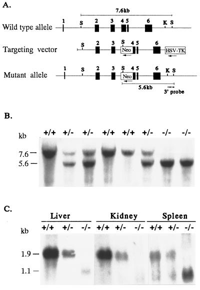
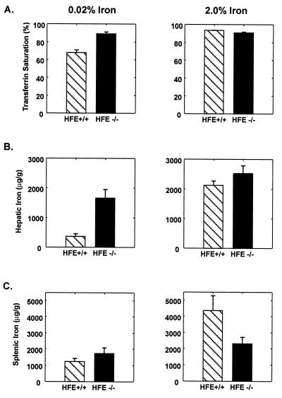
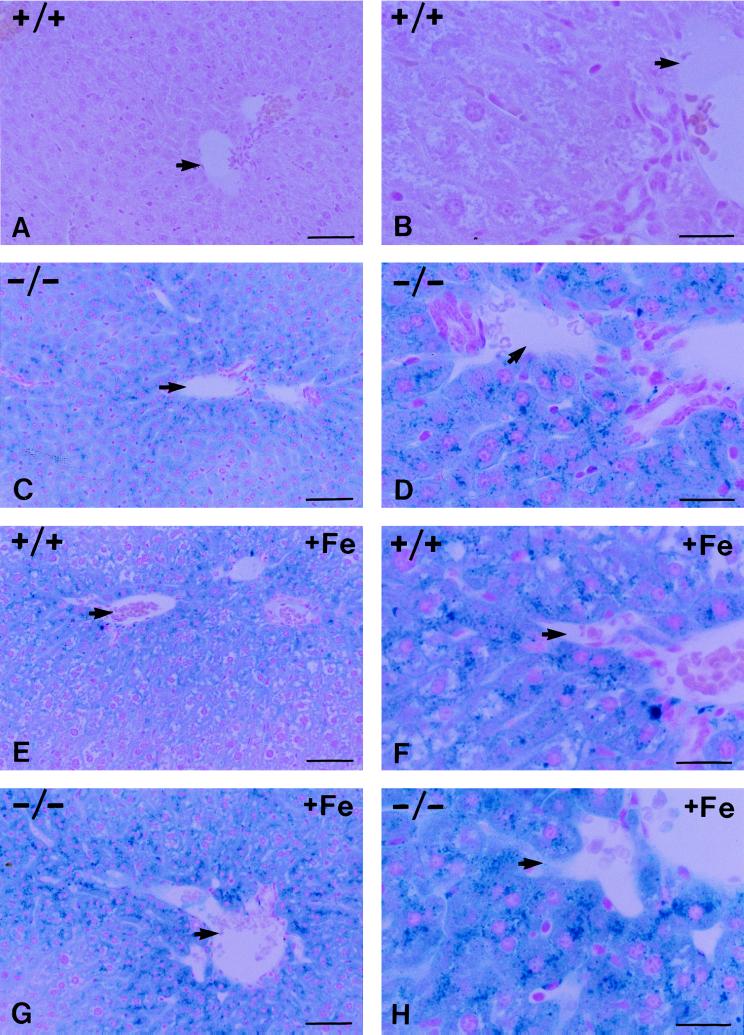
Hereditary hemochromatosis (HH) is a common autosomal recessive disease characterized by increased iron absorption and progressive iron storage that results in damage to major organs in the body. Recently, a candidate gene for HH called HFE encoding a major histocompatibility complex class I-like protein was identified by positional cloning. Nearly 90% of Caucasian HH patients have been found to be homozygous for the same mutation (C282Y) in the HFE gene. To test the hypothesis that the HFE gene is involved in regulation of iron homeostasis, we studied the effects of a targeted disruption of the murine homologue of the HFE gene. The HFE-deficient mice showed profound differences in parameters of iron homeostasis. Even on a standard diet, by 10 weeks of age, fasting transferrin saturation was significantly elevated compared with normal littermates (96 +/- 5% vs. 77 +/- 3%, P < 0.007), and hepatic iron concentration was 8-fold higher than that of wild-type littermates (2,071 +/- 450 vs. 255 +/- 23 microg/g dry wt, P < 0.002). Stainable hepatic iron in the HFE mutant mice was predominantly in hepatocytes in a periportal distribution. Iron concentrations in spleen, heart, and kidney were not significantly different. Erythroid parameters were normal, indicating that the anemia did not contribute to the increased iron storage. This study shows that the HFE protein is involved in the regulation of iron homeostasis and that mutations in this gene are responsible for HH. The knockout mouse model of HH will facilitate investigation into the pathogenesis of increased iron accumulation in HH and provide opportunities to evaluate therapeutic strategies for prevention or correction of iron overload.


Figures

Figure 1
Figure 1
Targeted disruption of the HFE gene. (A) Structure of the HFE gene (Top), the targeting construct (Middle), and the predicted structure of the disrupted HFE gene after homologous recombination (Bottom). Only the relevant restriction sites are shown: S, SacI site; K, KpnI site. The numbered solid boxes represent exons. Neo and HSV-TK (herpes simplex virus thymidine kinase) refer to the positive and negative selective markers, respectively. The position of the 3′ external probe is indicated. The horizontal lines show the positions of the 7.6-kb and 5.6-kb restriction fragments diagnostic for wild-type and properly targeted alleles, respectively. (B) Southern blot analysis of SacI-digested genomic DNA of eight mice from one litter resulting from a cross between two HFE+/− mice. The blot was hybridized with the 3′ KpnI–SacI probe. The wild-type and mutant alleles are indicated by 7.6- and 5.6-kb SacI fragments, respectively. (C) Northern blot analysis of liver, kidney, and spleen HFE mRNA from HFE+/+, HFE+/−, and HFE−/− mice. Twenty micrograms of total RNA from liver, kidney, or spleen RNA were analyzed by Northern blotting with mouse HFE 32P-labeled riboprobe. The 1.9-kb mRNA transcript present in +/+ mice was reduced in amounts in HFE+/− tissues, and absent in tissues from HFE−/− mutant mice.
Figure 2
Figure 2
Increased transferrin saturation and hepatic iron content in HFE−/− mice. Values represent means ± SE. (Left) The transferrin saturation (A), hepatic iron content (B), and splenic iron content (C) in HFE+/+ and HFE−/− mice fed 0.02% iron diets from weaning until sacrifice at 10 weeks of age. Both the transferrin saturation and hepatic iron content were increased in the HFE−/− mice (P < 0.001 and P < 0.01, respectively). (Right) The transferrin saturation (A), hepatic iron content (B), and splenic iron content (C) in HFE+/+ and HFE−/− mice fed control diets supplemented with 2% wt/wt carbonyl iron for the 14 days before sacrifice at age 10 weeks. HFE+/+ mice showed an increase in transferrin saturation from 68 ± 3 to 94 ± 0.2%, a 6-fold increase in hepatic iron (370 ± 87 to 2,124 ± 149 μg/g dry wt, P < 0.001), and a 3.5-fold increase in splenic iron (1,244 ± 191 to 4,361 ± 905 μg/g dry wt, P < 0.004) in response to dietary iron loading. The HFE−/− mice had no increase in the already high transferrin saturation with dietary iron loading. However, their 1.5-fold increase in hepatic iron (from 1,660 ± 286 to 2,523 ± 262 μg/g dry wt) was significant (P < 0.03). Splenic iron was not significantly increased in response to iron loading in −/− mice (P = 0.15).
Figure 3
Figure 3
Perls’ Prussian blue staining of liver sections from HFE+/+ and HFE−/− mice fed control diet (AD) or control diet supplemented with 2% (wt/wt) carbonyl iron (EH). Shown are low-power views (A, C, E, and G) and high-power views (B, D, F, and H) of sections. A and B show the absence of stainable iron in the +/− mouse liver fed the control diet. C and D show prominent stainable iron in hepatocytes with periportal predominance in liver from HFE−/− mice fed the control diet. E and F show iron accumulation in HFE+/+ mouse liver in response to iron loading. G and H show the stainable iron in the HFE−/− mice after 2 weeks of feeding with the iron-supplemented diet. The arrows indicate the location of branches of the portal vein. (Bars: A, C, E, and G = 50 μm in the low-power views; B, D, F, and H = 20 μm in the high-power views.)

Comment in

  • Targeted disruption of the HFE gene.
    Beutler E. Beutler E. Proc Natl Acad Sci U S A. 1998 Mar 3;95(5):2033-4. doi: 10.1073/pnas.95.5.2033. Proc Natl Acad Sci U S A. 1998. PMID: 9482831 Free PMC article. Review. No abstract available.

References

    1. Cartwright G E, Edwards C Q, Kravitz K, Skolnick M, Amos D B, Johnson A, Buskjaer L. N Engl J Med. 1979;301:175–179. - PubMed
    1. Borwein S T, Ghent C N, Flanagan P R, Chamberlain M J, Valberg L S. Clin Invest Med. 1983;6:171–179. - PubMed
    1. Edwards C Q, Griffen L M, Goldgar D, Drummond C, Skolnick M H, Kushner J P. N Engl J Med. 1988;318:1355–1362. - PubMed
    1. Bacon B R, Tavill A S. In: Hepatology: A Textbook of Liver Disease. Zakim D, Boyer T D, editors. Philadelphia: Saunders; 1996. pp. 1439–1472.
    1. Barton J C, Bertoli L F. Nat Med. 1996;2:394–395. - PubMed

Publication types

MeSH terms