. 1998 Mar 3;95(5):2503-8.
doi: 10.1073/pnas.95.5.2503.

Mice with type 2 and 3 Gaucher disease point mutations generated by a single insertion mutagenesis procedure

Affiliations

Mice with type 2 and 3 Gaucher disease point mutations generated by a single insertion mutagenesis procedure

Y Liu et al. Proc Natl Acad Sci U S A. .

Abstract

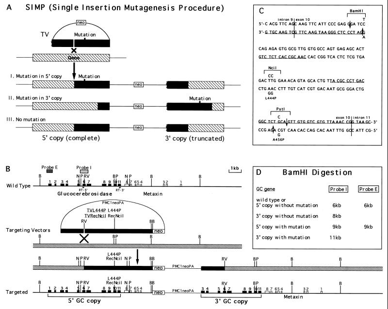
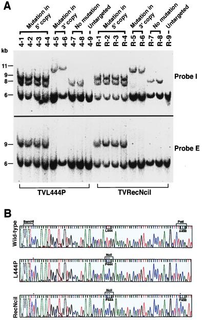
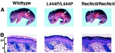
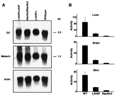
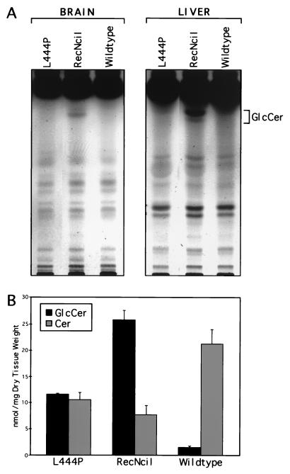
Gaucher disease is caused by mutations in the gene encoding the lysosomal enzyme glucocerebrosidase (GC). Three clinical types of Gaucher disease have been defined according to the presence (type 2 and 3) or absence (type 1) of central nervous system disease and severity of clinical manifestations. The clinical course of the disease correlates with the mutation carried by the GC gene. To produce mice with point mutations that correspond to the clinical types of Gaucher disease, we have devised a highly efficient one-step mutagenesis method-the single insertion mutagenesis procedure (SIMP)-to introduce human disease mutations into the mouse GC gene. By using SIMP, mice were generated carrying either the very severe RecNciI mutation that can cause type 2 disease or the less severe L444P mutation associated with type 3 disease. Mice homozygous for the RecNciI mutation had little GC enzyme activity and accumulated glucosylceramide in brain and liver. In contrast, the mice homozygous for the L444P mutation had higher levels of GC activity and no detectable accumulation of glucosylceramide in brain and liver. Surprisingly, both point mutation mice died within 48 hr of birth, apparently of a compromised epidermal permeability barrier caused by defective glucosylceramide metabolism in the epidermis.


Figures

Figure 1
Figure 1
Introduction of L444P and RecNciI human Gaucher disease mutations by the SIMP strategy. (A) A general scheme of the SIMP strategy. The ragged end of the gene in the targeting vector represents a truncation, filled bars represent the homology in the targeting vectors, hatched bars represent the endogenous allele. (B) The specific SIMP scheme for the introduction of the L444P and RecNciI mutations into the GC locus. The wild-type locus is shown on the top and the correctly targeted locus is shown on the bottom. The SIMP recombination step is shown in the middle. The filled boxes represent the exons of the GC gene, open boxes represent the exons of the nearby metaxin gene, stippled boxes represent probes used in the Southern analysis (probe I is a 0.7-kb KpnI fragment covering exon 6 and probe E is a 0.4-kb EcoRI fragment covering exon 1), filled bars represent the homology in the targeting vectors, hatched bars represent the endogenous allele. RT-5′, 5′ primer used in the reverse transcription (RT)-PCR primer (5′-CACGAATTCACATCACCCACTTGGCTCAAG-3′); RT-3′, 3′ primer used in the RT-PCR (5′-GTCGAATTCATGTCCATGCTAAGCCCAGGT-3′); B, BamHI; N, NciI; P, PstI; RV, EcoRV; Probe E, external probe; Probe I, internal probe. (C) The DNA sequence of the mouse GC exon 10 region. The vertical lines are exon/intron borders. The underlined sequences are oligonucleotides used in the RecA-assisted restriction endonuclease cleavage experiment. The mutations are indicated above and below the wild-type sequences. The mutated sequences between the two downward arrows were used as top strand of the mutation inserts. The mutated sequences between the two upward arrows were used as bottom strand of the mutation inserts. Both targeting vectors contained the change that eliminated the BamHI site and the L444P mutation. In addition, the RecNciI vector contain the L456P change. (D) The expected sizes of BamHI restriction fragments of wild-type and targeted GC genes detected by the I (internal) and E (external) probes.
Figure 2
Figure 2
Analysis of three different homologous insertion events by the two targeting vectors: TVL444P and TVRecNciI. (A) ES cell DNAs were digested with BamHI, and the filter was probed with probe I (Upper) and probe E (Lower), respectively. Clones 4-1 → 4-9, ES cell clones from TVL444P targeting experiment; Clones R-1 → R-9, ES cell clones from TVRecNciI targeting experiment. (B) Sequence analysis of the mouse GC transcripts from ES cells. cDNA clones, obtained by reverse transcription–PCR, of wild-type and targeted alleles were sequenced with an automated DNA sequencer (Applied Biosystems model 373A). Only mutated amino acids are labeled. Nucleotide mutations are indicated by black dots.
Figure 3
Figure 3
Phenotypes of RecNciI and L444P mice after birth. (A) Photographs of the mice within 12 hr after birth. The arrow denotes the presence of milk in the stomach. Note the smaller size and wrinkled skin of the RecNciI/RecNciI mouse. The skin of the L444P/L444P mouse is less severely wrinkled. The bracket indicates the sites where skin samples were taken for the photomicrographs in B. (B) Section of epidermis (×150). Stratum corneum (SC) of RecNciI/RecNciI and L444P/L444P mice are compacted and less eosinophillic compared with the loosely packed eosinophillic SC in the wild-type mouse. No significant differences are noted in the three other layers of the epidermis (EP) (stratum basale, stratum spinosum, and stratum granulosum) or the dermis (D).
Figure 4
Figure 4
Expression of GC mRNA and enzyme in RecNciI and L444P mice. (A) A Northern blot with 5 μg of brain poly(A)+ RNA from a RecNciI homozygote mouse, a L444P homozygote mouse, a L444P heterozygote mouse, and a wild-type mouse. The blot first was probed with a mouse GC cDNA. After stripping, it was reprobed with a mouse metaxin cDNA. Finally, it was stripped once more and probed with a human actin cDNA. (B) Acid β-glucosidase activity was determined in extracts of liver, brain, and skin from homozygote L444P and RecNciI mice. Tissues from three mice of each genotype were assayed. The assays were repeated three times. The activity (±SD) is expressed as nmol substrate cleaved/hr per mg of protein.
Figure 5
Figure 5
Sphingolipid accumulation in tissues from RecNciI and L444P mice. (A) Lipid profiles from brain and liver. Each lane represents a portion of the material derived from a single mouse (L444P, RecNciI, or wild type). The position of the glucosylceramide standard is indicated. (B) The levels of total ceramides (Cer) and glucosylceramides (GlcCer) in epidermis from RecNciI and L444P mice.

References

    1. Beutler E, Grabowski G A. In: The Metabolic and Molecular Basis of Inherited Disease. Scriver C R, Beaudet A L, Sly W S, Valle D, editors. New York: McGraw-Hill; 1995. pp. 2641–2670.
    1. Balicki D, Beutler E. Medicine (Baltimore) 1995;74:305–323. - PubMed
    1. Horowitz M, Zimran A. Hum Mutat. 1994;3:1–11. - PubMed
    1. Strasberg P M, Skomorowski M A, Warren I B, Hilson W L, Callahan J W, Clarke J T. Biochem Med Metab Biol. 1994;53:16–21. - PubMed
    1. Sidransky E, Tayebi N, Stubblefield B K, Eliason W, Klineburgess A, Pizzolato G P, Cox J N, Porta J, Bottani A, DeLozier-Blanchet C D. J Med Genet. 1996;33:132–136. - PMC - PubMed

Publication types

MeSH terms