. 1998 Mar 3;95(5):2515-9.
doi: 10.1073/pnas.95.5.2515.

Modulation of GTPase activity of G proteins by fluid shear stress and phospholipid composition

Affiliations

Modulation of GTPase activity of G proteins by fluid shear stress and phospholipid composition

S Gudi et al. Proc Natl Acad Sci U S A. .

Abstract

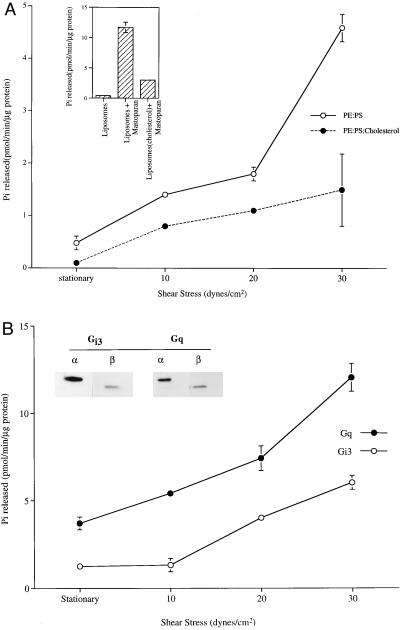
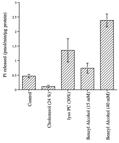
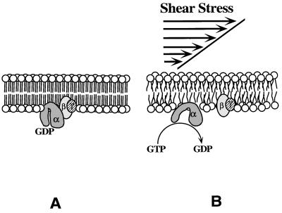
Mechanical forces arising from strain, pressure, and fluid shear stress are sensed by cells through an unidentified mechanoreceptor(s) coupled to intracellular signaling pathways. In vascular endothelial cells, fluid shear stress is transduced via pathway(s) involving heterotrimeric guanine nucleotide-binding proteins (G proteins) by molecular mechanisms that are unknown. In the present study, we investigated the activation of purified G proteins reconstituted into phospholipid vesicles. Vesicles containing G proteins were loaded with [gamma-32P]GTP and subjected to physiological levels of fluid shear stress in a cone-and-plate viscometer. Steady-state GTP hydrolysis was measured as an index of G protein function. Shear stress (0-30 dynes/cm2) activated G proteins in dose-dependent manner (0.48-4.6 pmol/min per microg of protein). Liposomes containing lysophosphatidylcholine (30 mol %) or treated with benzyl alcohol (40 mM), conditions that increase bilayer fluidity, exhibited 3- to 5-fold enhancement of basal GTPase activity. Conversely, incorporation of cholesterol (24 mol %) into liposomes reduced the activation of G proteins by shear. These results demonstrate the ability of the phospholipid bilayer to mediate the shear stress-induced activation of membrane-bound G proteins in the absence of protein receptors and that bilayer physical properties modulate this response.


Figures

Figure 1
Figure 1
Shear stress-stimulated GTPase activity of reconstituted G proteins. (A) G protein reconstituted liposomes loaded with [γ-32P]GTP were passed through Sephadex G-50 to remove external GTP. GTPase was stimulated when vesicles were subjected to shear stress (0–30 dynes/cm2) in a cone-and-plate viscometer (37°C, 1 min). This activity was attenuated by incorporation of cholesterol (24 mol %). (Inset) Mastoparan (900 μM) incubated with liposomes for 1 min (37°C) stimulated GTPase activity. (B) Affinity-purified Gαq and Gαi3, reconstituted into liposomes along with their respective βγ subunits (immunoblots for α and β are shown in Inset) were stimulated by shear stress. The values presented here are a mean ± SD of three measurements.
Figure 2
Figure 2
Effect of membrane fluidity modulators on G protein GTPase activity. The histogram shows basal GTPase activity (37°C, 1 min) of G proteins reconstituted with control (PE/PS; 6:4, mol/mol), cholesterol (PE/PS/cholesterol; 4.5:3:2.5, mol/mol), and LPC (PE/PS/LPC; 4.2:2.8:3, mol/mol). Benzyl alcohol was added to control liposomes (PE/PS, 6:4). The values presented here are mean ± SD of three measurements.
Figure 3
Figure 3
Brief proteolytic digestion of peripheral G proteins did not affect shear stress-stimulated GTPase activity in reconstituted G proteins. Proteolytic digestion was assessed by GTP[γ-S] binding efficiency (Inset). Integrity of inner leaflet G proteins in trypsinized vesicles was monitored by disrupting with Triton X-100, to make G proteins available for GTP[γ-S] binding.
Figure 4
Figure 4
Schematic representation of protein receptor-independent activation of G proteins by fluid shear stress. Increase in the rotational and translational mobility in the lipid bilayer and the accompanying decrease in microviscosity activate membrane-bound G proteins by facilitating exchange of GDP for GTP. The intrinsic GTPase activity of the α subunit hydrolyzes GTP to GDP, releasing inorganic phosphate (Pi), and α-GDP recombines with βγ, ending the activation cycle.

References

    1. Davies P F. Physiol Rev. 1995;75:519–560. - PMC - PubMed
    1. Olesen S P, Clapham D E, Davies P F. Nature (London) 1988;331:168–170. - PubMed
    1. Shepherd G M. Cell. 1991;67:845–851. - PubMed
    1. Gudi S, Clark C B, Frangos J A. Circ Res. 1996;79:834–839. - PubMed
    1. Berthiaume F, Frangos J A. Biochim Biophys Acta. 1994;1191:209–218. - PubMed

Publication types

MeSH terms

LinkOut - more resources