

doi: 10.1073/pnas.95.5.2603.
Bcl-xL protects adult septal cholinergic neurons from axotomized cell death
Affiliations
- PMID: 9482933
- PMCID: PMC19429
- DOI: 10.1073/pnas.95.5.2603
Item in Clipboard
Bcl-xL protects adult septal cholinergic neurons from axotomized cell death
Proc Natl Acad Sci U S A.
.
Abstract
Bcl-xL suppresses apoptotic cell death induced by diverse stimuli in cell lines in vitro. To examine the mechanism by which axotomized cholinergic neurons die in vivo, lentiviral vectors expressing Bcl-xL, human nerve growth factor (hNGF), or green fluorescent protein were injected into the septum 3 weeks before transection of the fimbria fornix. Three weeks after transection, Bcl-xL- and hNGF-injected animals showed significantly higher numbers of spared cholinergic neurons compared with control (green fluorescent protein) injected animals. These results provide evidence that adult axotomized cholinergic neurons die of apoptotic death that can be prevented by local delivery of hNGF or intracellular delivery of Bcl-xL.
Figures

Immunocytochemistry of PC12 cells survival and differentiation. (A) PC12 cells died when they were differentiated with hNGF and subsequently deprived of serum and hNGF. When infected with a lentiviral vector encoding Bcl-xL, PC12 cells differentiated with hNGF survived for an additional 3 weeks (B) (stained blue for tyrosine hydroxylase). PC12 cells not differentiated with hNGF and deprived of serum were dead after 3 days (C). However, PC12 cells infected with a vector containing Bcl-xL expressed Bcl-xL (green) and tyrosine hydroxylase (blue) and survived for 7 days (D).
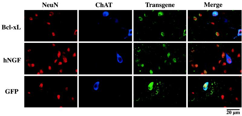
Immnohistochemical staining of coronal sections through the septal area. Confocal microscopic images show in vivo transduction of adult rat septal neurons expressing neuronal markers (NeuN; red), cholinergic markers (ChAT; blue), and the hNGF or Bcl-xL transgene (hNGF/Bcl-xL; green). The images obtained from each individual staining and from the merged images are shown. Representative fields of the area surrounding the injections site are shown with several cells triple labeled for hNGF, NeuN, and ChAT, as well as for Bcl-xL, NeuN, and ChAT.
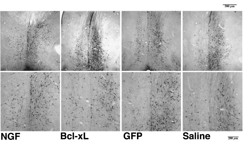
Coronal sections (50 μm) through the medial septum stained for ChAT immunoreactivity 6 weeks after hNGF, Bcl-xL, or GFP vector injection or saline control injection and 3 weeks after unilateral aspirative lesion of the fimbria fornix, ipsilateral to the injection. The medial septum was defined as the area lying above a line between the midportion of the anterior commissure, beneath the corpus callosum, and laterally limited by the ventricles. ChAT-positive cells displaying a round cell body and at least one process were counted for the lesioned and the unlesioned site of the septum.
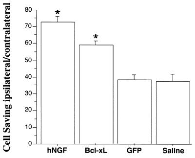
Percentage of ipsilateral/contralateral septal neurons quantified after intraseptal vector injections and lesion of the fimbria fornix. The percentage of ChAT-immunoreactive cells (mean ± SEM) are shown for hNGF-, Bcl-xL-, and GFP-expressing vector and saline control. hNGF and Bcl-xL viral vector-injected animals presented significantly higher numbers of surviving cholinergic septal neurons compared with GFP viral vector or saline-injected control animals (∗, P < 0.05, ANOVA, Fischer post hoc test).
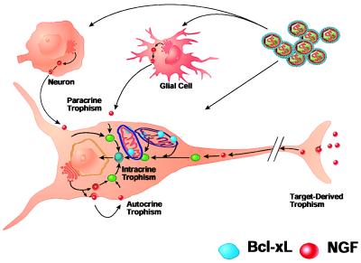
In vivo model for the role of hNGF and Bcl-xL in neuronal survival. Lentiviral transfer and expression of hNGF and Bcl-xL by lentiviral vectors is obtained in target and adjacent glial and neuronal cells. Bcl-xL stably expressed is localized within the target cell in mitochondrial and nuclear membranes; however, it is not secreted. Only infected cholinergic neurons can survive axotomy-induced cell death and trophic factor withdrawal by interfering with the cell death cascade by overexpression of Bcl-xL. Surrounding cells do not contribute to cell survival and, therefore, cell saving is less than that obtained with overexpression of hNGF. hNGF is expressed by the target neuron and adjacent glial and neuronal cells as a secreted protein; the autocrine and paracrine effects on the target cell allow for a higher number of cells saved by hNGF.
References
Publication types
MeSH terms
Substances
Grants and funding
LinkOut - more resources
Full Text Sources
Other Literature Sources
Research Materials
