. 1997 Jul;56(7):414-20.
doi: 10.1136/ard.56.7.414.

Human arthroplasty derived macrophages differentiate into osteoclastic bone resorbing cells

Affiliations

Human arthroplasty derived macrophages differentiate into osteoclastic bone resorbing cells

A Sabokbar et al. Ann Rheum Dis. 1997 Jul.

Abstract

Objective: In aseptic loosening, a heavy macrophage response to biomaterial wear particles is commonly found in arthroplasty tissues. The aim of this study was to discover if these cells contribute to the bone resorption of aseptic loosening by differentiating into osteoclasts.

Methods: Macrophages were isolated from the pseudocapsule and pseudomembrane of loose cemented and uncemented hip arthroplasties at the time of revision surgery and then co-cultured on glass coverslips and dentine slices with UMR 106 rat osteoblast-like cells, both in the presence and absence of 1,25 dihydroxyvitamin D3 [1,25(OH)2D3]. Macrophages isolated from the synovial membrane of patients with osteoarthritis (OA) undergoing hip replacements were similarly studied as a control group.

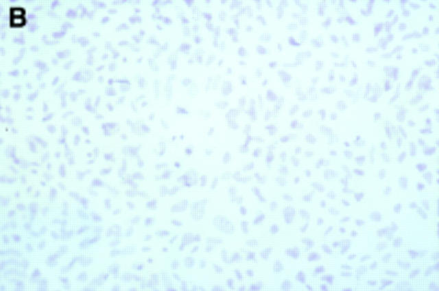
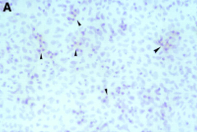
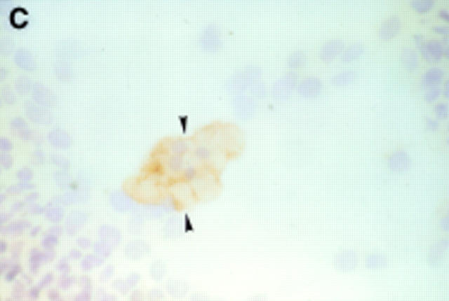
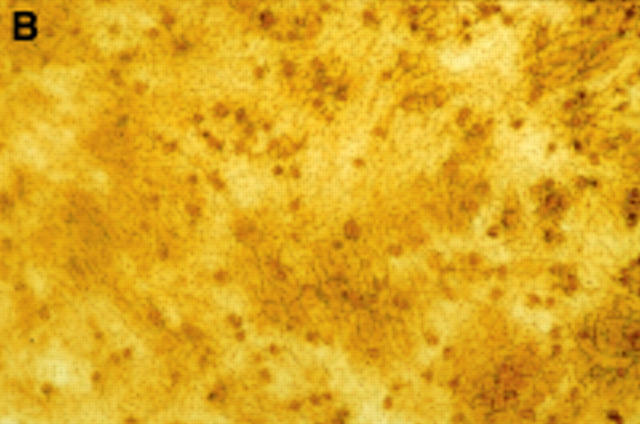
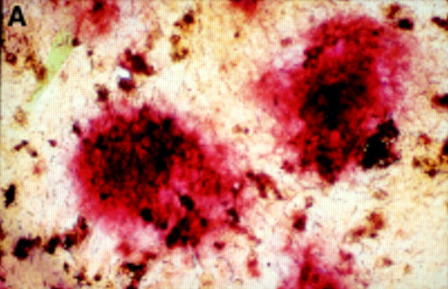
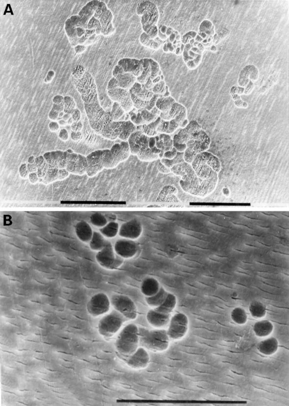
Results: After 24 hours incubation, most cells isolated from the above periprosthetic tissues strongly expressed macrophage (CD11b, CD14) but not osteoclast markers. However, after 14 days incubation, numerous multinucleated cells showing the phenotypic features of osteoclasts (that is, positive for tartrate resistant acid phosphatase, the vitronectin receptor, and capable of extensive lacunar resorption) formed in co-cultures of arthroplasty derived macrophages and UMR 106 cells, in the presence of 1,25(OH)2D3. The addition of an antibody to macrophage colony stimulating factor (M-CSF) considerably reduced macrophage-osteoclast differentiation and hence the lacunar resorption seen in these co-cultures. In contrast, OA synovial macrophage/UMR 106 co-cultures showed little or no evidence of macrophage-osteoclast differentiation and this was only seen when human M-CSF was added to the co-cultures.

Conclusion: This is the first report showing that human macrophages isolated directly from periprosthetic tissues surrounding loosened implants can differentiate into multinucleated cells showing all the functional and cytochemical characteristics of osteoclasts. In contrast with other macrophage populations, exogenous M-CSF is not required for this to occur. In the context of the heavy macrophage response to wear particles in periprosthetic tissues macrophage-osteoclast differentiation may represent an important cellular mechanism whereby osteolysis is effected in aseptic loosening.


Figures

Figure 1
Figure 1
Twenty four hour culture of arthroplasty derived macrophages incubated with UMR 106 cells (in the presence of 1,25(OH)2D3) stained by an indirect immunoperoxidase technique with monoclonal antibodies.(A) GRS 1 (anti-CD14) and (B) 23C6 (anti- VNR), showing reaction for CD14 (arrows) but not VNR on isolated cells. This indicates that these cells express a monocyte/macrophage but not osteoclast immunophenotype. (Original magnification × 200).
Figure 1
Figure 1
Twenty four hour culture of arthroplasty derived macrophages incubated with UMR 106 cells (in the presence of 1,25(OH)2D3) stained by an indirect immunoperoxidase technique with monoclonal antibodies.(A) GRS 1 (anti-CD14) and (B) 23C6 (anti- VNR), showing reaction for CD14 (arrows) but not VNR on isolated cells. This indicates that these cells express a monocyte/macrophage but not osteoclast immunophenotype. (Original magnification × 200).
Figure 2
Figure 2
Seven day culture of arthroplasty derived macrophages incubated with UMR 106 cells showing: (A) clusters of TRAP positive cells in the presence of 1,25(OH)2D3. (Original magnification × 200). (B) negative control for the TRAP staining (that is, in the absence of 1,25(OH)2D3. (Original magnification × 200). (C) A VNR positive multinucleated cell (arrow heads). (Original magnification × 400).
Figure 2
Figure 2
Seven day culture of arthroplasty derived macrophages incubated with UMR 106 cells showing: (A) clusters of TRAP positive cells in the presence of 1,25(OH)2D3. (Original magnification × 200). (B) negative control for the TRAP staining (that is, in the absence of 1,25(OH)2D3. (Original magnification × 200). (C) A VNR positive multinucleated cell (arrow heads). (Original magnification × 400).
Figure 3
Figure 3
(A) Fourteen day culture of arthroplasty derived macrophages incubated with UMR 106 cells on dentine slices. The cells have been removed to reveal evidence of lacunar resorption with the formation of numerous well defined resorption pits. (Black bar = 100 µm). (B) A high power view of individual resorption pits. (Black bar = 100 µm).
Figure 4
Figure 4
Effect of neutralising anti-M-CSF antibody on the mean number of lacunar pits formed on dentine slices from co-cultures of arthroplasty derived macrophages from case numbers 6 and 7. Results are expressed as mean (SD). Levels of significance using Student's paired t test: (*); p=0.024 and (***); p=0.00092.
Figure 5
Figure 5
Proposed cellular and humoral mechanism whereby human wear particle induced foreign body macrophages in the periprosthetic tissues differentiate into osteoclastic bone resorbing cells.

References

    1. Ann Rheum Dis. 1996 Nov;55(11):816-22 - PubMed
    1. Endocrinology. 1996 Sep;137(9):4058-60 - PubMed
    1. Calcif Tissue Int. 1982 May;34(3):285-90 - PubMed
    1. Curr Top Pathol. 1982;71:73-101 - PubMed
    1. Br Dent J. 1984 Mar 24;156(6):216-20 - PubMed

Publication types