

Review
doi: 10.1136/bjo.81.10.818.
The aging human lens: structure, growth, and physiological behaviour
Affiliations
- PMID: 9486018
- PMCID: PMC1722031
- DOI: 10.1136/bjo.81.10.818
Item in Clipboard
Review
The aging human lens: structure, growth, and physiological behaviour
Br J Ophthalmol.
1997 Oct.
No abstract available
Figures
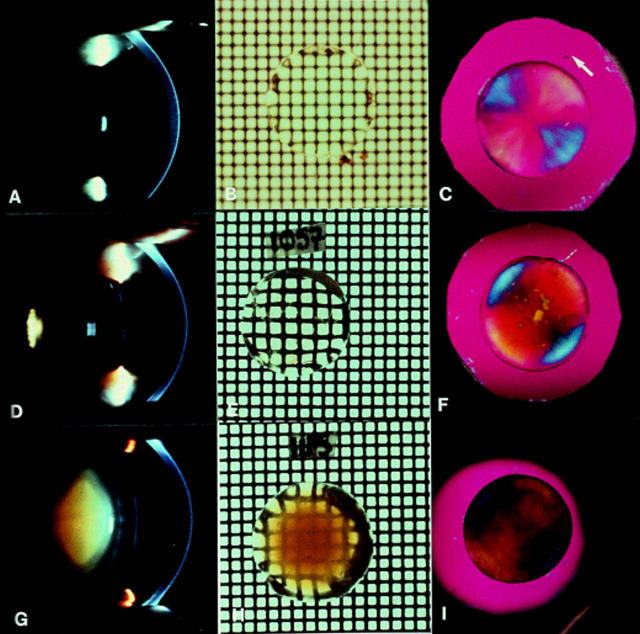
Images of normal human lenses (A, B, and C), posterior polar cataract (D, E, F), and pure nuclear cataract (G, H, I). Note that slit lamp camera images (A, D, G) all have a scattering reflect artefact (small white rectangle). The normal subject (A) was 40 years of age and the accompanying in vitro grid photographs (B) and polarising images (C) were obtained from a donor eye of similar age (42 years). The white arrow gives the direction of plane of polarisation of the major axis of the first order red plate.63 Note that (B) was photographed under fluid and so the grid is out of focus compared with (E) and (H), which were photographed in air. Also note that the polarising images of the cataractous lenses (F and I) maintain the blue/yellow radially symmetrical pattern of the normal lens (C) except where the opacities occur in highly localised polar cataract (F) and where the brunescence is strongest in the nuclear cataract (I). The images (D), (E), (G), (H) are taken from Marcantonio et al11 while the additional images are unpublished.

Brewster's model of the arrangement of fibre cells within the lens.15 He discovered that the fibre diameter varied throughout the lens and that the tips met in a series of suture lines on the optic axis. Hence, when viewed along the optic axis (as in Fig 1, C, F, and I), such an arrangement would give rise to positive birefringence such that the fibres lying along the direction of the first order red plate give a blue addition colour and those at right angles, a yellow subtraction colour.14 15 63
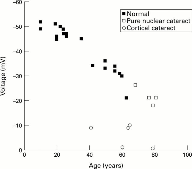
Human lens membrane voltage measured in eye bank and cataract lenses. Note that the pure nuclear cataracts follow the pattern of normal lenses, while the cortical cataracts have voltages considerably lower than normal lenses of a similar age. Data taken from Duncan and Hightower43 and Duncan et al.45

Relative cation permeability (PNa/PK) of human lenses as a function of age computed from measurements of ion concentration and electrical potential.45 The solid line shows the change with age of the mean optical density (OD) of the human lens measured at 490 nm.19
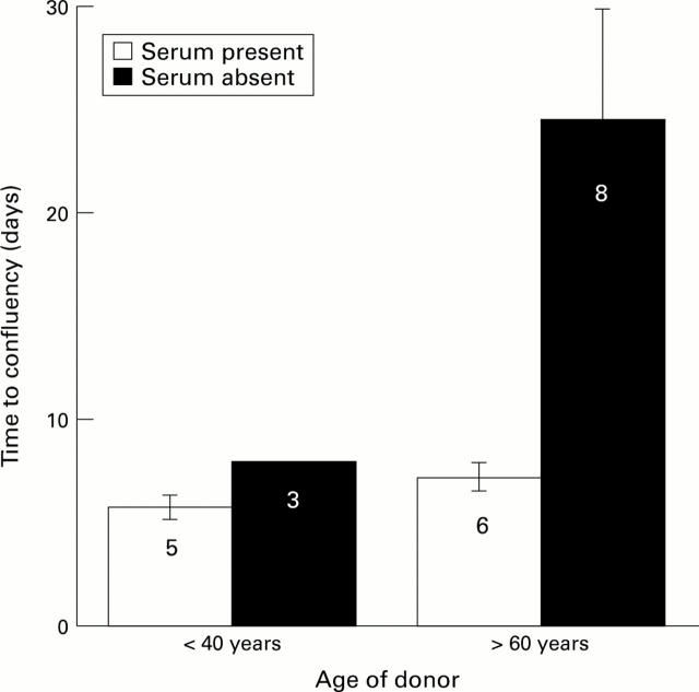
The effects of age and serum on time to confluence of cells on the human posterior capsule. Cell growth was observed using phase contrast microscopy. Confluency was taken at the point when 100% of the area on the posterior capsule, within the confines of the capsulorhexis, was covered by epithelial cells. Serum data were taken from Liu et al58 and serum free data from Wormstone et al.17 The numbers of different capsules used to obtain the data are given.
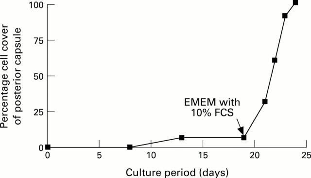
Cell cover on the posterior capsule from a 78-year-old donor lens as a function of time. The capsular bag was initially cultured in protein free medium where it ceased to grow after 12 days. The culture medium was then supplemented with serum after 19 days. The time to confluency (100%) was estimated as in Figure 5. Data taken from Wormstone et al.17
References
Publication types
MeSH terms
LinkOut - more resources
Full Text Sources
Medical