. 1998 Mar;9(3):623-35.
doi: 10.1091/mbc.9.3.623.

The Golgi and endoplasmic reticulum remain independent during mitosis in HeLa cells

Affiliations
Free PMC article

The Golgi and endoplasmic reticulum remain independent during mitosis in HeLa cells

S A Jesch et al. Mol Biol Cell. 1998 Mar.
Free PMC article

Abstract

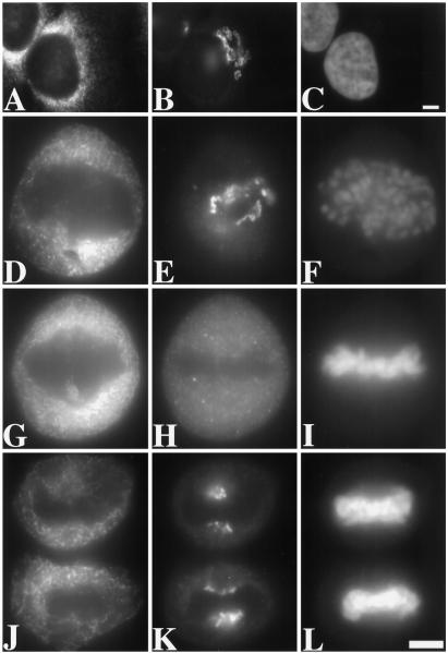
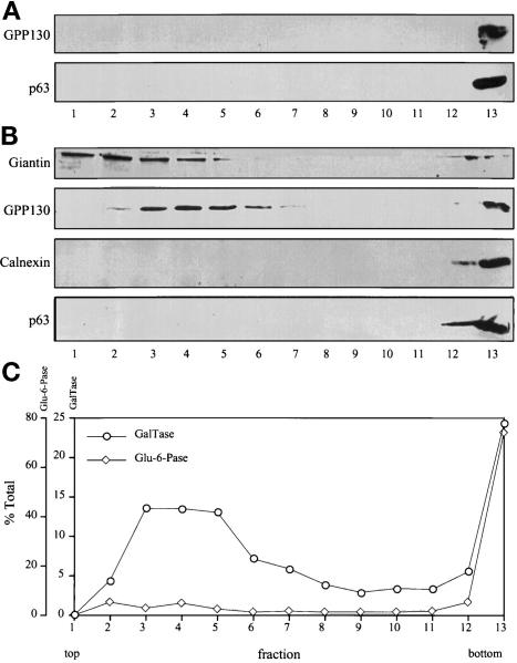
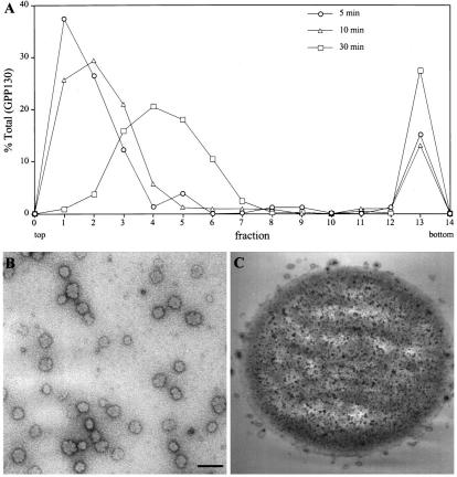
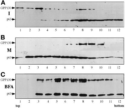
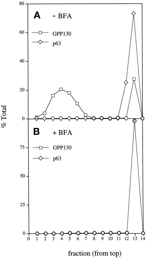
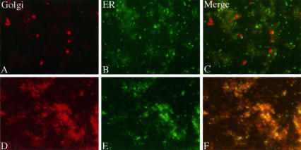
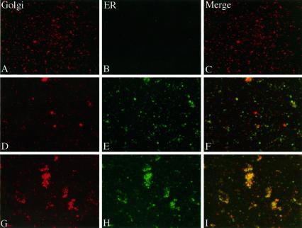
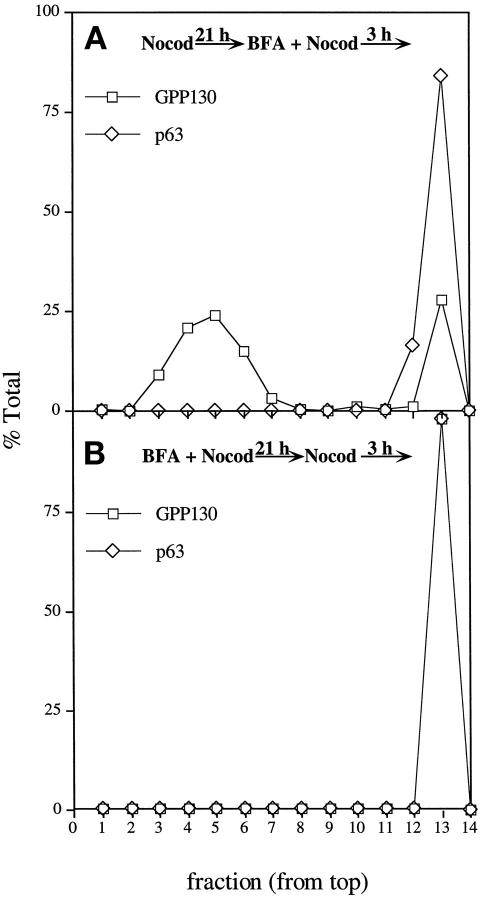
Partitioning of the mammalian Golgi apparatus during cell division involves disassembly at M-phase. Despite the importance of the disassembly/reassembly pathway in Golgi biogenesis, it remains unclear whether mitotic Golgi breakdown in vivo proceeds by direct vesiculation or involves fusion with the endoplasmic reticulum (ER). To test whether mitotic Golgi is fused with the ER, we compared the distribution of ER and Golgi proteins in interphase and mitotic HeLa cells by immunofluorescence microscopy, velocity gradient fractionation, and density gradient fractionation. While mitotic ER appeared to be a fine reticulum excluded from the region containing the spindle-pole body, mitotic Golgi appeared to be dispersed small vesicles that penetrated the area containing spindle microtubules. After cell disruption, M-phase Golgi was recovered in two size classes. The major breakdown product, accounting for at least 75% of the Golgi, was a population of 60-nm vesicles that were completely separated from the ER using velocity gradient separation. The minor breakdown product was a larger, more heterogenously sized, membrane population. Double-label fluorescence analysis of these membranes indicated that this portion of mitotic Golgi also lacked detectable ER marker proteins. Therefore we conclude that the ER and Golgi remain distinct at M-phase in HeLa cells. To test whether the 60-nm vesicles might form from the ER at M-phase as the result of a two-step vesiculation pathway involving ER-Golgi fusion followed by Golgi vesicle budding, mitotic cells were generated with fused ER and Golgi by brefeldin A treatment. Upon brefeldin A removal, Golgi vesicles did not emerge from the ER. In contrast, the Golgi readily reformed from similarly treated interphase cells. We conclude that Golgi-derived vesicles remain distinct from the ER in mitotic HeLa cells, and that mitotic cells lack the capacity of interphase cells for Golgi reemergence from the ER. These experiments suggest that mitotic Golgi breakdown proceeds by direct vesiculation independent of the ER.


Figures

Figure 1
Figure 1
Examination of triply stained interphase and mitotic HeLa cells by fluorescence microscopy. The staining pattern of the ER marker protein p63 (A, D, G, and J), the Golgi marker protein giantin (B, E, H, and K), and the DNA stain Hoechst 33258 (C, F, I, and L) is shown for cells at interphase (A–C), prometaphase (D–F), metaphase (G–I), and late anaphase (J–L). To allow direct comparison, each label was viewed at the same focal plane. Compared with the Golgi staining, the ER staining was significantly more reticular and was excluded from a larger area near the condensed chromosomes. The pictures shown are representative of hundreds of different cells examined at the different stages of division. Bars, 25 μm.
Figure 2
Figure 2
Distribution of ER and Golgi markers after velocity gradient sedimentation. (A) Interphase (nonsynchronized) HeLa cell postnuclear supernatants were fractionated on glycerol gradients and immunoblotted with antibodies against the Golgi marker protein GPP130 and the ER marker protein p63. Both Golgi and ER marker proteins were recovered on a sucrose cushion at the bottom of the gradient. (B) Mitotic HeLa cell postnuclear supernatants were fractionated and immunoblotted with antibodies against Golgi marker proteins (GPP130 and giantin) and ER marker proteins (calnexin and p63). The cells were blocked at mitosis with 0.5 μg/ml nocodazole and collected by shake-off. In contrast to interphase, a major portion of the mitotic Golgi was recovered in a slowly sedimenting position near the top of the gradient. These fractions lacked detectable ER. (C) Fractions resulting from separation of mitotic postnuclear supernatants were also assayed by enzymatic activity for the Golgi marker galactosyltransferase and the ER marker glucose-6-phophatase. By activity assay also, mitotic Golgi, but not mitotic ER, was recovered in a slowly sedimenting position. Centrifugation in these experiments was for 30 min at 150,000 × g; similar results were obtained under a variety of centrifugation conditions.
Figure 3
Figure 3
Analysis of the major mitotic Golgi breakdown product. (A) Mitotic postnuclear supernatants were fractionated on glycerol velocity gradients by centrifugation at 75,000 × g for 5 or 10 min or 150,000 × g for 30 min. The recovery of GPP130 in each fraction was determined by densitometry of immunoblots. The slowly sedimenting mitotic Golgi was recovered as a uniform peak of 115 S. (B) The 115 S mitotic Golgi peak was collected on a sucrose cushion and analyzed by electron microscopy after negative staining with uranyl acetate. A 60-nm diameter population of vesicles was present. To exclude potential artifacts, the vesicles were also treated with protease or detergent before analysis. Similar results were obtained for samples treated with protease, and no structures were visualized for samples treated with detergent. Bar, 150 nm. (C) Mitotic Golgi vesicles immunoisolated from 115 S vesicle peak fractions on anti-giantin–coated magnetic beads are shown after analysis by electron microscopy. No vesicles were observed when control magnetic beads were incubated with identical velocity gradient fractions.
Figure 4
Figure 4
Fractionation of interphase and mitotic ER and Golgi on flotation gradients. (A) The relative distribution of GPP130 and p63 flotation on a sucrose step gradient is shown for a postnuclear supernatant from a culture of nonsynchronized HeLa cells. (B) Identical analysis for cells blocked at mitosis with 0.5 μg/ml nocodazole and collected by shake-off. Note that the distribution of mitotic ER significantly differed from that of mitotic Golgi. (C) Identical analysis for cells in which brefeldin A was used to induce ER–Golgi fusion during progression to M-phase. Subconfluent cultures of HeLa cells were treated with 0.5 μg/ml nocodazole and 10 μg/ml brefeldin A for 24 h. Mitotic cells were obtained by shake-off, and the postnuclear supernatant was fractionated and analyzed as above. Unlike nontreated mitotic cells, cells that progressed to M-phase during brefeldin A treatment yielded equivalent ER and Golgi distributions.
Figure 5
Figure 5
Fractionation of mitotic ER and Golgi after treatment with brefeldin A during progression to M-phase. Subconfluent cultures of HeLa cells were treated with 0.5 μg/ml nocodazole in the absence (A) or presence (B) of 10 μg/ml brefeldin A for 24 h. Mitotic cells were obtained by shake-off, and the postnuclear supernatants were fractionated on glycerol velocity gradients as indicated above. The recovery, in each fraction, of the Golgi marker GPP130 and the ER marker p63 was determined by densitometry of immunoblots. Unlike nontreated mitotic cells, cells that progressed to M-phase during brefeldin A treatment lacked the 115 S major mitotic Golgi breakdown product. Instead, the mitotic Golgi was completely recovered at the bottom of the gradient together with the ER.
Figure 6
Figure 6
Examination of double-stained control and brefeldin A-treated cell extracts. Membrane vesicles present in a control HeLa cell homogenate were double-stained for the Golgi marker protein giantin (A), and the ER marker protein p63 (B). The Golgi and ER images are also shown after merging (C). Identical analysis of homogenates from brefeldin A-treated cells is also shown (D–F). Note that Golgi and ER staining patterns were coincident in the extracts from brefeldin A-treated, but not nontreated cells.
Figure 7
Figure 7
Examination of double-stained mitotic membrane fractions. Membrane vesicles present in the 115 S mitotic Golgi velocity gradient peak were double-stained for the Golgi marker protein giantin (A), and the ER marker protein p63 (B). The Golgi and ER images are also shown after merging (C). Identical analysis of membranes in the rapidly sedimenting mitotic Golgi fraction at the bottom of the velocity gradient is also shown (D–F). In addition, an identical analysis of membranes recovered at the bottom of the velocity gradient from cells treated with brefeldin A during progression to M-phase is shown (G–I). Note that Golgi and ER membrane-staining patterns were coincident in the rapidly sedimenting membrane fraction from brefeldin A-treated, but not nontreated, mitotic cells.
Figure 8
Figure 8
Fractionation of mitotic ER and Golgi after continuous brefeldin A treatment. (A) Subconfluent cultures of HeLa cells were incubated with 0.5 μg/ml nocodazole for 21 h followed by an additional incubation with 10 μg/ml brefeldin A and 0.5 μg/ml nocodazole for 3 h. Mitotic cells were collected by shake-off, and the postnuclear supernatant was fractionated on a 5–25% glycerol velocity gradient. The relative distribution of GPP130 and p63 was determined by immunoblotting. (B) Subconfluent cultures of HeLa cells were incubated with 0.5 μg/ml nocodazole and 10 μg/ml brefeldin A for 21 h followed by complete brefeldin A washout. Cultures were allowed to incubate with 0.5 μg/ml nocodazole for an additional 3 h and analyzed as above.

References

    1. Acharya U, McCaffery JM, Jacobs R, Malhotra V. Reconstitution of vesiculated Golgi membranes into stacks of cisternae: requirement of NSF in stack formation. J Cell Biol. 1995a;129:577–589. - PMC - PubMed
    1. Acharya U, Jacobs R, Peters J-M, Watson N, Farquhar MG, Malhotra V. The formation of Golgi stacks from vesiculated Golgi membranes requires two distinct fusion events. Cell. 1995b;82:895–904. - PubMed
    1. Aoki D, Appert HE, Johnson D, Wong SS, Fukuda MN. Analysis of the substrate binding sites of human galactosyltransferase by protein engineering. EMBO J. 1990;9:3171–3178. - PMC - PubMed
    1. Anhquyen L, Steiners JL, Ferrell GA, Shaker JC, Sifers RN. Association between calnexin and a secretion-incompetent variant of human alpha 1-antitrypsin. J Biol Chem. 1994;269:7514–7519. - PubMed
    1. Arosond NN, Touster O. Isolation of rat liver plasma membrane fragments in isotonic sucrose. Methods Enzymol. 1974;21:90–102. - PubMed

LinkOut - more resources