

Chlamydia trachomatis infection in the female reproductive tract of the rat: influence of progesterone on infectivity and immune response
- PMID: 9488372
- PMCID: PMC107992
- DOI: 10.1128/IAI.66.3.893-898.1998
Chlamydia trachomatis infection in the female reproductive tract of the rat: influence of progesterone on infectivity and immune response
Abstract
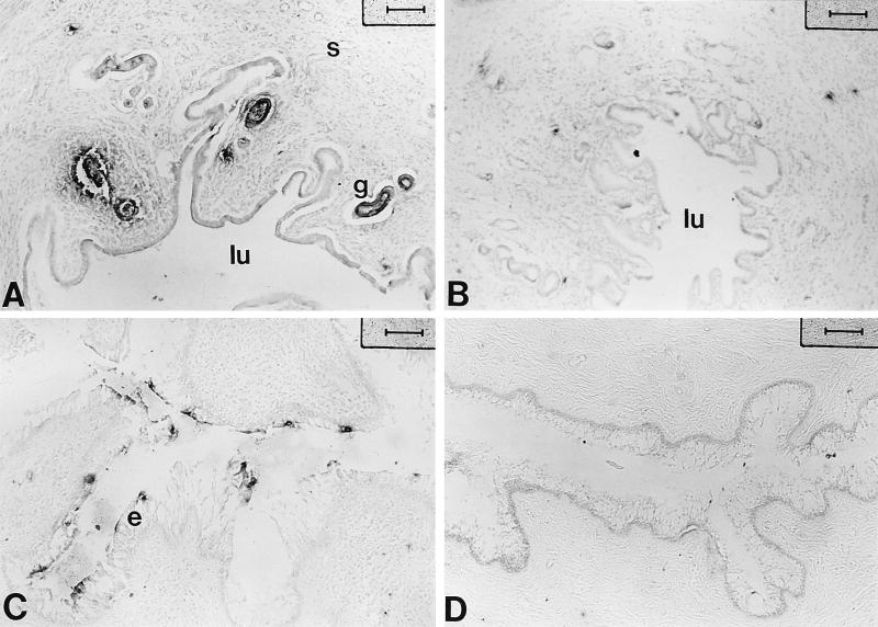
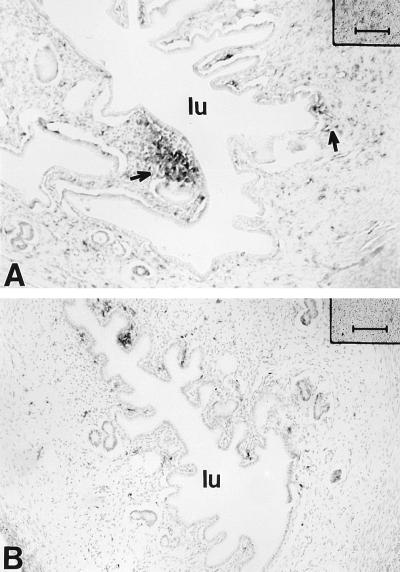
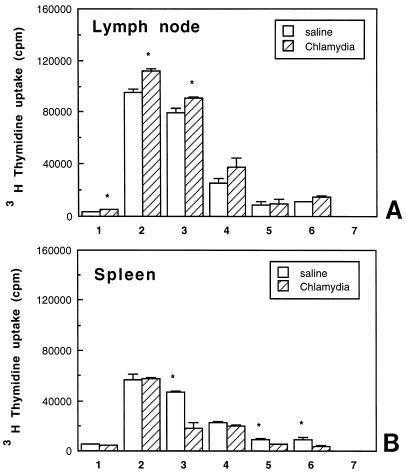
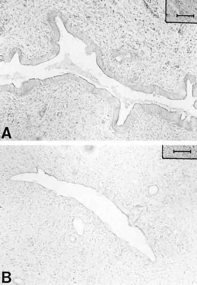
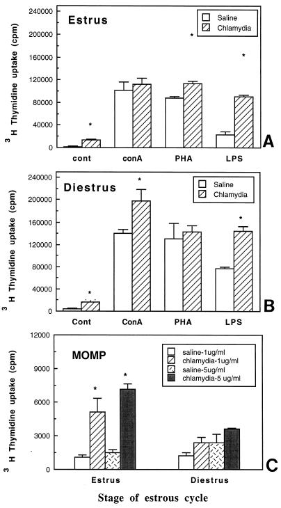
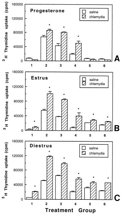
As the most common cause of sexually transmitted disease in women, chlamydial infections can lead to pelvic inflammatory disease, infertility, and ectopic pregnancy. To better understand the role played by sex hormones in modulating the immune response of the genital tract to microbial infections, we have developed a rat model to study Chlamydia trachomatis infection. Inbred female Lewis rats were primed with progesterone and inoculated by intrauterine instillation of C. trachomatis (mouse pneumonitis strain MoPn) into each uterine horn. When infected animals were examined for the presence of chlamydial antigens 14 days postinfection, both the uterus and vagina were found to be positive compared to those of saline-treated animals, which did not show specific staining. The involvement of local and systemic immune systems following chlamydial infection was determined by analyzing major histocompatibility complex (MHC) class II expression in the reproductive tract and lymphocyte proliferation in response to mitogenic and chlamydia-specific stimulation of cells from the spleen and lymph nodes (LN) draining the reproductive tract. Enhanced proliferation was observed in LN following mitogenic but not antigenic (MOMP [major outer membrane protein]) stimulation. In contrast, spleen cell proliferation was lower in chlamydia-infected rats than in saline-treated controls. MHC class II expression, an indicator of inflammatory responses, was upregulated in the uterus, on glandular epithelial cells, and adjacent to glands in response to chlamydial infection. In other experiments, when rats were infected at estrus and diestrus without prior progesterone priming, chlamydial inclusions were not detected in either the uterus or vagina. However, enhanced lymphocyte proliferation was observed in response to mitogenic and MOMP stimulation in the reproductive tract-draining LN from estrous and diestrous animals. These findings indicate that under appropriate endocrine conditions, the rat uterus is susceptible to C. trachomatis infection and that immune responses to this pathogen can be detected locally and systemically. Further, they suggest that clearance of the infection from the reproductive tract involves immune cells from the LN draining the reproductive tract.
Figures
References
-
- Athanassakis I, Grigoriou M, Galanopoulos V, Papamatheakis J. Induction of class II MHC antigens expression on murine placenta by 5-AzaC correlates with fetal abortion. Cell Immunol. 1990;128:438–449. - PubMed
-
- Baker D A, Plotkin S A. Enhancement of vaginal infection in mice by herpes simplex virus type Ii with progesterone. Proc Soc Exp Biol Med. 1978;158:131–134. - PubMed
-
- Crainie M, Semeluk A, Lee K C, Wegmann T G. Regulation of constitutive and lymphokine-induced Ia expression by murine alpha-fetoprotein. Cell Immunol. 1989;118:41–52. - PubMed
Publication types
MeSH terms
Substances
Grants and funding
LinkOut - more resources
Full Text Sources
Medical
Research Materials
